Abstract
Internal radiocarpal distraction plating is a versatile tool in the treatment of distal radius fractures that are not amenable to nonoperative treatment or operative fixation with standard volar or dorsal implants. Internal distraction plates may also be indicated in the setting of polytrauma or osteopenic bone. The plate functions as an internal fixator, using ligamentotaxis to restore length and alignment while providing relative stability for bony healing. The plate can be fixed to either the second or the third metacarpal, and anatomic and biomechanical studies have assessed the strengths and weaknesses of each strategy. This operative fixation technique leads to acceptable radiographic results and functional outcomes. Following fracture union, the plate is removed, and wrist range of motion is resumed.
Keywords: distal radius fracture, bridge plate, internal radiocarpal distraction plate, osteoporosis, spanning distraction plate
Introduction
Distal radius fractures are common orthopedic injuries resulting in significant morbidity and functional limitation.10,18 These injuries are frequently sustained by young patients after high-energy trauma and elderly patients with osteoporotic bone after low-energy falls.10 Simple extra-articular fractures may be amenable to cast immobilization, closed reduction and percutaneous fixation, or standard open reduction and internal fixation with volar or dorsal plating. However, displaced, dorsally comminuted distal radius fractures can be more difficult to treat and often require operative fixation due to the tendency of these fracture patterns to collapse, resulting in shortening and malalignment of the radiocarpal joint.
Highly comminuted, unstable distal radius fractures can be managed with external fixation, which allows for indirect reduction through ligamentotaxis and prevention of shortening due to spanning radiocarpal fixation.5,8,11,21 Several studies demonstrated that external fixation of the distal radius can result in adequate radiographic outcomes.7,27,36 However, external fixation has been reported to have a 23% to 62% complication rate, most commonly due to pin-site infection, malreduction, and patient discomfort.20,35,38,39 In addition, patients with highly comminuted fracture patterns may require prolonged periods of external fixation, further increasing the risk for pin-site infection or loosening.39
The use of internal radiocarpal distraction plating, or bridge plating, can serve as a viable alternative to external fixation for highly comminuted distal radius fractures at increased risk for loss of reduction, or in polytrauma patients whereby the spanning internal distraction plate may allow for early mobilization and forearm weight-bearing. Similar to external fixation, an internal radiocarpal distraction plate offers relative stability and an indirect reduction of the distal radius fracture, but with the convenience of internalized hardware eliminating the risk for pin-site infection. Furthermore, the internal distraction plate may also be better tolerated by the patient compared with external fixation.38
Development
In 1998, Burke and Singer described a technique for treating comminuted distal radius fractures using a dorsal internal radiocarpal distraction plate serving as an internal fixator.4 In this original technique, a dorsal incision was made over the radiocarpal joint and the fracture fragments were stabilized with Kirschner wires (K-wires) using supplemental bone graft as needed. Once alignment was obtained, a distraction force was applied to the fracture and a plate was applied as a neutralization construct, affixed to the third metacarpal and radial shaft.4 In the same year, Becton et al reported an alternative technique using antegrade placement of the plate onto the second metacarpal.2
Subsequent studies described modifications to these techniques. Two- and three-incision techniques over the second or third metacarpal, radial shaft, and optionally over the dorsal distal radius to mobilize extensor tendons and directly reduce the articular surface have been described in the literature.13,31 The plate may be applied in a retrograde or antegrade fashion below the extensor tendons of the dorsal forearm and hand.12,14 K-wires or interfragmentary screws may be used to augment the construct to stabilize articular fragments if necessary.13,31
Burke and Singer originally used a 3.5 mm steel dynamic compression plate or single- or double-stacked one-third tubular plates as neutralization constructs.4 Subsequently, the use of 2.4 mm and 3.5 mm reconstruction plates and 2.4 mm dynamic compression plates was reported.14,31,34 Other surgeons used a wrist arthrodesis plate as a bridging construct allowing for placement of smaller diameter screws in the metacarpal, with larger screws in the radial shaft.30
Most recently, internal radiocarpal distraction plates specific to the distal radius have been designed—these plates feature contoured ends to facilitate passage below extensor tendons, optional threaded holes for locked screw placement, and fewer central holes to increase stiffness.13,15,23 Similar to wrist arthrodesis plates, modern internal radiocarpal distraction plates have smaller holes for metacarpal fixation and a straight design. The increased length of distal radius bridge plates relative to arthrodesis plates provides increased stability for fractures with proximal extension. These plates are produced by multiple manufacturers and are available in 2.4, 2.7, 3.2, and 3.5 mm sizes.12,23
Indications
The use of an internal radiocarpal distraction plate was initially described for intra-articular, comminuted, and displaced (>2 mm) distal radius fractures (AO type C3)4,13 where the highly comminuted articular fragments are too small to be captured with a volar plate or fragment-specific fixation, and relative stability is likely required.13 Similar to external fixation, internal radiocarpal distraction plate fixation provides relative stability to highly comminuted fractures, but reduces the complications associated with external fixation. In addition, indirect reduction and a bridging construct preserve the periosteal blood supply and soft tissue and distribute strain, allowing for greater likelihood of fracture consolidation.
Since the introduction of internal radiocarpal distraction plating, the indications for its use have increased. In subsequent reports, internal distraction plating has been used for the treatment of distal radius fractures with metaphyseal-diaphyseal extension.13,15,37 Due to the proximal extension of these injuries, this fracture pattern requires a longer working length which decreases the rigidity provided by a stabilization construct such as external fixation.3 The extended duration of treatment required for these fractures with diaphyseal extension increases risks of pin-site complications if an external fixator is used.13,24
Internal radiocarpal distraction plating may also be indicated in polytrauma patients with distal radius fractures that could otherwise be treated with cast immobilization or standard volar or dorsal plating. This technique allows immediate weight-bearing through the forearm, thereby facilitating mobilization and rehabilitation of the polytrauma patient. For multiply injured patients, increased mobility can decrease medical complications such as pressure ulcers and thromboembolism, in addition to hospital costs, length of stay, and nonhome discharge.16,26,33 The plate can remain in place until the distal radius fracture and other injuries have healed.3,14
In addition, internal radiocarpal distraction plates can be effective in elderly patients with osteoporotic bone where highly comminuted fractures are notoriously difficult to treat. Low bone mineral density limits screw purchase with standard volar and dorsal plates, increasing the risk for subchondral collapse and loss of reduction.29 Internal radiocarpal distraction plates can avoid these challenges by bypassing the fracture site and anchoring the construct in stronger diaphyseal bone, while relying on ligamentotaxis to maintain fracture length and alignment.34 In addition, modern plates allow for the placement of locking screws, which decreases the risk of screw pullout.14
Lastly, internal radiocarpal distraction plates can be used as part of a combined fixation strategy with volar plates, fragment-specific plates, K-wires, and interfragmentary screws as an augment to decrease the likelihood of fracture collapse due to comminution.4,31 Apart from distal radius fractures, an internal radiocarpal distraction plate can be used as a neutralization plate for other injuries, such as radiocarpal dislocations, articular shear injuries, or as a salvage option for distal radius nonunions.3,28,32
Despite its multiple uses, internal radiocarpal distraction plating is contraindicated for distal radius fractures that are irreducible by ligamentotaxis, fractures with severe dorsal soft tissue compromise which would result in exposed hardware, associated ipsilateral fractures of the radial shaft or second/third metacarpals, severe preexisting arthritis which may benefit from primary arthrodesis, and patients who are unlikely to return for follow-up and plate removal.12,23,28,31
Biomechanical Studies
Several biomechanical studies have been performed to assess the stability of internal radiocarpal distraction plate constructs. A mechanical study by Chhabra et al used an acrylic rod model with one rod serving as the radius, and another as a metacarpal. The rods were separated by a 2-cm gap simulating an unstable distal radius fracture. The gap was spanned by either a 3.5 mm 10-hole pelvic reconstruction plate or a limited contact wrist fusion plate serving as an internal radiocarpal distraction plate or an external fixator.6 Internal radiocarpal distraction plate fixation had increased mean stiffness values in compression, tension, and lateral bending compared with external fixation.6 The mean stiffness values in compression and tension for the internal radiocarpal distraction plate were almost 10-fold greater than the stiffness of the external fixator system; however, this model is limited by the use of acrylic rods and the lack of an anatomically contoured internal radiocarpal distraction plate.6
A subsequent biomechanical study by Wolf et al compared a 2.4 mm locked internal radiocarpal distraction plate with an external fixator in cadaveric specimens.40 The authors loaded the constructs through the flexor and extensor tendons to measure fracture displacement, and found that an internal radiocarpal distraction plate is more stable than external fixation in both flexion and extension.40 In addition, this study showed that 3 screws proximal and 3 screws distal to the fracture are sufficient for stability.40 However, this model only allowed for motion in one plane, which may not accurately represent physiologic forces.
Huang et al performed a study on cadaveric specimens to compare volar locking plates with internal radiocarpal distraction plates in an axial compression model simulating crutch weight-bearing.17 Compared with volar locking plate fixation, a 2.4 mm internal radiocarpal distraction plate prevented collapse at the radiocarpal joint when subjected to axial force, but failed with wrist flexion resulting in plate bending.17
Alluri et al compared internal radiocarpal distraction plate fixation with the third metacarpal with fixation to the second metacarpal in cadaveric specimens.1 A 2.7 mm internal radiocarpal distraction plate was affixed to either the second or third metacarpal, and stiffness, displacement, and load to failure were measured. Distal fixation to the third metacarpal resulted in greater stiffness in flexion, with equivalent stiffness in extension and load to failure.1 These studies suggest that internal radiocarpal distraction plate fixation is biomechanically superior to external fixation in the treatment of comminuted distal radius fractures, and stiffness may be optimized by fixation to the third metacarpal.
Anatomic Studies
There is no consensus on whether fixation to the second or third metacarpal is optimal. With distal fixation to the second metacarpal, the plate is passed under the second extensor compartment.2 Alternatively, fixation to the third metacarpal requires plate placement under the fourth extensor compartment.14 Usually, the fracture site is not exposed during internal radiocarpal distraction plate fixation, leading to concern about damage to the structures dorsal to the radius. Lewis et al showed that plate fixation to the third metacarpal resulted in no entrapped nerves, but entrapped tendons of the first and third extensor compartments in 100% of cadaveric specimens, while fixation to the second metacarpal did not result in any entrapment.25
In a similar cadaveric study, Dahl et al showed that superficial branches of the radial nerve contacted the plate in all specimens with fixation to the second metacarpal, but none of the specimens with third metacarpal fixation.9 However, they noted an average of 10 cm of contact between the plate and the extensor digitorum communis (EDC) tendons and one case of tendon entrapment in the third metacarpal group, compared with no entrapment or EDC contact with the plate in the second metacarpal group.9 Both groups had contact between the extensor pollicis longus tendon and the plate, but this was significantly less with second metacarpal fixation.9
Clinical Studies
Internal radiocarpal distraction plate fixation has demonstrated success in treating comminuted intra-articular fractures of the distal radius. Several clinical studies using this technique have been described; however, these studies are primarily retrospective reviews and do not compare internal radiocarpal distraction plating to other modes of fixation. In addition, follow-up and reported outcomes are inconsistent between studies (Table 1).
Table 1.
Outcomes of Published Studies Reporting Internal Radiocarpal Distraction Plate Fixation of Distal Radius Fractures.
| Author | Number of patients | Technique | Mean flexion and extension | Mean pronation and supination | Mean palmar tilt | Mean radial inclination | Mean ulnar variance | Mean DASH (final) | Grip strength | Complications |
|---|---|---|---|---|---|---|---|---|---|---|
| Hanel et al14 | 62 (range of motion values for 1 patient case report) | second metacarpal, 2 incisions | 45°/35° | 70°/60° | All “at least neutral” | All “greater than 5” | All “within 5 mm of ulnar neutral” | Not specified | Not specified | 1 ECRL rupture, 1 hardware failure (after union), 3 patients unable to return to prior level of work due to wrist |
| Dodds et al12 | 25 | second metacarpal, 2 incisions, 6.6-month follow-up | 46°/42° | 76°/69° | 4.2° | 18.9° | 0.18 mm | Not specified | Not specified | 3 hardware failures (after union), 19 underwent tenolysis |
| Lauder et al22 | 18 | second metacarpal, 2 incisions, 32.4-month follow-up | 43°/46° | 66°/71° | Not specified | Not specified | Not specified | 16 | 86% of contralateral | 2 had surgical site pain |
| Burke and Singer4 | 1 | third metacarpal, 1 incision, 4-year follow-up | 45°/65° | 80°/75° | Not specified | Not specified | Not specified | Not specified | Not specified | None |
| Ruch et al37 | 22 | third metacarpal, 3 incisions, 24.8-month follow-up | 57°/65° | 77°/76° | 4.6° | Not specified | 0 mm | 11.5 | 69% of contralateral | 3 surgical site infections, 3 middle finger extensor lag, 2 did not have “excellent” or “good” Gartland-Werley results |
| Jain and Mavani19 | 20 | third metacarpal, three incisions, 24-month follow-up | 46°/50° | 79°/77° | 7° | 18° | 0.5 mm | 32 | 84% of contralateral | 2 delayed union, 3 wrist and finger stiffness, 1 CRPS, 2 middle finger extensor tendon lag, 3 did not have “excellent” or “good” Gartland-Werley results |
| Hanel et al15 | 134 | second or third metacarpal, two incisions, 9.9-month follow-up | Not specified | Not specified | Not specified | Not specified | Not specified | Not specified | Not specified | 2 minor wound healing complications, 5 hardware failures (after union), 2 malunion, 2 nonunion, 1 wound complication, 2 deep infection, 2 extensor tendon adhesions (required tenolysis), 1 EPL rupture |
| Richard et al34 | 33 | second metacarpal in 12 patients, third metacarpal in 21 patients, three incisions, 11.8-month follow-up | 46°/50° | 79°/77° | 5° | 20° | 0.6 mm | 32 | Not specified | 10 with digital stiffness (1 required tenolysis), 1 transient superficial radial neuritis, 1 CRPS, 1 infection/wound healing complications requiring skin graft |
Note. DASH = disabilities of the arm, shoulder, and hand; CRPS = complex regional pain syndrome; EPL = extensor pollicis longus; ECRL = extensor carpi radialis longus.
In the three studies describing fixation to the second metacarpal, 105 patients were included. There were no reports of nonunion or nerve injuries, but 1 ECRL rupture, 4 hardware failures, 19 patients requiring tenolysis, and 2 cases of wrist pain were reported.12,15,22 The studies by Hanel et al and Lauder et al reporting range of motion demonstrated that mean flexion, extension, pronation, and supination returned to functional levels.12,22 Dodds et al reported a mean palmar tilt of 4.2° and a mean ulnar variance of 0.18 mm, while Hanel et al stated that all patients had a palmar tilt of “at least neutral” and variance “within 5 mm of ulnar neutral.”12,14 Only Lauder et al reported grip strength and mean final Disabilities of the Arm, Shoulder, and Hand (DASH) scores. Grip strength returned to 86% of contralateral in the entire cohort, and 95% of contralateral in the patients with dominant-sided injury.22 Mean DASH score at 32.4-month follow-up was 16.22
Three studies consisting of 43 patients describe fixation to the third metacarpal.4,19,37 These patients had 2 delayed unions, 8 with stiffness or extensor tendon lags, and 1 patient with complex regional pain syndrome (CRPS).4,19,37 Mean range of motion was higher in these patients than the reported averages for second metacarpal fixation, but the results are from different studies with inconsistent statistical analysis, and thus cannot be directly compared. Mean palmar tilt was 4.6° and mean ulnar variance was 0 mm in the study by Ruch et al, while these values were 7° and 0.5 mm, respectively, in the report by Jain and Mavani.19,37 Ruch et al reported grip strength of 69% of the contralateral side, while Jain and Mavani found 84% of contralateral grip strength, though neither study stratified these results by hand dominance.19,37 Ruch et al reported a mean DASH score of 11.5 at 24.8 months, while Jain et al found a mean DASH score of 32 at 24-month follow-up.19,37
Despite the limitations of these studies, most notably lack of comparison with other methods of fixation, these reports show promising results after internal radiocarpal distraction plating for distal radius fractures. Although internal distraction plating of distal radius fractures has been used for over 2 decades, further prospective studies are needed to better define the expected outcomes of this technique and further elucidate potential limitations and complications.
Limitations
Although internal radiocarpal distraction plates are versatile, and several studies have demonstrated successful outcomes in the treatment of distal radius fractures, there are limitations associated with their use. Unlike locking volar plates or fragment-specific implants which may be retained indefinitely, or external fixators which may be removed in office, radiocarpal distraction plates require a subsequent surgery for hardware removal.14
In addition to the aforementioned limitations, there are notable complications. The most common complications reported in the literature are digital stiffness requiring tenolysis and hardware failure after union (Table 1). Infection and nonunion have also been reported. Due to the subtendinous placement of these plates, often without direct visualization, there is risk of tendon entrapment, rupture, or adhesions.15 The distraction required to achieve ligamentotaxis may result in loss of digital motion and development of CRPS, and should be limited to less than 5 mm.13 Although there are no reported cases of metacarpal fracture during internal radiocarpal distraction plate fixation, this is theoretically possible given the width of the screws compared with the metacarpal shaft. This may be mitigated by using smaller diameter screws for distal fixation. In addition, while not reported in the literature, the stiffness of these constructs when relative stability is required may predispose fractures to nonunion, particularly when locked screws are used. Complications from overly stiff constructs may become apparent as internal radiocarpal distraction plating increases in popularity.
Recommendations
Internal radiocarpal distraction plating for the treatment of distal radius fractures is a useful technique, but not suited for every distal radius fracture. Fractures that can be adequately treated nonoperatively or operatively with standard volar or dorsal plating are a contraindication to internal radiocarpal distraction plate application.13 When dorsal soft tissue is compromised, internal radiocarpal distraction plating risks hardware exposure and should be avoided13; in these cases, external fixation is preferable. For depressed articular fragments that are irreducible with ligamentotaxis, a separate incision for direct reduction and stabilization with a volar plate and/or screws may be necessary to avoid articular step off and resultant radiocarpal arthrosis.13
Authors’ Preferred Technique
The procedure is performed under tourniquet with the patient lying supine and the affected extremity on a radiolucent arm board. In severely comminuted fractures where articular alignment needs to be restored and cannot be achieved with ligamentotaxis alone, reduction is performed percutaneously or with a formal open reduction. Stabilization is achieved with K-wires, screws, and plates as needed prior to plating.
A two-incision technique is used with one 4-cm incision over the dorsal surface of the second or third metacarpal, and a second 5-cm incision over the dorsal radial shaft, approximately 4 cm proximal to the fracture. The second metacarpal is preferentially used based on the results from Lewis et al, unless the fracture is better aligned through placement of the plate at the third metacarpal.25 The internal radiocarpal distraction plate is placed in a retrograde fashion deep to the extensor tendons, and position is checked fluoroscopically. Using nonlocking cortical screws, the plate is affixed to the metacarpal distally and longitudinal traction is applied. Fracture reduction is assessed using fluoroscopy, followed by proximal fixation to the radius with nonlocking cortical screws. If locking screws are available, they can be used to increase construct stability, especially in osteoporotic bone. The distal radioulnar joint (DRUJ) is assessed and is stabilized with K-wires if indicated. After surgery, a small volar slab splint is placed until suture removal, and patients are instructed to perform digital range of motion exercises for the entire time the plate is in place to avoid tendon adhesions and stiffness. Patients may bear weight through the forearm with a platform walker or crutches in the case of polytrauma, but are restricted to a 5-pound lifting limit. The plate is removed after radiographic union, generally within 3 to 6 months.
Case 1
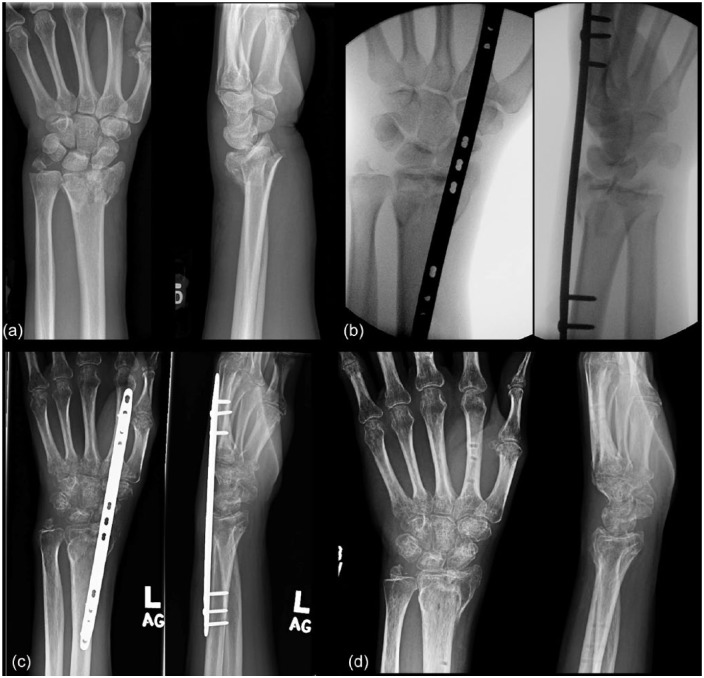
A 55-year-old right-hand-dominant male was involved in a bicycle accident and suffered a ground-level fall, resulting in an isolated dorsally angulated and displaced intra-articular distal radius fracture (Figure 1a). Given the intra-articular comminution, the decision was made to perform internal radiocarpal distraction plating. The plate was applied with fixation to the second metacarpal and radial shaft using one nonlocking and two locking 2.7 mm screws. Intraoperative imaging demonstrated restoration of length and alignment of the distal radius fracture (Figure 1b). Radiographic union occurred by 13 weeks after surgery (Figure 1c). At 14 weeks after surgery, the plate was removed uneventfully (Figure 1d). At final follow-up 6 months after the index surgery, the patient regained full functional range of motion, and returned to all prior activities. The patient subsequently did not return to clinic for follow-up.
Figure 1.
(a) Injury radiographs showing angulated, displaced intra-articular distal radius fracture. (b) Intraoperative fluoroscopy during internal radiocarpal distraction plate fixation. (c) Healed distal radius fracture. (d) Final postoperative radiograph after internal radiocarpal distraction plate removal (case 1).
Case 2
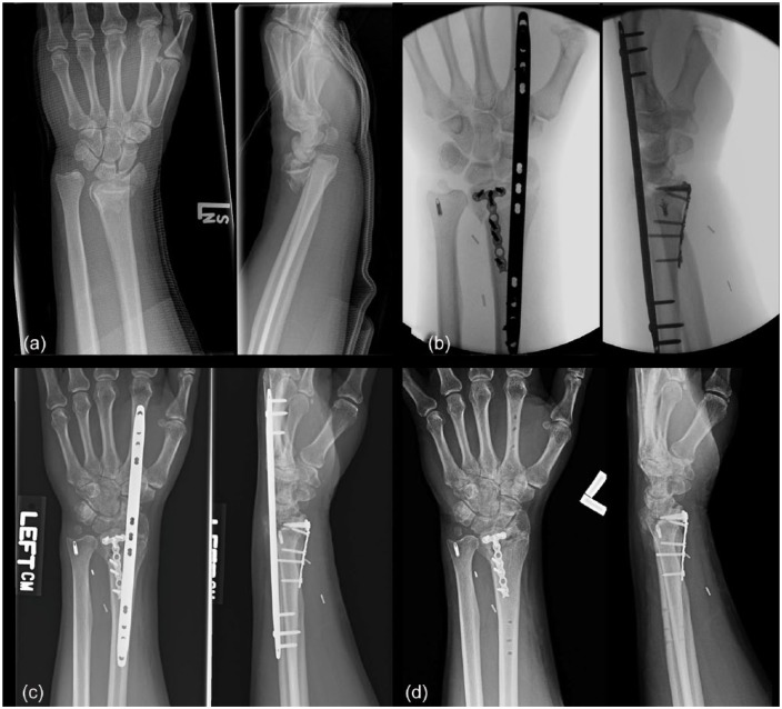
A 46-year-old right-hand-dominant male was involved in a motorcycle collision, which resulted in a left distal radius fracture with intra-articular involvement, comminution, and severe dorsal displacement (Figure 2a). Three days after the initial injury, an open reduction with internal radiocarpal distraction plating, volar fragment-specific plating, along with DRUJ repair was performed. An internal radiocarpal distraction plate was applied, with fixation to the second metacarpal and radial shaft with one nonlocking and two locking 2.7 mm screws on each side of the fracture. The lunate facet fragment remained malreduced; therefore, open reduction via a volar incision was performed, and a volar fragment-specific plate was applied. Last, the DRUJ was noted to be injured, and an open triangular fibrocartilage complex repair was performed using suture anchors. Intraoperative imaging demonstrated restored length and alignment of the distal radius fracture (Figure 2b). At 15 weeks, radiographic union was observed (Figure 2c). The patient complained of numbness and tingling in his left index and middle fingers that began about 2 months after surgery, with electromyography/nerve conduction testing showing mild injury to the median nerve. At 19 weeks, the internal radiocarpal distraction plate was removed, and a carpal tunnel release was performed (Figure 2d). At 6 months postoperatively, the patient had improving strength and range of motion and was able to perform all activities of daily living, but was subsequently lost to follow-up. At final follow-up, range of motion was 20° of flexion, 40° of extension, 65° of supination, and 60° of pronation.
Figure 2.
(a) Injury radiographs showing comminuted displaced intra-articular distal radius fracture. (b) Intraoperative fluoroscopy during internal radiocarpal distraction plate fixation, with additional volar fragment-specific fixation and DRUJ repair. (c) Healed distal radius fracture. (d) Final postoperative radiograph after internal radiocarpal distraction plate removal (case 2).
Conclusions
Internal radiocarpal distraction plates are a valuable tool in treating comminuted intra-articular distal radius fractures that are not amenable to nonoperative management or standard volar or dorsal plating. Through ligamentotaxis, this technique can restore length and alignment in fractures at high risk for collapse and loss of reduction or in polytrauma patients who may benefit from early mobilization through forearm weight-bearing. Initial biomechanical and clinical studies are promising, but further studies are needed to better understand the mechanical properties of internal distraction plating with various fracture patterns and fixation techniques. In addition, prospective clinical studies are still needed to further assess long-term outcomes and complications.
Footnotes
Ethical Approval: This study is a review article and did not require a review by our institutional review board.
Statement of Human and Animal Rights: The article does not contain research on human or animal subjects. The patients received standard care, and clinical information was used retrospectively.
Statement of Informed Consent: This article does not contain any identifying patient information. Informed consent was obtained to use clinical information for educational or research purposes.
Declaration of Conflicting Interests: The author(s) declared no potential conflicts of interest with respect to the research, authorship, and/or publication of this article.
Funding: The author(s) received no financial support for the research, authorship, and/or publication of this article.
ORCID iD: V Vakhshori  https://orcid.org/0000-0002-8509-7619
References
- 1. Alluri RK, Bougioukli S, Stevanovic M, et al. A biomechanical comparison of distal fixation for bridge plating in a distal radius fracture model. J Hand Surg Am. 2017;42:748.e1-748.e8. doi: 10.1016/j.jhsa.2017.05.010. [DOI] [PubMed] [Google Scholar]
- 2. Becton JL, Colborn GL, Goodrich JA. Use of an internal fixator device to treat comminuted fractures of the distal radius: report of a technique. Am J Orthop (Belle Mead NJ). 1998;27(9):619-623. [PubMed] [Google Scholar]
- 3. Brogan DM, Richard MJ, Ruch D, et al. Management of severely comminuted distal radius fractures. J Hand Surg. 2015;40(9):1905-1914. doi: 10.1016/j.jhsa.2015.03.014. [DOI] [PubMed] [Google Scholar]
- 4. Burke EF, Singer RM. Treatment of comminuted distal radius with the use of an internal distraction plate. Tech Hand Up Extrem Surg. 1998;2(4):248-252. [DOI] [PubMed] [Google Scholar]
- 5. Capo JT, Rossy W, Henry P, et al. External fixation of distal radius fractures: effect of distraction and duration. J Hand Surg Am. 2009;34(9):1605-1611. doi: 10.1016/j.jhsa.2009.07.010. [DOI] [PubMed] [Google Scholar]
- 6. Chhabra A, Hale JE, Milbrandt TA, et al. Biomechanical efficacy of an internal fixator for treatment of distal radius fractures. Clin Orthop. 2001;393:318-325. [DOI] [PubMed] [Google Scholar]
- 7. Chilakamary VK, Lakkireddy M, Koppolu KK, et al. Osteosynthesis in distal radius fractures with conventional bridging external fixator; tips and tricks for getting them right. J Clin Diagn Res. 2016;10(1):RC05- RC08. doi: 10.7860/JCDR/2016/16696.7048. [DOI] [PMC free article] [PubMed] [Google Scholar]
- 8. Cooney WP. External fixation of distal radial fractures. Clin Orthop. 1983;180:44-49. [PubMed] [Google Scholar]
- 9. Dahl J, Lee DJ, Elfar JC. Anatomic relationships in distal radius bridge plating: a cadaveric study. Hand (N Y). 2015;10(4):657-662. doi: 10.1007/s11552-015-9762-y. [DOI] [PMC free article] [PubMed] [Google Scholar]
- 10. Diaz-Garcia RJ, Oda T, Shauver MJ, et al. A systematic review of outcomes and complications of treating unstable distal radius fractures in the elderly. J Hand Surg Am. 2011;36(5):824-835.e2. doi: 10.1016/j.jhsa.2011.02.005. [DOI] [PMC free article] [PubMed] [Google Scholar]
- 11. Dicpinigaitis P, Wolinsky P, Hiebert R, et al. Can external fixation maintain reduction after distal radius fractures? J Trauma. 2004;57(4):845-850. [DOI] [PubMed] [Google Scholar]
- 12. Dodds SD, Save AV, Yacob A. Dorsal spanning plate fixation for distal radius fractures. Tech Hand Up Extrem Surg. 2013;17(4):192-198. doi: 10.1097/BTH.0b013e3182a5cbf8. [DOI] [PubMed] [Google Scholar]
- 13. Ginn TA, Ruch DS, Yang CC, et al. Use of a distraction plate for distal radial fractures with metaphyseal and diaphyseal comminution. Surgical technique. J Bone Joint Surg Am. 2006;88(suppl 1) (pt 1):29-36. doi: 10.2106/JBJS.E.01094. [DOI] [PubMed] [Google Scholar]
- 14. Hanel DP, Lu TS, Weil WM. Bridge plating of distal radius fractures: the Harborview method. Clin Orthop Relat Res. 2006;445:91-99. doi: 10.1097/01.blo.0000205885.58458.f9. [DOI] [PubMed] [Google Scholar]
- 15. Hanel DP, Ruhlman SD, Katolik LI, et al. Complications associated with distraction plate fixation of wrist fractures. Hand Clin. 2010;26(2):237-243. doi: 10.1016/j.hcl.2010.01.001. [DOI] [PubMed] [Google Scholar]
- 16. Hemmila MR, Jakubus JL, Maggio PM, et al. Real money: complications and hospital costs in trauma patients. Surgery. 2008;144(2):307-316. doi: 10.1016/j.surg.2008.05.003. [DOI] [PMC free article] [PubMed] [Google Scholar]
- 17. Huang JI, Peterson B, Bellevue K, et al. Biomechanical assessment of the dorsal spanning bridge plate in distal radius fracture fixation: implications for immediate weight-bearing. Hand (N Y). 2018;13(3):336-340. doi: 10.1177/1558944717701235. [DOI] [PMC free article] [PubMed] [Google Scholar]
- 18. Ikpeze TC, Smith HC, Lee DJ, et al. Distal radius fracture outcomes and rehabilitation. Geriatr Orthop Surg Rehabil. 2016;7(4):202-205. doi: 10.1177/2151458516669202. [DOI] [PMC free article] [PubMed] [Google Scholar]
- 19. Jain MJ, Mavani KJ. A comprehensive study of internal distraction plating, an alternative method for distal radius fractures. J Clin Diagn Res. 2016;10(12):RC14-RC17. doi: 10.7860/JCDR/2016/21926.9036. [DOI] [PMC free article] [PubMed] [Google Scholar] [Retracted]
- 20. Jorge-Mora AA, Cecilia-López D, Rodríguez-Vega V, et al. Comparison between external fixators and fixed-angle volar-locking plates in the treatment of distal radius fractures. J Hand Microsurg. 2012;4(2):50-54. doi: 10.1007/s12593-012-0072-0. [DOI] [PMC free article] [PubMed] [Google Scholar]
- 21. Kreder HJ, Agel J, McKee MD, et al. A randomized, controlled trial of distal radius fractures with metaphyseal displacement but without joint incongruity: closed reduction and casting versus closed reduction, spanning external fixation, and optional percutaneous K-wires. J Orthop Trauma. 2006;20(2):115-121. doi: 10.1097/01.bot.0000199121.84100.fb. [DOI] [PubMed] [Google Scholar]
- 22. Lauder A, Agnew S, Bakri K, et al. Functional outcomes following bridge plate fixation for distal radius fractures. J. Hand Surg. 2015; 40(8):1554-1562. doi: 10.1016/j.jhsa.2015.05.008. [DOI] [PubMed] [Google Scholar]
- 23. Lauder A, Hanel DP. Spanning bridge plate fixation of distal radial fractures. JBJS Rev. 2017;5(2):1-11. [DOI] [PubMed] [Google Scholar]
- 24. Lee DJ, Elfar JC. Dorsal distraction plating for highly comminuted distal radius fractures. J Hand Surg Am. 2015; 40(2):355-357. doi: 10.1016/j.jhsa.2014.10.005. [DOI] [PMC free article] [PubMed] [Google Scholar]
- 25. Lewis S, Mostofi A, Stevanovic M, et al. Risk of tendon entrapment under a dorsal bridge plate in a distal radius fracture model. J Hand Surg Am. 2015;40(3):500-504. doi: 10.1016/j.jhsa.2014.11.020. [DOI] [PubMed] [Google Scholar]
- 26. Linares HA, Mawson AR, Suarez E, et al. Association between pressure sores and immobilization in the immediate post-injury period. Orthopedics. 1987;10(4):571-573. [DOI] [PubMed] [Google Scholar]
- 27. Mellstrand Navarro C, Ahrengart L, Törnqvist H, et al. Volar locking plate or external fixation with optional addition of K-wires for dorsally displaced distal radius fractures: a randomized controlled study. J Orthop Trauma. 2016;30(4):217-224. doi: 10.1097/BOT.0000000000000519. [DOI] [PubMed] [Google Scholar]
- 28. Mithani SK, Srinivasan RC, Kamal R, et al. Salvage of distal radius nonunion with a dorsal spanning distraction plate. J Hand Surg Am. 2014;39(5):981-984. doi: 10.1016/j.jhsa.2014.02.006. [DOI] [PubMed] [Google Scholar]
- 29. Mudgal CS, Jupiter JB. Plate fixation of osteoporotic fractures of the distal radius. J Orthop Trauma. 2008;22(8 suppl):S106-S115. doi: 10.1097/BOT.0b013e31815e9fcd. [DOI] [PubMed] [Google Scholar]
- 30. Nourissat G, Mudgal CS, Ring D. Bridge plating of the wrist for temporary stabilization of concomitant radiocarpal, intercarpal, and carpometacarpal injuries: a report of two cases. J Orthop Trauma. 2008;22(5):368-371. doi: 10.1097/BOT.0b013e318177816d. [DOI] [PubMed] [Google Scholar]
- 31. Papadonikolakis A, Ruch DS. Internal distraction plating of distal radius fractures. Tech Hand Up Extrem Surg. 2005;9(1):2-6. [DOI] [PubMed] [Google Scholar]
- 32. Potter MQ, Haller JM, Tyser AR. Ligamentous radiocarpal fracture-dislocation treated with wrist-spanning plate and volar ligament repair. J Wrist Surg. 2014;3(4):265-268. doi: 10.1055/s-0034-1394134. [DOI] [PMC free article] [PubMed] [Google Scholar]
- 33. Pottier P, Hardouin JB, Lejeune S, et al. Immobilization and the risk of venous thromboembolism. A meta-analysis on epidemiological studies. Thromb Res. 2009;124(4):468-476. doi: 10.1016/j.thromres.2009.05.006. [DOI] [PubMed] [Google Scholar]
- 34. Richard MJ, Katolik LI, Hanel DP, et al. Distraction plating for the treatment of highly comminuted distal radius fractures in elderly patients. J Hand Surg Am. 2012;37(5):948-956. doi: 10.1016/j.jhsa.2012.02.034. [DOI] [PubMed] [Google Scholar]
- 35. Richard MJ, Wartinbee DA, Riboh J, et al. Analysis of the complications of palmar plating versus external fixation for fractures of the distal radius. J Hand Surg Am. 2011;36(10):1614-1620. doi: 10.1016/j.jhsa.2011.06.030. [DOI] [PubMed] [Google Scholar]
- 36. Roh YH, Lee BK, Baek JR, et al. A randomized comparison of volar plate and external fixation for intra-articular distal radius fractures. J Hand Surg Am. 2015;40(1):34-41. doi: 10.1016/j.jhsa.2014.09.025. [DOI] [PubMed] [Google Scholar]
- 37. Ruch DS, Ginn TA, Yang CC, et al. Use of a distraction plate for distal radial fractures with metaphyseal and diaphyseal comminution. J Bone Joint Surg Am. 2005;87(5):945-954. doi: 10.2106/JBJS.D.02164. [DOI] [PubMed] [Google Scholar]
- 38. Sanders RA, Keppel FL, Waldrop JI. External fixation of distal radial fractures: results and complications. J Hand Surg Am. 1991;16(3):385-391. [DOI] [PubMed] [Google Scholar]
- 39. Weber SC, Szabo RM. Severely comminuted distal radial fracture as an unsolved problem: complications associated with external fixation and pins and plaster techniques. J Hand Surg Am. 1986;11(2):157-165. [DOI] [PubMed] [Google Scholar]
- 40. Wolf JC, Weil WM, Hanel DP, et al. A biomechanic comparison of an internal radiocarpal-spanning 2.4-mm locking plate and external fixation in a model of distal radius fractures. J Hand Surg Am. 2006;31(10):1578-1586. doi: 10.1016/j.jhsa.2006.09.014. [DOI] [PubMed] [Google Scholar]


