Abstract
Background:
Currently, there are few data on the association between participation in soccer and the condition of the knee joints in adult professional players.
Hypothesis:
A high percentage of professional soccer players will have asymptomatic intra-articular changes of the knee.
Study Design:
Cross-sectional study; Level of evidence, 3.
Methods:
The condition of the intra-articular structures (osteophytes, cartilage, and menisci) in 94 knee joints of 47 adult professional soccer players (mean ± SD age, 25.7 ± 4.6 years; body mass index, 22.8 ± 1.4 kg/m2) was analyzed. A 1.5-T magnetic resonance imaging scanner was used to perform the imaging, and the anonymized data were analyzed by 2 experienced radiologists.
Results:
Cartilage of both knee joints was affected in 97.9% of soccer players. Meniscal lesions were detected in 97.8% of joints, affecting both joints in 93.6% of athletes. Grade 2 cartilage lesions were the most prevalent (36%-60% depending on the lesion site), and grade 4 lesions were detected in 12.7% of joints. The medial femoral condyle and medial tibial plateau were most frequently affected by cartilage lesions (85.1%). Among meniscal lesions, grade 2 lesions were the most prevalent, being detected in 71% of the cases. Grade 3 lesions were detected in 13.8% of the joints. The posterior horn of the lateral meniscus was the most common site of meniscal lesions (affected in 95.7% of the joints). Osteophytes were detected in 4.2% of joints.
Conclusion:
The prevalence of asymptomatic cartilage and meniscal lesions in the knees of adult professional soccer players is extremely high and is not associated with the reduction of sports involvement. This research should promote the correct interpretation of magnetic resonance imaging data obtained from soccer players with acute trauma and the reduction of the number of unwarranted surgical procedures.
Keywords: knee articular cartilage, knee meniscus, magnetic resonance imaging, football (soccer)
The prevalence of asymptomatic changes in the knee joint has been a topic of interest in the sports medicine literature. Related studies have included athletes of varying ages and competence performing in different sports, and studies on those who do not regularly participate in sports have also been published. These studies mostly focus on participants aged 40 years and older. For instance, even in participants younger than 45 years, the prevalence of asymptomatic meniscal injuries is estimated at 13% to 37%.4,7,31 An important aspect of these studies is that they alerted sports physicians, orthopaedic trauma surgeons, and radiologists to the high prevalence of asymptomatic lesions of the large joints in all the population groups. This information should help them to correctly interpret the magnetic resonance imaging (MRI) results of patients with acute joint injuries and choose therapy accordingly.
Among the published literature were 2 reviews analyzing the prevalence of asymptomatic changes of the knee joint in athletes of different performance levels. Beals et al3 summarized the results of 14 studies on the prevalence of asymptomatic meniscal injuries in amateur and professional athletes (N = 295; mean age, 31.2 years) and found changes of meniscal tissue in 31.1% of the participants. A study by Flanigan et al8 analyzed 11 studies (N = 931) on the prevalence of changes in tissue of menisci and cartilages in groups of adult elite athletes. Depending on the study, full-thickness cartilage defects were detected in 2.4% to 75% of the cases (mean prevalence, 36%). In 14% of cases, these changes were detected in athletes who had no symptoms of cartilage injury. Meniscal injuries of varying degrees were detected in 47% of the cases. Thus, a high prevalence of various asymptomatic changes in the knee joints of athletes was detected in the analyzed studies.
Soccer has been reported to increase the frequency of lesions of large joints of the lower limb.2,16 This primarily stems from the macrotrauma of the cartilage and soft tissues of the knee joint that develops during practice and games.24 This results in a statistically significant increase in the frequency of osteoarthritis of the knee joint in former soccer players as compared with the general population, which may negatively affect the quality of their lives.1,15,21 At the same time, although soccer is one of the most popular sports in the world and the number of professional soccer players is about 200,000,10 few studies on the prevalence of asymptomatic changes of the knee joints in soccer players have been performed to date. We were able to find only 3 studies analyzing junior players.18,27,29
We were unable to find any studies where MRI was used to assess intra-articular changes of the knee joint in professional adult soccer players. An analysis of the prevalence of asymptomatic changes of intra-articular structures in adult professional soccer players thus presents an important issue, and this was the aim of our study. Our hypothesis was that a high percentage of professional soccer players will have asymptomatic intra-articular changes of the knee.
Methods
This study was approved by the local ethics committee of the research facility where it was performed.
Patients
This controlled cohort study was performed from December 2014 to January 2019. In total, 47 professional male elite soccer players (age, 25.7 ± 4.6 years [mean ± SD]; body mass index, 22.8 ± 1.4) who underwent medical examination before signing a contract with the leading Russian Premier League soccer clubs were enrolled in the study. No one refused to participate.
All the participants had played soccer for 6 to 7 years, had been or were members of junior or adult national teams of their countries, and had performed in 80 or more matches as members of professional leagues of their countries. The criteria for inclusion in the study were as follows:
Age 18 years and older
No complaints or abnormal findings regarding knee joints at the time of physical examination
No medical history of knee surgery or any other surgery of lower limb joints
No medical history of intra-articular puncture of knee joints
No history of knee joint trauma or surgery for 6 months after the examination, as confirmed by all the medical documentation from the clubs
Signing a contract with a club on the results of the full medical examination, including MRI of the knee joints
The exclusion criteria were as follows:
Medical history of knee joint surgery or any surgery of any other lower limb joint
Began systematic soccer training at 8 years old or older
Participated in a soccer game 5 days or less before MRI
A total of 94 knee joints were analyzed with MRI.
Magnetic Resonance Tomography
The images were analyzed to estimate the presence of joint effusion, bone marrow edema (BME), and meniscal or cartilage lesions. Synovitis was interpreted as the presence of >5 mL of synovial fluid in the suprapatellar bursa.13 BME was interpreted as an area of low or unchanged signal intensity on T1-weighted images and an area of high signal intensity on proton density–weighted images. The presence of intra-articular osteophytes was also evaluated.
All MRI scans were performed with 1.5-T MRI scanners (Philips Ingenia and Siemens Magnetom). Images in the sagittal, axial, and coronal planes were obtained for analysis with standard pulse sequences (short tau inversion recovery images, repetition time/echo time [TR/TE]) and fast spin echo T1-weighted images (TR/TE) in the sagittal plane. The layer thickness was 3 mm.
A total of 6 articular surfaces were evaluated, including those of the patella, medial condyle of femur, lateral condyle of femur, medial condyle of tibia, lateral condyle of tibia, and trochlear surface of femur. A modified Noyes and Stabler system was used to grade the articular cartilage lesions19:
Grade 0: normal thickness and signal
Grade 1: normal thickness with an altered signal
Grade 2: superficial partial-thickness cartilage defect (<50% of the total cartilage thickness affected)
Grade 3: deep partial-thickness cartilage defect (>50% of the total cartilage thickness affected)
Grade 4: full-thickness chondral defect with exposure of subchondral bone
Meniscal lesions were graded separately with the system described by Stoller et al28:
Grade 0: normal signal
Grade 1: one or several punctate signal intensities that do not reach the surface of the meniscus
Grade 2: linear signal intensity that does not reach the surface of the meniscus
Grade 3: signal intensity that reaches the surface of the meniscus
Image Analysis
All images were processed with eFilm Workstation (Version 4.2.2; IBM) and saved for later analysis. All images were independently analyzed by 2 radiologists with at least 7 years of experience working with athletes (A.V.G., E.Y.S.). They were blinded to the participants’ ages, their type and level of physical activity, and whether the images of the right and left extremities belonged to the same person. If there was disagreement between the radiologists, the final decision was made by a third independent radiologist (A.P.S.).
Statistical Analysis
Data were stored in a Microsoft Excel spreadsheet and analyzed with SPSS (v 23.0; IBM). Results were considered statistically significant at P ≤ .05. In the search for correlations in the localization of various intra-articular lesions, frequency analysis and Spearman rank correlation methods were used.
Results
The right leg was dominant in 78.7% of the participants, the left leg in 17%, and no distinct dominant foot could be identified in 4.3%. One or more lesions were found in all 94 joints. BME was detected in 10.6% and effusion-synovitis in 16% of all joints examined. No significant difference was found in the incidence of cartilage and meniscal lesions in the dominant versus nondominant limbs. At the same time, the signal from the posterior horn of the medial meniscus was most frequently seen in both limbs (95.6%), and most often the lesion was grade 2 (73.3% in the dominant limb, 75.6% in the nondominant limb).
Hyaline Cartilage Lesions
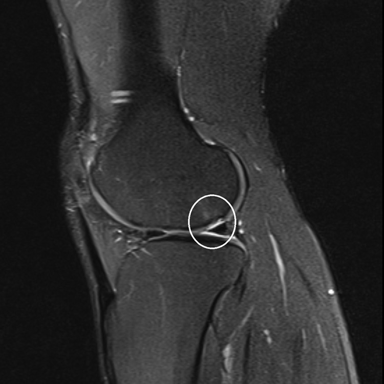
Cartilage lesions in both joints were detected in 97.9% of the examined soccer players. At least 1 lesion was detected in 100% of the participants; 6.3% of the soccer players had 2 to 5 lesions; 93.7% had 5 or more lesions; and 53.2% had 10 to 12 lesions. Grade 2 lesions located at the medial condyle of tibia and lateral condyle of femur were the most frequent (found in 59.6% and 52.1% of the participants, respectively). The medial condyles of the femur and tibia were the most frequent lesion sites (85.1% of all joints) (Table 1). Grade 4 lesions (Figure 1) were detected in 12 (12.8%) joints of 8 (17.0%) soccer players. Of these, 6 players had only 1 grade 4 lesion site, while the remaining 2 players had 2 and 4 lesions that were grade 4. These lesions were most often located at the medial or lateral condyle of femur, patellar surface of femur (25% each), lateral condyle of tibia (16.6%), or medial condyle of tibia (8.4%). The presence of BME was significantly associated with the presence of cartilage lesions grade 3 to 4 on the same leg (P < .05).
TABLE 1.
Prevalence, Localization, and Grade of Asymptomatic Hyaline Cartilage Lesions of the Knee Joint in Adult Professional Soccer Playersa
| Lesion site | Grade 1 | Grade 2 | Grade 3 | Grade 4 |
|---|---|---|---|---|
| Patella | 23 | 44.7 | 8.5 | — |
| Condyle of femur | ||||
| Medial | 11.7 | 36.2 | 34 | 3.2 |
| Lateral | 11.7 | 52.1 | 7.4 | 3.2 |
| Condyle of tibia | ||||
| Medial | 14.9 | 59.6 | 9.6 | 1.1 |
| Lateral | 22.3 | 47.9 | 3.2 | 2.1 |
| Patellar surface of femur | 11.7 | 41.5 | 4.3 | 3.2 |
aValues are presented as percentages.
Figure 1.

Axial magnetic resonance image obtained from an asymptomatic 31-year-old soccer player. A grade 4 lesion of the lateral condyle of femur with the subchondral edema is present (circle).
Meniscal Lesions
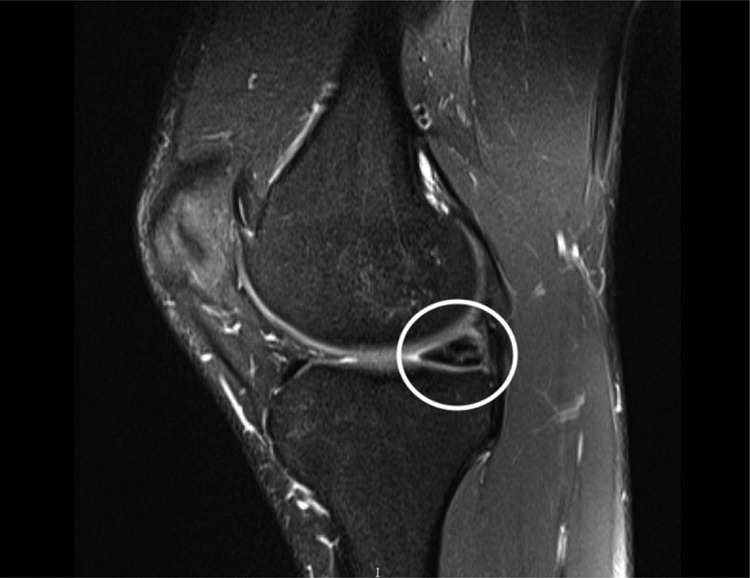
In 93.6% of the examined soccer players, both joints were affected by meniscal lesions. In 4.2%, meniscal lesions were detected in 1 joint only, and 2.2% were unaffected. Overall, 27.7% of the participants had 1 to 3 lesions; 40.4% had 4 to 7 lesions; and 31.9% had 8 lesions, with grade 2 meniscal lesions being predominant. The posterior horn of the lateral meniscus was the most frequent lesion site (95.7% of all the joints) (Table 2). Grade 3 lesions (Figure 2) were found in 13 (13.8%) joints of 10 (21.3%) soccer players. Eight participants had 1 lesion site that was grade 3, while 2 participants had 2 lesion sites. These lesions were most often found at the posterior horn of the medial meniscus (53.8%). The anterior horn of the lateral meniscus was affected in 30.8% of the cases, while the posterior horn of the lateral meniscus and the anterior horn of the medial meniscus were affected in 7.7% of the cases each.
TABLE 2.
Prevalence, Localization, and Grade of Asymptomatic Meniscal Lesions in Adult Professional Soccer Playersa
| Lesion Site | Absent | Grade 1 | Grade 2 | Grade 3 |
|---|---|---|---|---|
| Horn of the lateral meniscus | ||||
| Anterior | 44.7 | 12.8 | 38.3 | 4.3 |
| Posterior | 37.2 | 12.8 | 48.9 | 1.1 |
| Horn of the medial meniscus | ||||
| Anterior | 37.2 | 18.1 | 43.6 | 1.1 |
| Posterior | 4.3 | 17 | 71.3 | 7.4 |
aValues are presented as percentages.
Figure 2.

Sagittal magnetic resonance image obtained from an asymptomatic 21-year-old soccer player. A grade 3 lesion of the posterior horn of the medial meniscus is present (circle).
Correlation Between Localization of Various Intra-articular Lesions
The degree of patellar injury was significantly and positively correlated with the degree of injury of the medial and lateral condyles of femur, the medial and lateral condyles of tibia, and the patellar surface of femur (P < .001) (Table 3).
TABLE 3.
Localizations of Intra-articular Lesions: P Values and Correlationsa
| P Value (R Value) | |||||||||
|---|---|---|---|---|---|---|---|---|---|
| Condyle of Femur | Condyle of Tibia | Patellar Surface of Femur | Horn of the Lateral Meniscus | Anterior Horn of the Medial Meniscus | |||||
| Patella | Medial | Lateral | Medial | Lateral | Anterior | Posterior | |||
| Condyle of femur | |||||||||
| Medial | <.001 (0.397) | ||||||||
| Lateral | <.001 (0.385) | <.001 (0.465) | |||||||
| Condyle of tibia | |||||||||
| Medial | <.001 (0.373) | <.001 (0.583) | <.001 (0.506) | ||||||
| Lateral | <.001 (0.434) | <.001 (0.643) | <.001 (0.736) | <.001 (0.670) | |||||
| Patellar surface of femur | <.001 (0.673) | <.001 (0.539) | <.001 (0.497) | <.001 (0.410) | <.001 (0.483) | ||||
| Horn of the lateral meniscus | |||||||||
| Anterior | .587 | .094 | .783 | .430 | .388 | .082 | |||
| Posterior | .127 | .008 (–0.272) | .164 | .254 | .097 | .003 (–0.301) | <.001 (0.685) | ||
| Horn of the medial meniscus | |||||||||
| Anterior | .443 | .024 (–0.233) | .253 | .288 | .112 | .008 (–0.273) | <.001 (0.666) | <.001 (0.757) | |
| Posterior | .363 | .840 | .407 | .682 | .179 | .222 | .003 (0.304) | <.001 (0.434) | <.001 (0.366) |
aR value shown only for entries with a P value <.05. Dark gray shading indicates a statistically significant positive correlation; light gray shading indicates a statistically significant negative correlation.
The degree of injury of the medial condyle of femur was significantly and positively correlated with the degree of injury of the lateral condyle of femur, the lateral and medial condyles of tibia, and the patellar surface of femur (P < .001). It was also significantly and negatively correlated with the degree of deformation of the posterior horn of the lateral meniscus (P = .008) and the anterior horn of the medial meniscus (P = .024). The degree of injury of the lateral condyle of femur was significantly and positively correlated with the degree of injury of the medial and lateral condyles of tibia and patellar surface of femur (P < .001).
The degree of injury of the patellar surface of femur was significantly and negatively correlated with the degree of deformation of the posterior horn of the lateral meniscus (P = .003) and the anterior horn of the medial meniscus (P = .008). The degree of deformation of the anterior horn of the lateral meniscus was significantly and positively correlated with the degree of injury of the posterior horn of the lateral meniscus (P < .001) and the posterior (P < .001) and anterior (P = .003) horns of the medial meniscus. The degree of injury of the posterior horn of the lateral meniscus was significantly and positively correlated with the degree of deformation of the anterior and posterior horns of the medial meniscus (P < .001). The degree of injury of the anterior horn of the medial meniscus was significantly and positively correlated with the degree of injury of the posterior horn of the medial meniscus (P < .001).
Discussion
The study results indicate a high prevalence of abnormal cartilage and MRI signals of meniscal lesions in adult asymptomatic professional soccer players, affecting 100% and 97.8% of the examined joints, respectively. Grade 1 or 2 meniscal signal changes were common, although these findings are not thought to represent meniscal tears. Similarly, minor articular cartilage findings were frequently seen. Full-thickness cartilage lesions and grade 3 meniscal lesions were detected in 12.7% and 13.8% of the joints, respectively. Synovitis of the knee joint was also relatively common (16%). At the same time, BME occurred in only 10.6% of the joints. Many of the results obtained in this study do not correspond with the data previously published by other authors. Several explanations may exist for this phenomenon—for example, different types of MRI (0.35-T and 3-T MRI in other studies versus 1.5-T MRI in our study), differences in the level of sports qualification of the examined athletes, and difference in the length of the observation period for athletes after the end of the study (6-12 months after MRI in our study). This study is the first to analyze the condition of the knee joints of professional soccer players who not only have long training experience but have also played at an elite level for many years.
When the asymptomatic changes in the knee joints of soccer players are considered, the study by Soder et al27 is the one mentioned the most frequently. This study was carried out on 14 adolescent soccer players (28 knee joints) aged 14 to 15 years who had at least 3 years of systematic practice (3-3.5 hours, 5 times a week). The control group consisted of 14 adolescents (28 knee joints) with age and body mass index comparable with those of the study group, who did not engage in sports regularly. The knee joints were examined with 0.35-T MRI. The authors established a high prevalence of various intra-articular changes in both groups (48.2%). Notably, the prevalence of changes in soccer players was higher than that in the control group (64.3% vs 32.1%, respectively). BME (50% of the knee joints) and Hoffa fat pad edema (35.7%) were the most prevalent findings in the soccer players. In the control group, however, only 1 joint was affected by BME (3.6%). Hoffa fat pad edema was the most prevalent MRI finding among the controls, occurring in 25% of the knee joints. The authors found no meniscal or cartilage lesions in the examined joints.
The Soder et al27 study benefits from having a control group of comparable age and body weight. At the same time, the group of adolescent players was relatively small, and the length of required systematic training used as an inclusion criterion was short (3 years). The training program of the participants warrants attention as well, as 3 to 3.5 hours of training for 5 days a week constitutes a very rigorous program for adolescents aged 14 to 15 years. Therefore, a possible explanation for the high incidence of bone edema is that it was caused by extensive training during a period of rapid growth of the musculoskeletal system. Another drawback of that study is the usage of a low-field MRI scanner, as it does not allow one to detect the early changes of cartilage tissue.
The association of systematic soccer training with the condition of intra-articular structures was also the subject of study by Matiotti et al.18 The authors analyzed the 3-T MRI scans of the knee joints in 23 Brazilian junior soccer players aged 14 to 17 years. The participants were engaged in soccer practice 2 to 6 times a week (mean training time, 10 hours a week) for a minimum of 2 years and had participated in regional or national soccer tournaments. The control group was composed of volunteers comparable by age and body weight whose engagement in any physical activity did not exceed 100 minutes per week. Medical history of knee trauma or knee surgery was selected as an exclusion criterion. In the soccer players, 67.4% of the joints had at least 1 change detected by MRI, as opposed to 48.4% in the control group. BME was the most prevalent finding (41.3% and 7.3% of the joints in case and control groups, respectively). These findings correspond well to the results obtained by Soder et al.27
The Soder et al27 study detected synovitis in 19% of the joints in the case group, implicit cartilage lesions in 8.7% of the joints, and lesions of the posterior horn of the medial meniscus in 10.8% of the joints. Joint effusion was the most prevalent finding in the control group (19.4% of the joints). Hoffa fat pad edema was detected in 9.8% of the joints, and no cartilage or meniscal lesions were found. Compared with Soder et al,27 the increase in the incidence of cartilage and meniscal lesions in the soccer players analyzed by Matiotti et al can be attributed to either the older age of the participants, which would result in a longer period of systematic soccer training, or the usage of 3-T MRI, which provides better sensitivity and specificity for the detection of cartilage lesions as compared with the low-field 0.35-T MRI.
A number of articles have since been published analyzing basketball players, swimmers, long-distance runners, gymnasts, football players, and kangoo jumpers.11,14,17,22,25,26,28,29 Notably, most of the studies concerning asymptomatic changes of the large joints have primarily analyzed amateurs. Only a few studies have been performed on elite athletes. Brunner et al6 can be considered the first to study asymptomatic changes of the knee joints in competitive athletes. Using MRI, the authors discovered that meniscal changes had developed in 50% of the basketball players who participated in the study.
Walczak et al30 included professional basketball players in their study. They found asymptomatic meniscal tissue lesions in 54% of the athletes. On the basis of MRI data obtained from scanning 40 knee joints of professional basketball players, Kaplan et al11 reported a 20% rate of meniscal injury, noting that the injuries of the medial meniscus were by far the most frequent. MRI is a precise method that allows one to detect articular cartilage injury, meniscal tears, ligament injury, and BME.5,9,23 For the diagnosis of intra-articular injury, the usage of 1.5-T or 3-T MRI scanners is preferable, as they allow higher sensitivity, specificity, and precision for the detection of cartilage injury in comparison with low-field MRI.12 Using 1.5-T MRI, Wacker et al29 analyzed 21 right knee joints of junior soccer players. The control group consisted of 12 adolescents who did not regularly participate in any sports. The authors found meniscal lesions in 38.1% of the joints in the soccer players and 16.7% of the joints in the control group. No cases of synovitis were detected in either group.
The prevalence of asymptomatic lesions in the knee joints of adult professional athletes was also assessed by Pappas et al.20 The authors used 3-T MRI to analyze the condition of knee joints in 24 young adult elite basketball players (12 women and 12 men aged 18-22 years) before the beginning of the basketball season and after its end. Changes in intrameniscal signal intensity were found in 50% of the joints before the season and in 62% of the joints after the season end. However, no high-grade lesions were detected. BME was observed in 75% and 86% of knee joints before and after the season, respectively, and cartilage lesions were detected in 71% and 81% of knee joints before and after the season. The authors concluded that basketball, being a high-intensity game, can exert a potentially harmful effect on articular cartilage.
The study by Pappas et al20 involved more mature and experienced professional athletes as compared with the research performed in adolescent soccer players.18,27 This might explain the observed high prevalence of cartilage and meniscal lesions. The high prevalence of bone edema can be explained by the specifics of basketball, as jumps are the essential component of the game. This assumption is confirmed by Polat et al,22 who found bone edema to be highly prevalent in kangoo jumpers.
The prevalence of various asymptomatic lesions of the joints in athletes of varying age and ability performing in different sports was found to be high in all the aforementioned studies.∥ ∥ Heterogeneity of the studied groups makes it impossible to detect any consistent patterns regarding the influence of any specific type of strain on the condition of knee joints in adult athletes.
The results of the current assessment of the impact of soccer on the joints of athletes will allow us to correctly interpret data obtained during magnetic resonance tomography and avoid unwarranted surgical procedures. We believe that this information will help sports doctors and traumatologists adequately compare MRI data, medical history, complaints, and clinical tests to make a correct diagnosis. Future studies should focus on studying the effect of age, experience, and leg dominance on the prevalence and severity of intra-articular changes in the knee joints of not only soccer players but other sports athletes. Future studies should also address whether these asymptomatic findings will affect future performance or lead to long-term degenerative changes. The limitations of this study were that (1) there was no control group composed of individuals of comparable age and body mass index who never performed in sports systematically and never underwent knee surgery; (2) only 1.5-T MRI was used; and (3) there were no intra- or interobserver correlations calculated for the radiological analysis. Additionally, there is no follow-up reported on these abnormal findings to see if any become symptomatic over time. Finally, professional athletes undergoing a physical examination before signing a contract may have an incentive to minimize knee symptoms.
Conclusion
This study showed an extremely high frequency of abnormal MRI findings of the meniscus and cartilage in the knee joints among asymptomatic professional soccer players.
Footnotes
The authors declared that there are no conflicts of interest in the authorship and publication of this contribution. AOSSM checks author disclosures against the Open Payments Database (OPD). AOSSM has not conducted an independent investigation on the OPD and disclaims any liability or responsibility relating thereto.
Ethical approval for this study was obtained from Sechenov First Moscow State Medical University.
References
- 1. Arliani G, Astur D, Yamada R, et al. Early osteoarthritis and reduced quality of life after retirement in former professional soccer players. Clinics (Sao Paulo). 2014;69(9):589–594. [DOI] [PMC free article] [PubMed] [Google Scholar]
- 2. Baker P, Reading I, Cooper C, Coggon D. Knee disorders in the general population and their relation to occupation. Occup Environ Med. 2003;60(10):794–797. [DOI] [PMC free article] [PubMed] [Google Scholar]
- 3. Beals CT, Magnussen RA, Graham WC, Flanigan DC. The prevalence of meniscal pathology in asymptomatic athletes. Sports Med. 2016;46(10):1517–1524. [DOI] [PubMed] [Google Scholar]
- 4. Boden SD, Davis DO, Dina TS, et al. A prospective and blinded investigation of magnetic resonance imaging of the knee: abnormal findings in asymptomatic subjects. Clin Orthop Relat Res. 1992;282:177–185. [PubMed] [Google Scholar]
- 5. Bredella MA, Tirman PF, Peterfy CG, et al. Accuracy of T2-weighted fast spin-echo MR imaging with fat saturation in detecting cartilage defects in the knee: comparison with arthroscopy in 130 patients. AJR Am J Roentgenol. 1999;172(4):1073–1080. [DOI] [PubMed] [Google Scholar]
- 6. Brunner MC, Flower SP, Evancho AM, Allman FL, Apple DF, Fajman WA. MRI of the athletic knee: findings in asymptomatic professional basketball and collegiate football players. Invest Radiol. 1989;24(1):72–75. [DOI] [PubMed] [Google Scholar]
- 7. Englund M, Guermazi A, Gale D, et al. Incidental meniscal findings on knee MRI in middle-aged and elderly persons. N Engl J Med. 2008;359(11):1108–1115. [DOI] [PMC free article] [PubMed] [Google Scholar]
- 8. Flanigan DC, Harris JD, Trinh TQ, Siston RA, Brophy RH. Prevalence of chondral defects in athletes knees: a systematic review. Med Sci Sports Exerc. 2010;42(10):1795–1801. [DOI] [PubMed] [Google Scholar]
- 9. Hodler J, Buess E, Rodriguez M, Imhoff A. Magnetic resonance tomography (MRT) of the knee joint: meniscus, cruciate ligaments and hyaline cartilage. Rofo. 1993;159(2):107–112. [PubMed] [Google Scholar]
- 10. Junge A, Dvorak J. Soccer injuries: a review on incidence and prevention. Sports Med. 2004;34(13):929–938. [DOI] [PubMed] [Google Scholar]
- 11. Kaplan LD, Schurhoff MR, Selesnick H, Thorpe M, Uribe JW. Magnetic resonance imaging of the knee in asymptomatic professional basketball players. Arthroscopy. 2005;21(5):557–561. [DOI] [PubMed] [Google Scholar]
- 12. Kijowski R, Blankenbaker DG, Davis KW, Shinki K, Kaplan LD, De Smet AA. Comparison of 1.5- and 3.0-T MR imaging for evaluating the articular cartilage of the knee joint. Radiology. 2009;250(3):839–848. [DOI] [PubMed] [Google Scholar]
- 13. Kolman BH, Daffner RH, Sciulli RL, Soehnlen MW. Correlation of joint fluid and internal derangement on knee MRI. Skeletal Radiol. 2004;33:91-95. [DOI] [PubMed] [Google Scholar]
- 14. Krampla W, Mayrhofer R, Malcher J, Kristen KH, Urban M, Hruby W. MR imaging of the knee in marathon runners before and after competition. Skeletal Radiol. 2001;30:72–76. [DOI] [PubMed] [Google Scholar]
- 15. Kuijt MT, Inklaar H, Gouttebarge V, Frings-Dresen MH. Knee and ankle osteoarthritis in former elite soccer players: a systematic review of the recent literature. J Sci Med Sport. 2012;15(6):480–487. [DOI] [PubMed] [Google Scholar]
- 16. Lee HH, Chu CR. Clinical and basic science of cartilage injury and arthritis in the football (soccer) athlete. Cartilage. 2012;3(1 Suppl):63S–68S. [DOI] [PMC free article] [PubMed] [Google Scholar]
- 17. Ludman CN, Hough DO, Cooper TG, Gottschalk A. Silent meniscal abnormalities in athletes: magnetic resonance imaging of asymptomatic competitive gymnasts. Br J Sports Med. 1999;33:414–416. [DOI] [PMC free article] [PubMed] [Google Scholar]
- 18. Matiotti SB, Soder RB, Becker RG, Santos FS, Baldisserotto M. MRI of the knees in asymptomatic adolescent soccer players: a case-control study. J Magn Reson Imaging. 2017;45(1):59–65. [DOI] [PubMed] [Google Scholar]
- 19. Noyes FR, Stabler CL. A system for grading articular cartilage lesions at arthroscopy. Am J Sports Med. 1989;17:505-513. [DOI] [PubMed] [Google Scholar]
- 20. Pappas GP, Vogelsong MA, Staroswiecki E, Gold GE, Safran MR. Asymptomatic knees in collegiate basketball players: the effect of one season of play. Clin J Sport Med. 2016;26(6):483–489. [DOI] [PMC free article] [PubMed] [Google Scholar]
- 21. Paxinos O, Karavasili A, Delimpasis G, Stathi A. Prevalence of knee osteoarthritis in 100 athletically active veteran soccer players compared with a matched group of 100 military personnel. Am J Sports Med. 2016;44(6):1447–1454. [DOI] [PubMed] [Google Scholar]
- 22. Polat B, Aydın D, Polat AE, Gürpınar T, Özmanevra R, Dirik MA. Evaluation of the knees of asymptomatic kangoo jumpers with MR imaging [published online January 31, 2019]. Magn Reson Med Sci. doi:10.2463/mrms.mp.2018-0094 [DOI] [PMC free article] [PubMed] [Google Scholar]
- 23. Potter HG, Linklater JM, Allen AA, Hannafin JA, Haas SB. Magnetic resonance imaging of articular cartilage in the knee: an evaluation with use of fast spin-echo imaging. J Bone Joint Surg Am. 1998;80(9):1276–1284. [DOI] [PubMed] [Google Scholar]
- 24. Roos H. Are there long-term sequelae from soccer? Clin Sports Med. 1998;17(4):819–831. [DOI] [PubMed] [Google Scholar]
- 25. Smith TO, Drew BT, Toms AP, Donell ST, Hing CB. Accuracy of magnetic resonance imaging, magnetic resonance arthrography and computed tomography for the detection of chondral lesions of the knee. Knee Surg Sports Traumatol Arthrosc. 2012;20(12):2367–2379. [DOI] [PubMed] [Google Scholar]
- 26. Soder RB, Mizerkowski MD, Petkowicz R, Baldisserotto M. MRI of the knee in asymptomatic adolescent swimmers: a controlled study. Br J Sports Med. 2012;46(4):268–272. [DOI] [PubMed] [Google Scholar]
- 27. Soder RB, Simões JD, Soder JB, Baldisserotto M. MRI of the knee joint in asymptomatic adolescent soccer players: a controlled study. AJR Am J Roentgenol. 2011;196(1):61–65. [DOI] [PubMed] [Google Scholar]
- 28. Stoller DW, Martin C, Crues JV, 3rd, Kaplan L, Mink JH. Meniscal tears: pathologic correlation with MR imaging. Radiology. 1987;163:731-735. [DOI] [PubMed] [Google Scholar]
- 29. Wacker F, König H, Felsenberg D, Wolf KJ. MRI of the knee joint of young soccer players: are there early changes of the internal structures of the knee due to competitive sports? Rofo. 1994;160(2):149–153. [DOI] [PubMed] [Google Scholar]
- 30. Walczak BE, McCulloch PC, Kang RW, Zelazny A, Tedeschi F, Cole BJ. Abnormal findings on knee magnetic resonance imaging in asymptomatic NBA players. J Knee Surg. 2008;21(1):27–33. [DOI] [PubMed] [Google Scholar]
- 31. Zanetti M, Pfirrmann CW, Schmid MR, Romero J, Seifert B, Hodler J. Patients with suspected meniscal tears: prevalence of abnormalities seen on MRI of 100 symptomatic and 100 contralateral asymptomatic knees. AJR Am J Roentgenol. 2003;181(3):635–641. [DOI] [PubMed] [Google Scholar]


