Abstract
Melanoma is a type of aggressive cancer. Recent studies have indicated that blue light has an inhibition effect on melanoma cells, but the effect of photobiomodulation (PBM) parameters on the treatment of melanoma remains unknown. Thus, this study was aimed to investigate B16F10 melanoma cells responses to PBM with varying irradiance and doses, and further explored the molecular mechanism of PBM. Our results suggested that the responses of B16F10 melanoma cells to PBM with varying irradiance and dose were different and the inhibition of blue light on cells under high irradiance was better than low irradiance at a constant total dose (0.04, 0.07, 0.15, 0.22, 0.30, 0.37, 0.45, 0.56 or 1.12 J/cm2), presumably due to that high irradiance can produce more ROS, thus disrupting mitochondrial function.
1. Introduction
Melanoma is one of the most dangerous cancers in the world [1]. Malignant melanomas usually produce melanin in large quantities. It was reported that the melanin contents of the human melanoma cells were 4.2 ± 0.3 µg/106 cells and 11.3 ± 0.6 µg/106 cells when the melanoma cells grew to exponential and stationary phases respectively [2]. Clinical treatment options for malignant melanoma include radiotherapy, surgery, immunotherapy, chemotherapy and biological therapy. However, these conventional treatments have not been effective [3]. Melanoma is highly resistant to chemotherapy drugs and its effectiveness against traditional chemotherapy drugs is less than 20%. Particularly, because there is no effective treatment for metastatic melanoma, the patients’ median survival time is only 6 to 9 months and the 5-year survival rate is less than 5% [4]. Thus, novel treatments are required to treat the malignant melanoma.
Photobiomodulation (PBM), also named as low-level light therapy (LLLT), is one of the most critical methods for medical cure [5]. At present, the light sources commonly used in photobiomodulation are laser and Light Emitting Diode (LED). The laser is coherent light and the LED device emits non-coherent light. Overall, Laser and LED therapy are similarly effective for superficial tissue. The ability of the laser to penetrate the tissue is stronger than that of LED, which can reach deeper lesions, but LED devices are cheaper than laser devices and can illuminate large areas of tissue, even can be made into a wearable device. Both lasers and LEDs achieve biological regulation through low level of dose [6]. With higher anti-inflammatory effect and healing simulation, PBM was widely used in clinical applications, such as thyroiditis, psoriasis, and alleviation of muscle soreness [7]. Jeong et al. [8] observed the blue light could induce apoptotic cell death by activating the mitochondria-mediated pathway and inhibit the growth of melanoma cells. Most of the blue light is used to inhibit cell proliferation and inactivate important wound pathogenic bacteria [9], while red light is used to promote cell proliferation, such as enhancing the angiogenic effect of mesenchymal stem cells [10] and could treat hair loss effectively [11]. Besides, PBM is non-invasive and does not cause much damage to the body like traditional therapy [12]. Unfortunately, the light parameters for treating melanoma are still unclear. So, we studied the illumination parameters for treating B16F10 melanoma cells in this paper and aimed at providing a reference for clinical treatment, and further analyzed the mechanism of PBM.
The fundament of PBM is the light parameters. The parameters of the light include irradiation time, irradiance (intensity, power density), wavelength, dose (energy density, fluence), and light mode (continuous wave, pulsed light). As we all know, if incorrect parameters are applied, then treatment may not be effective. PBM has a biphasic dose response. It will lead to no significant effect or the opposite effect when dose (J/cm2) and irradiance (mW/cm2) are either too low or too high [13]. Dyson et al. [14] treated macrophages with the same dose (J/cm2) but with varying levels of irradiance(mW/cm2), and observed different results between the two parameters, 400 mW/cm2 promoted cell proliferation better than 800 mW/cm2 at a low dose (2.4 J/cm2), but at a high dose (7.2 J/cm2), 800 mW/cm2 promoted cell proliferation better than 400 mW/cm2. Raymond et al. [15] used a fixed dose of 5 J/cm2 and variable irradiance, ranging from 0.7 to 40 mW/cm2, observed that only with 8 mW/cm2 could be adequate for treating pressure ulcers in the mice. These studies demonstrated the importance of illumination parameters in clinical applications and there may be no treatment effect if incorrect light parameters are applied.
In our study, we used three different wavelengths of light (418 nm,457 nm, 630 nm) to treat B16F10 melanoma cells and aimed to study the effects of different wavelengths of light on B16F10 melanoma cells. Besides, we treated B16F10 melanoma cells by PBM with the same dose but with different irradiance to study whether the effect of irradiance on cells was different under the same dose. The results showed that blue light (418 nm and 457 nm) can inhibit the growth and migration of B16F10 melanoma cells and the inhibited effect of 457 nm was better than 418 nm. In addition, the effect of high irradiance on cell inhibition was better than low irradiance at the same doses (0.04, 0.07, 0.14, 0.22, 0.30, 0.37, 0.45, 0.56 or 1.12 J/cm2). Kleinpenning et al. demonstrated that blue light did not cause photo-ageing or DNA damage, thus the utilization of blue light (short-term) in treating melanoma is safe [16]. Given that malignant melanoma is a very harmful cancer, it is imperative to study the light parameters of PBM aimed at providing a reference for clinical treatment.
2. Materials and methods
2.1. Cell Culture
B16F10 melanoma cells were purchased from Cellcook (Guangzhou, China) and cultured in RPMI 1640 medium (Gibco, Logan, USA), supplemented with 1% streptomycin and penicillin (HyClone, Logan, USA) and 10% fetal bovine serum (Gibco, Logan, USA). The cells were maintained in 5% CO2 in a 37℃, humidified incubator (Qiqian, Shanghai, China), Subculturing after approximately 2 days.
2.2. LED irradiation and their technical parameters
One day before light treatment, B16F10 melanoma cells were transferred to 96 well culture plates containing 150 µL of medium/well seeded at a density of 5×10³ cells/well after 24 hours. The irradiation of PBM was performed by using two blue continuous waves (wavelength: 418 nm, 457 nm) and a red continuous wave (630 nm) in a light humidified incubator (LightEngin technology, Shanghai, China) (Fig. 1). In this experiment, we used three kinds of light parameters (Table 1). Irradiance was measured by an optical power meter (Thorlabs, Newtown, USA). Firstly, three different wavelengths of light were used to treat B16F10 melanoma cells at different times (0 s, 450 s, 900 s, 1800 s) respectively under the same irradiance conditions (2.48 mW/cm2) (Table 1, Parameter 1). Then, we treated B16F10 melanoma cells with different irradiance for the same amount of time (450 s) (Table 1, Parameter 2). Finally, the B16F10 melanoma cells were treated by the irradiance with 0.31, 0.62 and 0.93 mW/cm2 in the conditions of same dose to further investigate the effects of low- irradiance treat on B16F10 melanoma cells (Table 1, Parameter 3).
Fig. 1.
Light humidified incubator and spectrum of the respective LED light source. (A) Light humidified incubator. (B) The spectra of blue light (418 nm,457 nm) and red light (630 nm) used in this study. The LEDs light sources are on top of the incubator and can be replaced.
Table 1. Experimental conditions.
| Parameter 1 | ||||||||
|---|---|---|---|---|---|---|---|---|
| Wavelength | 418 nm | 457 nm | 630 nm | |||||
| Irradiance(mW/cm2) | 2.48 | |||||||
| Time (s) | 0 | 450 | 900 | 1800 | 3600 | |||
| Parameter 2 | ||||||||||||
|---|---|---|---|---|---|---|---|---|---|---|---|---|
| Wavelength | 457 nm | |||||||||||
| Time (s) | 450 | |||||||||||
| Irradiance(mW/cm2) | 0 | 0.31 | 0.62 | 0.93 | 1.24 | 1.55 | 1.86 | 2.17 | 2.48 | 4.96 | 9.92 | 19.84 |
| Parameter 3 | ||||||||||
|---|---|---|---|---|---|---|---|---|---|---|
| Wavelength | 457 nm | |||||||||
| Irradiance(mW/cm2) | 0.31 | |||||||||
| Time (s) | 0 | 120 | 240 | 480 | 720 | 960 | 1200 | 1440 | 1800 | 3600 |
| Dose (J/cm2) | 0 | 0.04 | 0.07 | 0.15 | 0.22 | 0.30 | 0.37 | 0.45 | 0.56 | 1.12 |
| Irradiance(mW/cm2) | 0.62 | |||||||||
| Time (s) | 0 | 60 | 120 | 240 | 360 | 480 | 600 | 720 | 900 | 1800 |
| Dose (J/cm2) | 0 | 0.04 | 0.07 | 0.15 | 0.22 | 0.30 | 0.37 | 0.45 | 0.56 | 1.12 |
| Irradiance(mW/cm2) | 0.93 | |||||||||
| Time (s) | 0 | 40 | 80 | 160 | 240 | 320 | 400 | 480 | 600 | 1200 |
| Dose (J/cm2) | 0 | 0.04 | 0.07 | 0.15 | 0.22 | 0.30 | 0.37 | 0.45 | 0.56 | 1.12 |
Dose (J/cm2) = Irradiance (mW/cm2) x Time of irradiation (s)/1000. The results shall be quoted in two decimal places.
2.3. Cell viability and inhibition
B16F10 melanoma cells inhibition rate was tested by CCK-8 assay (Dojindo, Shanghai, China) as initially mentioned [17]. The CCK8 solution (2-(2-methoxy-4-nitrophenyl)-3-(4-nitrophenyl)-5-(2,4-disulfophenyl)-2H-tetrazolium) can be reduced to formazan dye with high water solubility by dehydrogenase in cells under the action of 1-methoxy-5-methylphenazinium dimethyl sulfate (1-methoxy PMS). The number of the generated formazan dye was directly proportional to the number of living cells [18]. In brief, B16F10 melanoma cells were seeded in 96-well plates (Corning Incorporated, Corning, USA). After 24 h, the 96-well plates were treated with light for the above conditions. In the detection of cell viability, we used the medium with 10% serum. In order to further studied the effect of different irradiance on the inhibition of melanoma cells, the serum-free medium was used to eliminate the effect of serum on B16F10 melanoma cells before the light treatment. 24 hours after the end of the light treatment, cell viability and inhibition rate were measured with 10 µL/well CCK-8 solution and 100 µL/well medium, and then were incubated for 1.5 h in a humidified incubator. Ultimately, the absorbance at 450 nm was measured by a microplate reader (Thermo Scientific, Waltham, USA). The control group was also placed in the light incubator, but the light source was turned off. The negative group was medium and CCK-8 solution without cells. The cell inhibition rate was determined by using the formula:
(As: the light-treated group, Ac: the control group, Ab: the negative group).
2.4. Temperature measurement
The temperature of the cell medium may change during irradiation, the temperature changes of medium during irradiation at 418 nm, 457 nm and 630 nm (2.48 mW/cm2,60 min) (the maximum dose in this study) were measured by thermal resistance thermometer (Shimaden, Tokyo, Japan).
2.5. Cell migration
B16F10 melanoma cells were seeded in 12-well plates. Cells were cultured until 90% confluence and then wounded with a sterile 20 µL pipette tip. Cells were washed with PBS and cultured in serum-free medium. Subsequently, the cells were treated for 450 s at 2.48 mW/cm2 (418 nm,457 nm and 630 nm). The wounds were photographed at baseline and 24 h later under microscope bright field (Olympus BX53, Tokyo, Japan). The width of scratches was analyzed by Image J (NIH, Bethesda, USA).
2.6. Measurement of intracellular ROS
The level of intracellular ROS was measured using the fluorescence probe (DCFH-DA) (Beyotime, Shanghai, China). The B16F10 melanoma cells were treated by the irradiance with 0.31, 0.62 and 0.93 mW/cm2 in the dose (1.12 J/cm2) and 24 hours later ROS was detected, then incubated with 20 µM/mL DCFH-DA in serum-free medium at 37°C for 30 min, the cells were washed with PBS and observed with a fluorescence microscope at an emission wavelength of 500 nm (Olympus BX53, Tokyo, Japan).
2.7. Measurement mitochondrial membrane potential (mt.ΔΨ)
Mitochondrial membrane potential kit with JC-1 (Jiancheng, Nanjing, China) was used to detect the mt.ΔΨ. The B16F10 melanoma cells were treated by the irradiance with 0.31, 0.62 and 0.93 mW/cm2 in the dose (1.12 J/cm2) and 24 hours later MMP was detected, then incubated with JC-1 fluorescent probe at 37°C for 25 min. Cells were washed three times with JC-1(1X) buffer and observed with a fluorescence microscope at an emission wavelength of 510 nm and 580 nm (Olympus BX53, Tokyo, Japan).
2.8. Statistical analysis
All experiments were performed in triplicate. The results were analyzed by using the two-tailed Student’s t-test or Mann-Whitney test by GraphPad prism (Version 6.02, GraphPad software, San Diego, USA). P < 0.05 was designated to be significance.
3. Results
3.1. Blue light inhibited the growth of B16F10 melanoma cells
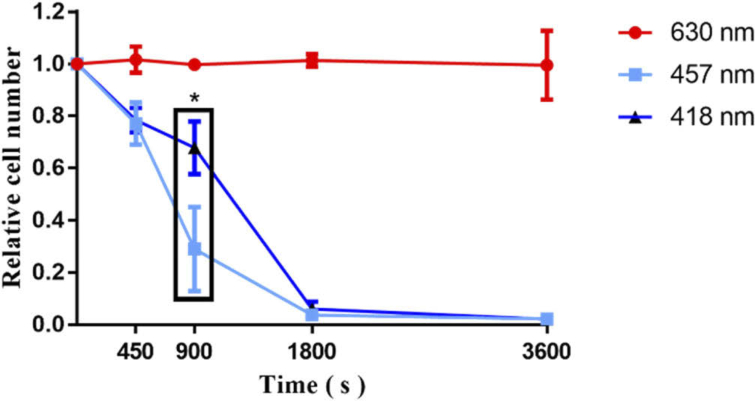
Under parameter 1 of light conditions (418 nm, 457 nm, 630 nm light treatment of cells for 450 s, 900 s, 1800 s, 3600 s at 2.48 mW/cm2), the CCK-8 assay for viability showed that the red light (630 nm) had no effects on cell activity, but the blue light at 418 nm and 457 nm had a significant light cytotoxicity on cells and the effect of killing cells increased with increased exposure time. When blue light (418 nm, 457 nm) was treated for 1800 s, blue light almost killed the cells and the effect between the results of 1800 s and 3600 s did not indicate any significant differences. When treated at 418 nm and 457 nm for 900 s, the 457 nm blue light inhibited cell activity was stronger than 418 nm significantly (p < 0.05) (Fig. 2). Therefore, the blue light (457 nm) was chosen to treat melanoma cells in order to observe the effect of cells responses to PBM with varying irradiance and dose in subsequent experiments.
Fig. 2.
B16F10 melanoma cells were treated with light at 418 nm and 457 nm of blue, and 630 nm of red. The 630 nm had no effect on cells, 418 nm and 457 nm inhibited cells. At 900 s, 457 nm inhibited cells were better than 418 nm(p < 0.05).
3.2. Blue light inhibited the migration of B16F10 melanoma cells
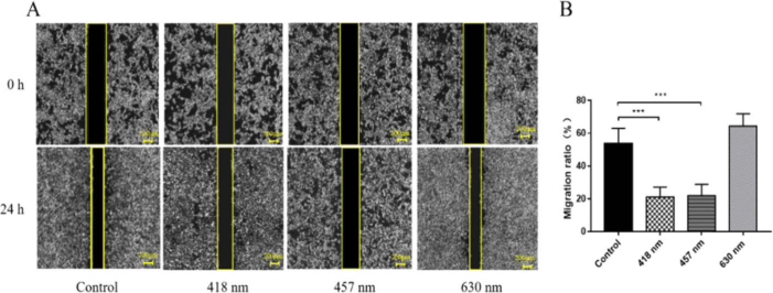
Figure 3 shows that at irradiance of 2.48 mW/cm2 and an irradiation time of 450 s,418 nm and 457 nm significantly inhibited cell migration compared with the control group (p < 0.05), while the effect of 630 nm on cells was not statistically different. This result demonstrated that 418 nm and 457 nm inhibited the migration of B16F10 melanoma cells.
Fig. 3.
Cell migration results after irradiation for 450 s at 2.48 mW/cm2 (418 nm, 457 nm, and 630 nm). (A) Photograph of cell migration, the area between the two yellow lines was the wound area (B) Statistical analysis of migration ratio,*** p < 0.001
3.3. Temperature changes
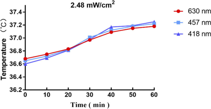
Figure 4 shows under the condition of irradiance (2.48 mW/cm2), the temperature of the three wavelengths (418 nm,457 nm and 630 nm) irradiation medium led to a slight increase, kept at about 37℃±0.4℃, there was no significant difference in temperature changes produced by the three wavelengths. This result demonstrated that the effect of LED (418 nm,457 nm and 630 nm) on melanoma cells proliferation and migration was independent of the temperature change.
Fig. 4.
In vitro cell culture temperature
3.4. The cell inhibition ratio increased with increased irradiance and dose
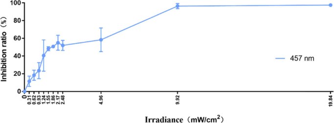
Under parameter 2 of light conditions, we treated the cells for 450 seconds under different irradiance conditions with a 457 nm light source. We aimed to study the effect of different irradiance on cell inhibition ratio under the same irradiation time. When the irradiance was from 0.31 to 1.55 mW/cm2, the cell inhibition ratio increased with increased irradiance. There was no decrease in the inhibition rate of blue light with the increased of the irradiance and dose, that indicated the B16F10 melanoma cells did not follow the biphasic dose response (stronger stimuli will achieve an opposite response). However, when the irradiance was from 1.55 to 4.96 mW/cm2, the cell inhibition ratio was not remarkably increased. The effect of cell inhibition was highest when the irradiance was 19.84 mW/cm2(Fig. 5).
Fig. 5.
Treatment of melanoma cells with various irradiance (0.31-19.84 mW/cm2) for 450 seconds, the inhibition ratio of the control group was 0.
3.5. Irradiance had an important effect on PBM
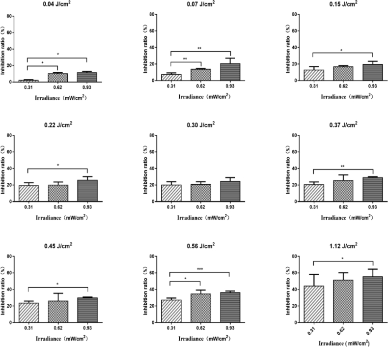
To further investigate the effect of low irradiance on B16F10 melanoma cells inhibition, we chose three low irradiance (0.31, 0.62, 0.93 mW/cm2) to treat cells at constant dose (Table 1, Parameter 3). Under the condition of parameter 3, we treated cells with three low irradiance under different doses (0, 0.04, 0.07, 0.15, 0.22, 0.30, 0.37, 0.45, 0.56 or 1.12 J/cm2) to further investigate the effects of low irradiance on cells. We found that under the same dose, high irradiance had better inhibition rate than low irradiance (Fig. 6,7).
Fig. 6.
The inhibition ratio of cells treated with three different irradiance (0.31,0.62,0.93 mW/cm2) at the same doses (0, 0.04, 0.07, 0.15, 0.22, 0.30, 0.37, 0.45, 0.56 or 1.12 J/cm2), the inhibition ratio of the control group was 0.
Fig. 7.
The inhibition ratio of cells treated with three different irradiance (0.31,0.62,0.93 mW/cm2) at the same doses (0, 0.04, 0.07, 0.15, 0.22, 0.30, 0.37, 0.45, 0.56 or 1.12 J/cm2), the inhibition ratio of the control group was 0. * p < 0.05, ** p < 0.01, *** p < 0.001
3.6. PBM increased ROS production
Many researchers have found that PBM can increase intracellular ROS in vitro [19,20]. However, it is still unclear about the ROS content produced by different irradiance at the same dose. Therefore, we detected ROS content produced by three different irradiance (0.31, 0.62, 0.93mW/cm2) at dose of 1.12 J/cm2. Interestingly, we found that different irradiance produced different levels of ROS at the same dose and the high irradiance produced more reactive oxygen than low irradiance. 0.31, 0.62, 0.93mW/cm2 gave a robust increase in ROS production (p < 0.05) (Fig. 8).
Fig. 8.
Effect of 457 nm irradiation on the production of ROS in B16F10 melanoma cells. (A) Fluorescence of cellular reactive oxygen species (B) Statistical analysis of the average fluorescence intensity of reactive oxygen species. * p < 0.05, *** p < 0.001
3.7. PBM resulted in mitochondrial dysfunction
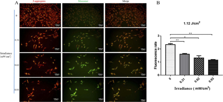
Since mitochondria are the primary place of ROS generation [21], we further studied the effects of three different irradiance (0.31, 0.62, 0.93 mW/cm2) on mitochondrial function at the dose of 1.12 J/cm2. Interestingly, the results are consistent with ROS production. When the mitochondrial membrane potential is high, JC-1 mainly exists as aggregates with red-fluorescent. When the mitochondrial membrane potential is low, JC-1 mainly exists as monomer with green-fluorescent, so the mitochondrial membrane potential can be measured by the relative ratio of red-green fluorescence [22]. With the increased of irradiance, the role of PBM in inhibiting mitochondrial function was more pronounced. The inhibition effects of 0.31, 0.62 and 0.93 mW/cm2 on mitochondrial function were more and more obvious (p < 0.05) (Fig. 9).
Fig. 9.
Effect of 457 nm irradiation on MMP in B16F10 melanoma cells. The higher the irradiance, the more obvious the loss of MMP (mt.ΔΨ) (A) Fluorescence of MMP (B) Statistical analysis of the fluorescence rate of MMP * p < 0.05, ** p < 0.01
4. Discussion
The findings demonstrated that blue light can inhibit the growth of melanoma. But it has still not been fully understood that the effectiveness of PBM with the different kinds of wavelengths and irradiance on the inhibition of melanoma. With regard to the irradiation could cause temperature rise, in order to address this problem, we used accurate thermal resistance thermometer and measured the temperature during the light irradiation. When the irradiance was at 2.48 mW/cm2 for 60 min (the maximum dose in this study), the temperature gain remained negligible, which showed that the influence of temperature on the results can be ignored.
From a comparison of the effects of three wavelengths (418 nm and 457 nm of blue light, and 630 nm of red light) under the condition of the same irradiance (2.48 mW/cm2), it was found that 418 nm and 457 nm of blue light can inhibit the growth of melanoma cells, which was consistent with the study by Ohara et al. [23]. Ottaviani et al. [24] discovered that laser treatment (200 mW/cm2,800 nm and 970 nm,6 J/cm2) could inhibit tumor growth and invasion. Sparas et al. [25] found that blue light (450 nm,10 J/cm2 and 20 J/cm2) was phototoxic for B16F10 Murine Melanoma. Their works only studied the effects of a kind of light parameter on melanoma cells, while we studied the effects of three light parameters (wavelength, dose, and irradiance) on B16F10 melanoma cells. Intriguingly, we further found that the inhibition effect of 457 nm was better than 418 nm at 900 s. Therefore, the optimum absorption wavelength of B16F10 melanoma cells photoreceptors was proposed to be around 457 nm. We speculated that this may be due to the best wavelength of B16F10 melanoma cells photoreceptor absorption is near 457 nm. Opsins (OPNs) are potential receptors for PBM, and the optimal absorption wavelength of OPN3 is 460 nm [26], which suggested that OPN3 may be one of the receptors for the blue light to inhibit melanoma growth. In addition to OPN3, other chromophores such as flavins (λabs = 400–500 nm) and metal-free porphyrins (λabs = 400–650 nm) can also absorb blue light and produce ROS, which suggested that blue light may inhibit melanoma cell growth by activating multiple photoreceptors [26,27].
The mechanism of PBM is that the light stimulates photoreceptors in cells, further activating signaling cascades and downstream molecular mechanisms that lead to the cellular responses [28]. Currently, the molecular targets of PBM include reactive oxygen species (ROS) [29], cytochrome c oxidase (CCO) [30], flavoproteins [31], light-activated calcium channels [32], nitrosated proteins [33]and Opsins [34]. ROS, nitrosated proteins and Opsins are associated with the blue light [22,33,34]. In this study, the wavelength of 457 nm was chosen to investigate the effect of cells responses to PBM with varying irradiance and dose.
The effect of PBM on B16F10 melanoma cells is not only related to wavelength, but also influenced by various irradiance and dose according to the subsequent experimental data. In this study, the cells were treated for 450 seconds under different irradiance (0.31-19.84 mW/cm2) with a 457 nm LED light source, and then demonstrated that the effects of inhibition were dose-dependent. For comparing the effects of different irradiance of PBM on cells, the B16F10 melanoma cells were treated with different irradiance (0.31, 0.62 and 0.93 mW/cm2) at the same dose. Consequently, we discovered that the inhibited ratio of high irradiance was highly effective compared to the low irradiance. This result was consistent with previous studies. For example, Young et al. [14] observed different results when macrophages were treated with the same dose (J/cm2) but with different irradiance (mW/cm2). Peter et al. [35] compared the effects of different irradiance (1.5 and 15 mW/cm2) of LLLT with a diode laser (daily dose 5 J/cm2) on healing skin wound, and he also discovered the optimal irradiance was 15 mW/cm2. Raymond et al. [15] also illustrated varying irradiance to achieve the same dose could affect phototherapy outcomes in Murine Pressure Ulcer model. All the above results followed the Arndt–Shulz law [13] in which insufficient irradiance will have no impact on the pathology and too much irradiance may have inhibitory effects. We did not observe the opposite effects when we treated the cells at a higher irradiance and dose, the inhibition of blue light on B16F10 melanoma cells increased with the increased irradiance and dose.
Nevertheless, this biphasic curve does not apply to all kinds of cell types, especially those with a spectrum of phenotypical responses, such as microglia. Sharma et al. presented that neurons did not follow the Arndt–Shulz law, in which the production of NO and ROS in response to PBM showed a dual maximum peak in expression [36]. We also discovered that B16F10 melanoma cells may not apply to the Arndt–Shulz law. No matter how high the irradiance is, it will not appear the biphasic curve. When analyzing the relationship between the efficiency of PBM and optical parameters, we proposed that total delivered dose was not only the most important consideration but also the irradiance and illumination time were the determining factors [37].
In order to further study the molecular mechanisms about the effects of different irradiance on cells. We examined the ROS levels and MMP (mt.ΔΨ) after PBM. Our results indicated the amount of ROS generated at lower irradiance (0.31 mW/cm2) was lower than generated at higher irradiance (0.93 mW/cm2). And MMP decreased with increased irradiance, higher irradiance was more likely to cause mitochondrial dysfunction. At present, many researchers have discovered that high dose of light have deleterious effects on cells by producing large amounts of ROS [38]. However, there are few studies on cellular reactive oxygen species levels caused by different irradiance. Our results demonstrated that different irradiance can affect the cells by altering the ROS levels and mitochondrial membrane potential. After the injury of excessive ROS, the respiration of mitochondria was reduced and the MMP was decreased, which may lead to cell apoptosis [39]. In the future, we need to study the effects of different irradiance on cell inhibition by using ROS scavengers (such as ascorbic acid and N-acetylcysteine) which can prevent ROS-induced oxidative stress and cell damage, and further verify whether it may lead to apoptosis.
At present, lots of studies only changed the dose and wavelength to study the effect of PBM on cell viability [40,41]. However, these studies ignored the influence of irradiance, while we demonstrated that the irradiance has an important influence on PBM treatment of melanoma. In order to better understand and verify the effect of irradiance on photobiomodulation, in vivo animal studies and clinical trials are necessary. We will further verify the role of irradiance in photobiomodulation through animal experiments in the future. We believe that our findings will have a potential clinical significance. These results will provide a reference for the clinical selection of appropriate PBM parameters.
5. Conclusions
Our findings demonstrated that blue light inhibited the growth of the B16F10 melanoma cells, and we also discovered that the inhibition of blue light on cells in high irradiance was better than low irradiance at a constant irradiance (0,0.04, 0.07, 0.15, 0.22, 0.30, 0.37, 0.45, 0.56 or 1.12 J/cm2). This may be because high irradiance at the same dose will produce more ROS than low irradiance, and excessive ROS are more likely to cause mitochondrial dysfunction and cell death. In a constant total dose, the irradiance was a major factor which determined the quality and quantity of the response. However, as the dose increased, the inhibition of cells did not show a decreasing trend, which failed to follow the “Arndt-Schulz Law” (stronger stimuli will achieve a negative response).
Acknowledgments
The present study was supported by the National Key Research and Development Program of China (grant no. 2017YFB0403805).
Funding
National Key Research and Development Program of China10.13039/501100012166 (2017YFB0403805).
Disclosures
The authors declare no conflicts of interest.
References
- 1.Lens M. B., Dawes M., “Global perspectives of contemporary epidemiological trends of cutaneous malignant melanoma,” Br. J. Dermatol. 150(2), 179–185 (2004). 10.1111/j.1365-2133.2004.05708.x [DOI] [PubMed] [Google Scholar]
- 2.Bustamante J., Guerra L., Bredeston L., Mordoh J., Boveris A., “Melanin content and hydroperoxidemetabolism in human melanoma cells,” Exp. Cell Res. 196(2), 172–176 (1991). 10.1016/0014-4827(91)90247-R [DOI] [PubMed] [Google Scholar]
- 3.Putzer B. M., Steder M., Alla V., “Predicting and preventing melanoma invasiveness: advances in clarifying E2F1 function,” Expert Rev. Anticancer Ther. 10(11), 1707–1720 (2010). 10.1586/era.10.153 [DOI] [PubMed] [Google Scholar]
- 4.Thompson J. F., Scolyer R. A., Kefford R. F., “Cutaneous melanoma,” Lancet 365(9460), 687–701 (2005). 10.1016/S0140-6736(05)70937-5 [DOI] [PubMed] [Google Scholar]
- 5.Hamblin M. R., “PBM, Photomedicine, and laser surgery: a new leap forward into the light for the 21(st) century,” Photobiomodulation, Photomed., Laser Surg. 36(8), 395–396 (2018). 10.1089/pho.2018.29011.mrh [DOI] [PubMed] [Google Scholar]
- 6.Salehpour F., Mahmoudi J., Kamari F., Sadigh-Eteghad S., Rasta S. H., Hamblin M. R., “Brain photobiomodulation therapy: a narrative review,” Mol. Neurobiol. 55(8), 6601–6636 (2018). 10.1007/s12035-017-0852-4 [DOI] [PMC free article] [PubMed] [Google Scholar]
- 7.Hamblin M. R., “Mechanisms and applications of the anti-inflammatory effects of PBM,” AIMS Biophys. 4(3), 337–361 (2017). 10.3934/biophy.2017.3.337 [DOI] [PMC free article] [PubMed] [Google Scholar]
- 8.Oh P. S., Na K. S., Hwang H., Jeong H. S., Lim S., Sohn M. H., Jeong H. J., “Effect of blue light emitting diodes on melanoma cells: involvement of apoptotic signaling,” J. Photochem. Photobiol., B 142(1), 197–203 (2015). 10.1016/j.jphotobiol.2014.12.006 [DOI] [PubMed] [Google Scholar]
- 9.Dai T., Gupta A., Murray C. K., Vrahas M. S., Tegos G. P., Hamblin M. R., “Blue light for infectious diseases: Propionibacterium acnes, Helicobacter pylori, and beyond?” Drug Resist. Updates 15(4), 223–236 (2012). 10.1016/j.drup.2012.07.001 [DOI] [PMC free article] [PubMed] [Google Scholar]
- 10.Avci P., Gupta G. K., Clark J., Wikonkal N., Hamblin M. R., “Low-level laser (light) therapy (LLLT) for treatment of hair loss,” Lasers Surg. Med. 46(2), 144–151 (2014). 10.1002/lsm.22170 [DOI] [PMC free article] [PubMed] [Google Scholar]
- 11.Kim K., Lee J., Jang H., Park S., Na J., Myung J. K., Kim M. J., Jang W. S., Lee S. J., Kim H., Myung H., Kang J., Shim S., “PBM enhances the angiogenic effect of mesenchymal stem cells to mitigate radiation-induced enteropathy,” Int. J. Mol. Sci. 20(5), 1131 (2019). 10.3390/ijms20051131 [DOI] [PMC free article] [PubMed] [Google Scholar]
- 12.DeFreitas L. F., Hamblin M. R., “Proposed mechanisms of PBM or low-level light therapy,” IEEE J. Sel. Top. Quantum Electron. 22(3), 348–364 (2016). 10.1109/JSTQE.2016.2561201 [DOI] [PMC free article] [PubMed] [Google Scholar]
- 13.Huang Y. Y., Sharma S. K., Carroll J., Hamblin M. R., “Biphasic dose response in low level light therapy - an update,” Dose-Response 9(4), 602–618 (2011). 10.2203/dose-response.11-009.Hamblin [DOI] [PMC free article] [PubMed] [Google Scholar]
- 14.Young S., Bolton P., Dyson M., Harvey W., Diamantopoulos C., “Macrophage responsiveness to light therapy,” Lasers Surg. Med. 9(5), 497–505 (1989). 10.1002/lsm.1900090513 [DOI] [PubMed] [Google Scholar]
- 15.Lanzafame R. J., Stadler I., Kurtz A. F., Connelly R., Peter Sr T. A., Brondon P., Olson D., “Reciprocity of exposure time and irradiance on energy density during photoradiation on wound healing in a murine pressure ulcer model,” Lasers Surg. Med. 39(6), 534–542 (2007). 10.1002/lsm.20519 [DOI] [PubMed] [Google Scholar]
- 16.Kleinpenning M. M., Smits T., Frunt M. H., van Erp P. E., van De Kerkhof P. C., Gerritsen R. M., “Clinical and histological effects of blue light on normal skin,” Photodermatol., Photoimmunol. Photomed. 26(1), 16–21 (2010). 10.1111/j.1600-0781.2009.00474.x [DOI] [PubMed] [Google Scholar]
- 17.Niu T., Tian Y., Mei Z., Guo G., “Inhibition of autophagy enhances curcumin united light irradiation-induced oxidative stress and tumor growth suppression in human melanoma cells,” Sci. Rep. 6(1), 31383 (2016). 10.1038/srep31383 [DOI] [PMC free article] [PubMed] [Google Scholar]
- 18.Brady A. J., Kearney F., Tunney M. M., “Comparative evaluation of 2,3-bis [2-methyloxy-4-nitro-5-sulfophenyl]-2H-tetrazolium -5-carboxanilide (XTT) and 2-(2-methoxy-4-nitrophenyl)-3-(4-nitrophenyl)-5-(2, 4-disulfophenyl)-2H-tetrazolium, monosodium salt (WST-8) rapid colorimetric assays for antimicrobial susceptibility testing of staphylococci and ESBL-producing clinical isolates,” J. Microbiol. Methods 71(3), 305–311 (2007). 10.1016/j.mimet.2007.09.014 [DOI] [PubMed] [Google Scholar]
- 19.Lubart R., Eichler M., Lavi R., Friedman H., Shainberg A., “Low-energy laser irradiation promotes cellular redox activity,” Photomed. Laser Surg. 23(1), 3–9 (2005). 10.1089/pho.2005.23.3 [DOI] [PubMed] [Google Scholar]
- 20.Zhang J., Xing D., Gao X., “Low-power laser irradiation activates Srctyrosine kinase through reactive oxygen species-mediated signaling pathway,” J. Cell. Physiol. 217(2), 518–528 (2008). 10.1002/jcp.21529 [DOI] [PubMed] [Google Scholar]
- 21.Zhu Y., He H., “Molecular response of mitochondria to a short-duration femtosecond-laser stimulation,” Biomed. Opt. Express 8(11), 4965–4973 (2017). 10.1364/BOE.8.004965 [DOI] [PMC free article] [PubMed] [Google Scholar]
- 22.Lynnyk A., Lunova M., Jirsa M., Egorova D., Kulikov A., Kubinová Š., Lunov O., Dejneka A., “Manipulating the mitochondria activity in human hepatic cell line Huh7 by low-power laser irradiation,” Biomed. Opt. Express 9(3), 1283–1300 (2018). 10.1364/BOE.9.001283 [DOI] [PMC free article] [PubMed] [Google Scholar]
- 23.Ohara M., Kobayashi M., Fujiwara H., Kitajima S., Mitsuoka C., Watanabe H., “Blue light inhibits melanin synthesis in B16 melanoma 4A5 cells and skin pigmentation induced by ultraviolet B in guinea-pigs,” Photodermatol., Photoimmunol. Photomed. 20(2), 86–92 (2004). 10.1111/j.1600-0781.2004.00077.x [DOI] [PubMed] [Google Scholar]
- 24.Ottaviani G., Martinelli V., Rupel K., Caronni N., Naseem A., Zandonà L., Perinetti G., Gobbo M., Di Lenarda R., Bussani R., Benvenuti F., Giacca M., Biasotto M., Zacchigna S., “Laser therapy inhibits tumor growth in mice by promoting immune surveillance and vessel normalization,” IEEE J. Sel. Top. Quantum Electron. 11(7), 165–172 (2016). 10.1016/j.ebiom.2016.07.028 [DOI] [PMC free article] [PubMed] [Google Scholar]
- 25.Sparsa A., Faucher K., Sol V., Durox H., Boulinguez S., Doffoel-Hantz V., Calliste C. A., Cook-Moreau J., Krausz P., Sturtz F. G., Bedane C., Jauberteau-Marchan M. O., Ratinaud M. H., Bonnetblanc J. M., “Blue light is phototoxic for B16F10 murine melanoma and bovine endothelial cell lines by direct cytocidal effect,” Anticancer research. 30(1), 143–147 (2010). [PubMed] [Google Scholar]
- 26.Garza Z. C. F., Born M., Hilbers P. A. J., van Riel N. A. W., Liebmann J., “Visible light therapy: molecular mechanisms and therapeutic opportunities,” Curr. Med. Chem. 25(40), 5564–5577 (2019). 10.2174/0929867324666170727112206 [DOI] [PubMed] [Google Scholar]
- 27.Plavskii V. Y., Mikulich A. V., Tretyakova A. I., Leusenka I. A., Plavskaya L. G., Kazyuchits O. A., Dobysh I. I., Krasnenkova T. P., “Porphyrins and flavins as endogenous acceptors of optical radiation of blue spectral region determining photoinactivation of microbial cells,” J. Photochem. Photobiol., B 183(6), 172–183 (2018). 10.1016/j.jphotobiol.2018.04.021 [DOI] [PubMed] [Google Scholar]
- 28.Ohara M., Kawashima Y., Katoh O., Watanabe H., “Blue light inhibits the growth of B16 melanoma cells,” Jpn. J. Cancer Res. 93(5), 551–558 (2002). 10.1111/j.1349-7006.2002.tb01290.x [DOI] [PMC free article] [PubMed] [Google Scholar]
- 29.Yoshida A., Shiotsu-Ogura Y., Wada-Takahashi S., Takahashi S. S., Toyama T., Yoshino F., “Blue light irradiation-induced oxidative stress in vivo via ROS generation in rat gingival tissue,” J. Photochem. Photobiol., B 151(2), 48–53 (2015). 10.1016/j.jphotobiol.2015.07.001 [DOI] [PubMed] [Google Scholar]
- 30.Karu T., “Primary and secondary mechanisms of action of visible to near-IR radiation on cells,” J. Photochem. Photobiol., B 49(1), 1–17 (1999). 10.1016/S1011-1344(98)00219-X [DOI] [PubMed] [Google Scholar]
- 31.Porter M. L., “Beyond the Eye: Molecular Evolution of Extraocular Photoreception,” Integr. Comp. Biol. 56(5), 842–852 (2016). 10.1093/icb/icw052 [DOI] [PubMed] [Google Scholar]
- 32.Wang Y., Huang Y. Y., Wang Y., Lyu P., Hamblin M. R., “PBM (blue and green light) encourages osteoblastic-differentiation of human adipose-derived stem cells: role of intracellular calcium and light-gated ion channels,” Sci. Rep. 6(1), 33719 (2016). 10.1038/srep33719 [DOI] [PMC free article] [PubMed] [Google Scholar]
- 33.Oplander C., Deck A., Volkmar C. M., Kirsch M., Liebmann J., Born M., van Abeelen F., van Faassen E. E., Kröncke K. D., Windolf J., Suschek C. V., “Mechanism and biological relevance of blue-light (420-453 nm)-induced nonenzymatic nitric oxide generation from photolabile nitric oxide derivates in human skin in vitro and in vivo,” Free Radical Biol. Med. 65(2), 1363–1377 (2013). 10.1016/j.freeradbiomed.2013.09.022 [DOI] [PubMed] [Google Scholar]
- 34.Castellano-Pellicena I., Uzunbajakava N. E., Mignon C., Raafs B., Botchkarev V. A., Thornton M. J., “Does blue light restore human epidermal barrier function via activation of Opsin during cutaneous wound healing?” Lasers Surg. Med. 51(4), 370–382 (2019). 10.1002/lsm.23015 [DOI] [PubMed] [Google Scholar]
- 35.Gál P., Mokrý M., Vidinský B., Kilík R., Depta F., Harakalová M., Longauer F., Mozes S., Sabo J., “Effect of equal daily doses achieved by different power densities of low-level laser therapy at 635 nm on open skin wound healing in normal and corticosteroid-treated rats,” Lasers Med Sci. 24(4), 539–547 (2009). 10.1007/s10103-008-0604-9 [DOI] [PubMed] [Google Scholar]
- 36.Sharma S. K., Kharkwal G. B., Sajo M., Huang Y. Y., De Taboada L., McCarthy T., Hamblin M. R., “Dose response effects of 810 nm laser light on mouse primary cortical neurons,” Lasers Surg. Med. 43(8), 851–859 (2011). 10.1002/lsm.21100 [DOI] [PMC free article] [PubMed] [Google Scholar]
- 37.Azevedo L. H., de Paula Eduardo F., Moreira M. S., de Paula Eduardo C., Marques M. M., “Influence of different power densities of LILT on cultured human fibroblast growth: a pilot study,” Lasers Med Sci. 21(2), 86–89 (2006). 10.1007/s10103-006-0379-9 [DOI] [PubMed] [Google Scholar]
- 38.Brookes P. S., “Mitochondrial H (+) leak and ROS generation: an odd couple,” Free Radical Biol. Med. 38(1), 12–23 (2005). 10.1016/j.freeradbiomed.2004.10.016 [DOI] [PubMed] [Google Scholar]
- 39.Wu S., Xing D., Gao X., Chen W. R., “High fluence low-power laser irradiation induces mitochondrial permeability transition mediated by reactive oxygen species,” J. Cell. Physiol. 218(3), 603–611 (2009). 10.1002/jcp.21636 [DOI] [PubMed] [Google Scholar]
- 40.Frigo L., Cordeiro J. M., Favero G. M., Maria D. A., Leal-Junior E. C. P., Joensen J., Bjordal J. M., Roxo D. C., Marcos R. L., Lopes-Martins R. A. B., “High doses of laser phototherapy can increase proliferation in melanoma stromal connective tissue,” Lasers Med Sci. 33(6), 1215–1223 (2018). 10.1007/s10103-018-2461-5 [DOI] [PubMed] [Google Scholar]
- 41.Tani A., Chellini F., Giannelli M., Nosi D., Zecchi-Orlandini S., Sassoli C., “Red (635 nm), near-infrared (808 nm) and violet-blue (405 nm) Photobiomodulation potentiality on human osteoblasts and mesenchymal stromal cells: a morphological and molecular in vitro study,” Int. J. Mol. Sci. 19(7), 1946 (2018). 10.3390/ijms19071946 [DOI] [PMC free article] [PubMed] [Google Scholar]


