

Veterinary dentistry has become well-established as one of the most important aspects of small animal primary care practice. It is well known that periodontitis is the most common disease of cats and dogs with a prevalence reported from 80% to 85% by the age of 3 years (1–3). Traumatic dentoalveolar injuries (TDI) can be defined as injuries to the anatomic structures of the teeth including the enamel, dentin, cementum, and pulp as well as their supporting structures including the alveolar bone and periodontal ligament. Traumatic dentoalveolar injuries are also very common in cats and dogs, being reported in 25% of patients in a large retrospective study (4). When considering the prevalence of periodontitis and TDI alone, and not including other oral and maxillofacial ailments, one can understand the potential serious consequences and negative effects on such a large proportion of the pet population. The value of dental radiography as a tool for diagnosis and treatment planning has been well-established in veterinary patients (5,6). Without dental radiography, veterinarians are not able to provide the care needed to alleviate the pain and suffering that is a result of these common dental problems.
Dental education is lacking in veterinary curricula across North America, the Caribbean, the United Kingdom, and Australia (7–9). A study to evaluate veterinary dental education in North America reported that most schools offered only 1 to 4 hours of lecture and laboratory based learning (7). Only 40% of the schools in this study, which included most veterinary schools in North America, reported having a faculty member credentialed in dentistry and the definition of dentistry faculty included those with a keen interest in the subject (i.e., no credible qualifications). Additionally, about 25% of the veterinary schools did not have a dentistry and oral surgery service in the teaching hospital, to offer experiential learning opportunities in the clinical year of study. This study also reported that only about half the schools with wet labs in dentistry offered instruction in dental radiography and only half the schools with a dentistry and oral surgery service offered instructional supervision in dental radiographic techniques and interpretation (7). One may surmise that this lack of inclusion of dentistry and dental radiography in veterinary curricula could lead to poor proficiency in performing and interpreting dental radiographs, which in turn could lead to inadequate diagnosis and treatment of patients.
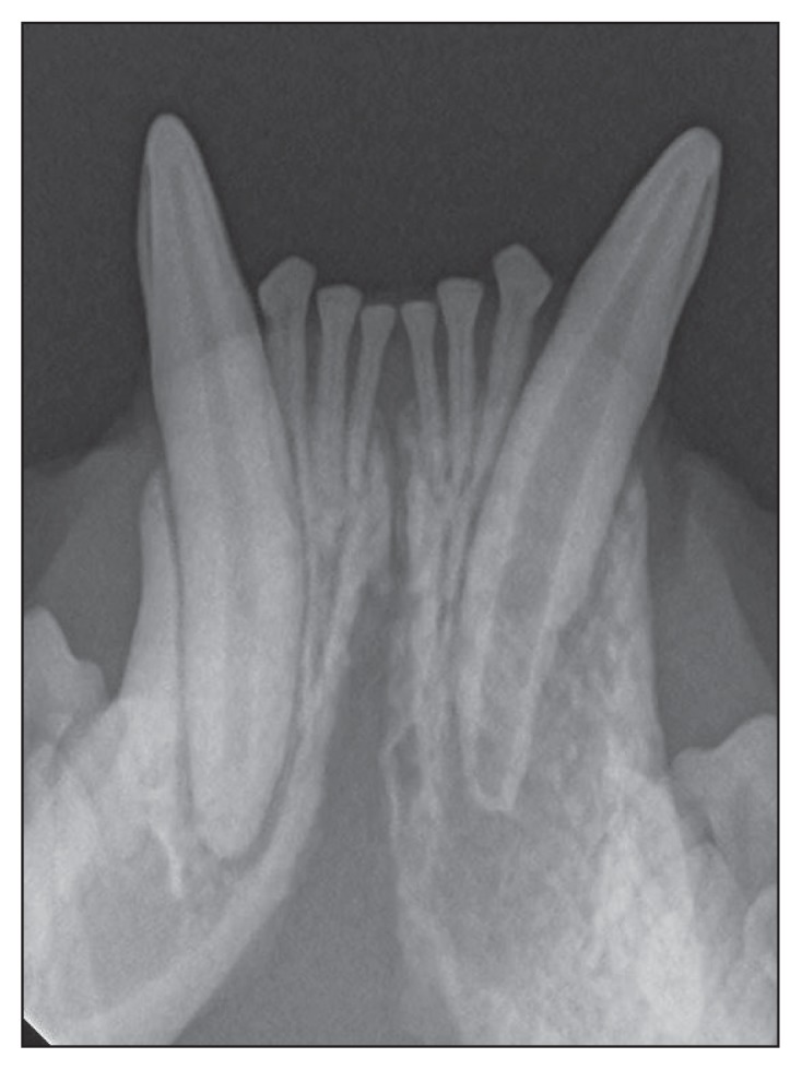
The diagnostic value of full-mouth radiography in cats and dogs was well-established over 2 decades ago (5,6). In these studies evaluating the merits of veterinary dental radiography, justification for routine use of full-mouth radiography in our patients was made based on not only confirmation of the clinical presenting problem, but also high likelihood of finding additional, clinically significant dental problems. In a study of 226 dogs, radiographs of teeth without lesions provided clinically important information in almost 1/3 of the cases. In the same study, radiographs of the teeth with clinical lesions uncovered additional information about half the time (5). The value of full-mouth radiography in dogs and cats has also been demonstrated to increase with patient age (5,6,10). Non-vital teeth are some of the more common unexpected, but clinically relevant findings on full-mouth radiographs. These teeth can appear clinically normal but are identified radiographically as having a wider pulp space than the same tooth on the opposite arc, due to the cessation of dentin formation by impaired or non-vital odontoblasts in the pulp (Figure 1). Supernumerary tooth roots and fused tooth roots are also typically incidental, clinically important findings on dental radiography in veterinary patients (Figures 2, 3). The previously mentioned study reported an incidence of supernumerary roots over 20% in large dogs (5). Without the radiographic evidence of supernumerary roots and fused tooth roots, one can easily envision difficulties in dental extractions including inadvertently missing the additional root and excess alveolar bone removal with fused roots. Dens invaginatus (DI), also known as dens in dente or tooth within a tooth is an uncommon developmental dental malformation found in teeth in which there is an infolding of enamel into dentin. These malformations may occur below the gingival margin and be small enough that they are not noted on clinical examination; therefore, diagnosis often requires the use of dental radiography (Figure 4). These invaginations result in incomplete covering of the dentin with enamel, ultimately exposing the endodontic system, which can lead to endodontic disease (Figure 5). Treatment options for DI include regular radiographic monitoring, surgical extraction, or endodontic therapy. One case report describes the endodontic treatment of a maxillary canine tooth with DI (11); however, it is the author’s experience and it has been previously reported that DI is more common in the mandibular 1st molar (12). Missing teeth are also a concern in veterinary patients and dental radiography is required to confirm this diagnosis. In a study of 226 dogs evaluated with dental radiography, 123 (54%) had a clinical finding of missing teeth, about 25% of which had retained tooth roots and 4% had impacted teeth (5). In a similar study, 87 of 115 cats (76%) were reported to have missing teeth and 72% had retained tooth roots. These are clinically relevant findings, as impacted teeth can lead to the formation of dentigerous cysts (Figure 6) and retained tooth roots commonly result in radiographically evident periapical pathology in cats and dogs (5,6,13; Figure 7). In a study of extraction sites of the maxillary 4th premolar in cats and dogs, the authors reported retained tooth root incidences of 93% and 82%, respectively (13). All of the extraction cases in that study were reported to be successful by the primary care veterinarian and about half of those patients had pre- and post-operative radiographs taken according to medical records (13). This study also pointed out that the high level of extraction failure could be explained by the fact that they were performed in a state in which technicians are legally permitted to perform dental extractions and these cases could not be identified for exclusion, as the medical records did not identify operators. In addition to the unexpected radiographic findings discussed here, several other oral and dental pathologies are also found including tooth/root resorption, root dilaceration, odontodysplasia, and neoplasia.
Figure 1.
Although 304 appears clinically similar to 404, this radiograph demonstrates cessation in dentinogenesis, external inflammatory root resorption and periapical bone loss; all indications of endodontic disease.
Figure 2.

This radiograph of the maxillary 3rd premolar reveals an additional root, which may be missed if extraction was performed without the use of radiographs.
Figure 3.

The left mandibular 2nd premolar (306) normally has 2 divergent tooth roots; however, in this radiograph, the roots are fused to form 1 large, abnormally shaped root.
Figure 4.

Dens invaginatus of 409 with no evidence of periapical disease.
Figure 5.

Dens invaginatus of 309 with radiographic evidence of periapical disease, indicating pulp necrosis.
Figure 6.

This impacted 405 caused dentigerous cyst formation in a mature dog. The expansion of the cyst results in loss of alveolar bone, damage to adjacent teeth, and could undergo neoplastic transformation.
Figure 7.

These retained tooth roots resulted in purulent discharge from a fistulous tract, indicating periapical abscess.
The importance of dental radiography cannot be overstated with the knowledge that significant pathology is commonly discovered only when full-mouth radiographs are performed in dogs and cats. Performing dental radiographs of diagnostic value and interpretation of those radiographs should be a core component of veterinary education. This is absolutely needed in order to provide veterinarians with the necessary tools required to diagnose and treat patients with dental ailments. As veterinarians we swear an oath which states, “to do no harm.” If we are performing dental evaluations and procedures without dental radiography, we are neglecting a large amount of pathology. Are we not then failing to abide by our sworn oath and thus performing malpractice?
Footnotes
Use of this article is limited to a single copy for personal study. Anyone interested in obtaining reprints should contact the CVMA office (hbroughton@cvma-acmv.org) for additional copies or permission to use this material elsewhere.
References
- 1.Stepaniuk K. Periodontology. In: Lobprise HB, Dodd JR, editors. Wiggs’s Veterinary Dentistry: Principles and Practice. 2nd ed. New York, New York: Wiley; 2019. pp. 81–108. [Google Scholar]
- 2.Harvey CE. Periodontal disease in dogs: Etiopathogenesis, prevalence and significance. Vet Clin North Am Small Anim Pract. 1998;28:1165–1188. doi: 10.1016/s0195-5616(98)50105-2. [DOI] [PubMed] [Google Scholar]
- 3.Kortgaard HE, Eriksen T, Baelum V. Periodontal disease in research beagles: An epidemiological study. J Small Anim Pract. 2008;49:610–616. doi: 10.1111/j.1748-5827.2008.00609.x. [DOI] [PubMed] [Google Scholar]
- 4.Soukup JW, Hetzel S, Paul A. Classification and epidemiology of traumatic dentoalveolar injuries in dogs and cats: 959 injuries in 660 patient visits (2004–2012) J Vet Dent. 2015;32:6–14. doi: 10.1177/089875641503200101. [DOI] [PubMed] [Google Scholar]
- 5.Verstraete FJM, Kass PH, Terpak CH. Diagnostic value of full-mouth radiography in dogs. Am J Vet Res. 1998;59:686–691. [PubMed] [Google Scholar]
- 6.Verstraete FJM, Kass PH, Terpak CH. Diagnostic value of full-mouth radiography in cats. Am J Vet Res. 1998;59:692–695. [PubMed] [Google Scholar]
- 7.Anderson JG, Goldstein G, Boudreaux K, Ilkiw JE. The state of veterinary dental education in North America, Canada and the Caribbean: A descriptive study. J Vet Med Educ. 2017;42:358–363. doi: 10.3138/jvme.1215-204R. [DOI] [PubMed] [Google Scholar]
- 8.Perry R. Final year veterinary students’ attitudes towards small animal dentistry: A questionnaire-based survey. J Small Anim Pract. 2014;55:457–464. doi: 10.1111/jsap.12258. [DOI] [PubMed] [Google Scholar]
- 9.Clark WT, Kane l, Arnold PK, Robertson ID. Clinical skills and knowledge used by veterinary graduates during their first year in small animal practice. Aust Vet J. 2002;80:37–40. doi: 10.1111/j.1751-0813.2002.tb12830.x. [DOI] [PubMed] [Google Scholar]
- 10.Kim CG, Lee SY, Kim JW, Park HM. Assessment of dental abnormalities by full-mouth radiography in small breed dogs. J Am Anim Hosp Assoc. 2013;49:23–30. doi: 10.5326/JAAHA-MS-5830. [DOI] [PubMed] [Google Scholar]
- 11.Coffman CR, Visser CJ, Visser L. Endodontic treatment of dens invaginatus in a dog. J Vet Dent. 2009;26:220–225. doi: 10.1177/089875640902600409. [DOI] [PubMed] [Google Scholar]
- 12.Stein KE, Manfra SM. Dens invaginatus of the mandibular first molars in a dog. J Vet Dent. 2005;22:21–25. doi: 10.1177/089875640502200103. [DOI] [PubMed] [Google Scholar]
- 13.Moore JI, Niemiec B. Evaluation of extraction sites for evidence of retained tooth roots and periapical pathology. J Am Anim Hosp Assoc. 2014;50:77–82. doi: 10.5326/JAAHA-MS-5977. [DOI] [PubMed] [Google Scholar]