Abstract
Objectives
This study aims to evaluate the anti-arthritic effect of orally administered niclosamide (NCL) on collagen-induced arthritis (CIA) in rats.
Materials and methods
The study included 35 Sprague Dawley rats (age range, 3 to 4 months; average weight, 100±10 g) of which seven were used as a negative control group (group A) whereas 28, in which arthritis was induced by injection of collagen type II emulsified by incomplete Freund's adjuvant and which were considered as CIA rats, were randomly divided equally into four groups and treated for 28 days with: normal saline (group B), low-dose NCL (group C), high-dose NCL (group D), and diclofenac sodium (group E). Body weight, arthritis index, ankle swelling, and footpad thickness were monitored before and after treatment in all groups. At the end of the treatment period, serum levels of tumor necrosis factor alpha (TNF-α), interleukin 1 beta (IL-1β), and IL-6 were measured together with a collection of articular synovial tissue to evaluate the pathological changes.
Results
After four weeks of treatment period, a high dose of orally administered NCL significantly reduced the arthritis index, footpad thickness, and ankle swelling. Significantly decreased serum levels of inflammatory biomarkers including TNF-α, IL-1β, and IL-6 were observed in rats treated with high-dose oral NCL or intramuscular injection of diclofenac sodium, compared with groups B and C. Histopathological examination revealed that a high dose of NCL significantly reduced the infiltration of inflammatory cells, synovial hyperplasia, and bone and cartilage destruction.
Conclusion
Niclosamide can effectively decrease the clinical scores, joint swelling, inflammatory markers, and pathological changes in arthritic rats.
Keywords: Collagen-induced arthritis, niclosamide, rheumatoid arthritis
Introduction
Rheumatoid arthritis (RA) is a long-lasting autoimmune disease that mainly affects the joints. It is characterized by inflammation of the synovium, destruction of joints, muscle atrophy, and bone erosion. Other organs of the body which may be involved include the respiratory system, eyes, vascular systems, and skin.[1] According to the treatment guidelines, management of RA includes several classes of agents, including non-steroidal anti-inflammatory drugs (NSAIDs), non-biologic and biologic disease-modifying antirheumatic drugs (DMARDs), immunosuppressants, and corticosteroids.[2,3] However, all the above medications have a wide range of risky side effects and some of them are costly, thus their use is limited. As a result, the development of new therapies with fewer side effects and lower costs should be a continuous effort to treat RA.
During the last decade, many researches have been performed regarding niclosamide (NCL), which is an old anthelmintic drug approved by the Food and Drug Administration (FDA), exploring its biological activity against multiple inflammatory diseases.[4] The drug has fewer side effects with a wide margin of safety even when used for long periods of time.[5]
The anti-inflammatory effect of NCL is mainly due to its ability in reducing cytokine expression and release from tumor necrosis factor alpha (TNF-α)-induced human RA fibroblast-like synoviocytes (FLS) in a dose-dependent manner. NCL treatment inhibited serum-induced synoviocyte migration and invasion and produced alterations in the filamentous-actin cytoskeletal network in these cells. It exerts this effect by decreasing TNF-α-stimulated mitogen-activated protein (MAP) kinase and I kappa B kinase/nuclear factor- kappa B (IKK/NF-κB) signaling activity in synoviocytes.[6]
Accordingly, this animal experimental study was designed to investigate the efficacy of orally administered NCL and compare it with the efficacy of diclofenac sodium injection. The collagen-induced arthritis (CIA) model is considered as an essential instrument used in the testing and development of new drugs that have an anti-inflammatory effect, such as those that target TNF-α, a cytokine produced by macrophages and T cells which are dominant inflammatory mediators in the pathogenesis of RA.[7]
Non-steroidal anti-inflammatory drugs such as diclofenac sodium, ibuprofen, and celecoxib act rapidly to suppress inflammation, thereby reducing pain and swelling. They may be useful as a ‘bridging therapy’ to control symptoms in the first few weeks after diagnosis while the slower-acting DMARDs are taking effect.[8] In this study, we used diclofenac sodium injection as standard therapy to compare the response of animals to different doses of orally administered NCL. Therefore, in this study, we aimed to evaluate the anti-arthritic effect of orally administered NCL on CIA in rats.
Patients and Methods
Fifty Sprague Dawley rat pups (age range, 3 to 4 months; average weight, 100±10 g) were included in the study, which was conducted at Department of Clinical Pharmacology, College of Medicine, Al-Mustansiriyha University between July 2017 and November 2017. The rats were housed in plastic cages containing hardwood chips for bedding. The bedding was changed weekly to ensure a clean environment. The animals were maintained in an air-conditioned room at 25±2°C, with 14/10-hour daily light/dark cycle and housed under standard laboratory conditions with food and water provided ad libitum. After seven days of acclimatization, the animals were evenly randomized into two groups as CIA model group (n=40) and negative control group (n=10). Animal experiments were performed in accordance with the guide for the care and use of laboratory animals and approved by the Al-Mustansiriyha Medical College scientific committee.
Immunization grade bovine type II collagen and incomplete Freund's adjuvant (IFA) were purchased from Chondrex Inc., (Redmond, WA, USA). NCL was obtained from Alexandria Co. For Pharmaceuticals and Chemical Industries (Alexandria, Egypt). Normal saline 0.9% solution was obtained from Pioneer Co. (Sulaymaniyah, Iraq). C-reactive protein (CRP), serum interleukin-1 beta (IL-1β), serum IL-6, and serum TNF-α rats enzyme-linked immunosorbent assay (ELISA) kits, were purchased from Chondrex, Inc. (Redmond, WA, USA). Ketamine solution was also utilized Hikma Pharmaceuticals PLC (Amman, Jordan).
Arthritis was induced in 40 rats using the CIA model by injection of 0.2 mL of prepared collagen-IFA emulsion at the base of the tail for each rat. A second booster dose was used after one week to increase the incidence of arthritis. Rats in the negative control group were injected with an equal volume of normal saline.[9]
Niclosamide solution was freshly prepared daily to be used by oral gavage. First, NCL tablet (500 mg) was triturated into powder using mortar and pestle. Then, the powder was transferred into the plain tube to prepare a stock solution by 10 mL of normal saline. From this stock solution, the different dose could be obtained by dilution with normal saline.
The evaluation of RA in rats was started on day 0 before the induction of arthritis and once weekly over 28 days. The assessment was performed by measuring the thickness of palm pads of both forelimbs and footpads of both hindlimbs. A Vernier caliper was used for measuring technique. Arthritis index (AI) was also determined qualitatively using Chondrex scoring system according to the following interpretation: 0: normal, 1: mild swelling and redness limited to single joint, 2: moderate swelling and redness limited to ankle and wrist, 3: severe redness and swelling of wrist or ankle, and 4: severe inflammation and swelling of the entire limb affecting multiple joints.[10]
Two weeks after the start of experiment, 28 rats with successfully developed RA in the CIA group were randomly divided into four groups of seven rats in each (groups B, C, D, and E) while group A included seven rats as the negative control group. All groups were treated for 28 days with the following regimes: group A: received no treatment, group B: treated with oral normal saline, group C: treated with NCL orally (50 mg/kg), group D: treated with NCL orally (100 mg/kg), and group E: treated with diclofenac sodium (10 mg/kg) intramuscular (IM). The tail of the rats in each group was marked with different color inks and the body weights were measured each week to demonstrate the changes exerted by treatment regimens.
At the end of the study, all the following parameters were measured and compared with their value before and after the experiment to demonstrate the effect of NCL on the treatment of RA.
Arthritis severity was determined at baseline and after the treatment period (after 20 days) according to the AI scoring mentioned above.[9] All measurements were obtained blindly by two Master of Science students and the average was taken for each assessment.
Ankle and paw swelling were determined by measuring the hind footpad and ankle thickness using Vernier caliper for the measuring technique. Paw swelling was expressed as the average thickness of both hind paws in millimeters.
The rats were anesthetized using ketamine (50 mg/kg, intraperitoneal [IP]). Then, blood sample of a volume of 3-5 mL was collected from individual animals at baseline and at time of sacrifice by cardiac puncture into plain tubes and left to coagulate at room temperature for at least 30 minutes and centrifuged for 10 minutes at 4,000 rpm to obtain serum. Using ready-made ELISA kits, the resultant serum was utilized for the measurement of CRP, TNF-α, IL-1β, and IL-6 (Shanghai Biological Science Inc., Shanghai, China).
Evaluation of the pathological changes in the joints was performed at the Department of Pathology, Al-Mustansiriyah University, College of Medicine. The ankle joints of each rat were fixed in 10% formalin for 14 days, decalcified with CalEx decalcification solution (Fisher Scientific, Ontario, Canada) for 35 days, and processed in paraffin. The fixed tissues of the ankle joints were longitudinally cut into 5 μm sections and stained with hematoxylin and eosin. Grading of cellular infiltration, synovial hyperplasia, pannus formation, joint space narrowing, and cartilage and bone erosion of the ankle joints was blindly investigated by a pathologist.
Statistical analysis
Data were analyzed using the IBM SPSS version 20.0 (IBM Corp., Armonk, NY, USA) and presented as mean ± standard deviation. Paired Student's t-test and one-way analysis of variance followed by Dunnett’s post hoc test were used. P values of 0.05 or lower were considered as statistically significant.
Results
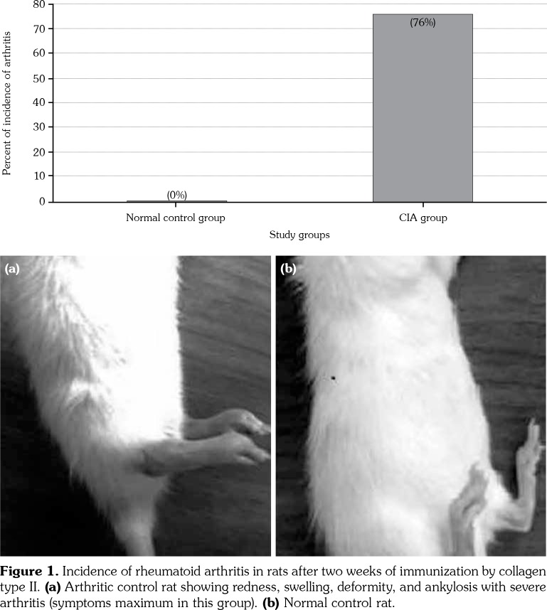
The incidence of RA in the two groups was calculated after two weeks. Figure 1 demonstrates the percent of RA incidence after induction by collagen which was very high in the CIA group (n=28). Group A showed no incidence of RA (0%) with a highly significant difference (p<0.001) compared to the CIA group. Only rats with AI scores of 4-5 were included in the CIA group. Severe signs of arthritis were observed clearly in the CIA group including swelling, redness, deformity, and ankylosis in the hind paws, forelimbs and ankle joints (Figure 1a). The hind paw of normal rats showed a significant difference from the hind paw of arthritic control rats with normal thickness and color (Figure 1b). A significant change was observed in the palm and footpad’s thickness of both left and right hindlimbs of CIA rats which were clearly thicker than pads of normal control rats (p<0.05) (Table 1). Change in ankle swelling was also studied; Table 1 shows a highly significant increase in the ankle thickness of all four limbs of CIA rats after two weeks of RA induction which were thicker than the corresponding group A (p<0.001).
Table 1. Changes in palms, footpads, ankles of forelimbs and hindlimbs after induction of arthritis.
| Study groups | ||||||
| Normal control group | CIA group | |||||
| Baseline | After 2 weeks | Baseline | After 2 weeks | |||
| Mean±SD | Mean±SD | p | Mean±SD | Mean±SD | p | |
| Right palm | 2.1±0.1 | 2.3±0.2 | 0,35 | 2.5±0.2 | 2.8±0.2 | 0.013* |
| Left palm | 2.1±0.1 | 2.3±0.2 | 0,12 | 2.5±0.2 | 3.2±0.3 | 0.015* |
| Right footpad | 3.0±0.0 | 3.2±0.1 | 0,12 | 3.7±0.1 | 5.8±0.4 | 0.001† |
| Left footpad | 3.1±0.1 | 3.2±0.1 | 0,44 | 3.9±0.2 | 5.7±0.4 | 0.004* |
| Ankle thickness of right forelimb | 3.7±0.3 | 3.7±0.4 | 0,54 | 3.7±0.4 | 4.2±0.5 | 0.001† |
| Ankle thickness of left forelimb | 3.7±0.4 | 3.7±0.3 | 0,26 | 3.7±0.4 | 4.2±0.5 | 0.001† |
| Ankle thickness of right hindlimb | 5.9±0.7 | 6.1±0.7 | 0,46 | 5.6±0.7 | 7.6±1.4 | 0.001† |
| Ankle thickness of left hindlimb | 5.8±0.7 | 6.1±1.2 | 0,45 | 5.7±0.6 | 7.7±1.4 | 0.001† |
| CIA: Collagen-induced arthritis; SD: Standard deviation; * p<0.05; † p<0.001. | ||||||
Figure 1. Incidence of rheumatoid arthritis in rats after two weeks of immunization by collagen type II. (a) Arthritic control rat showing redness, swelling, deformity, and ankylosis with severe arthritis (symptoms maximum in this group). (b) Normal control rat.
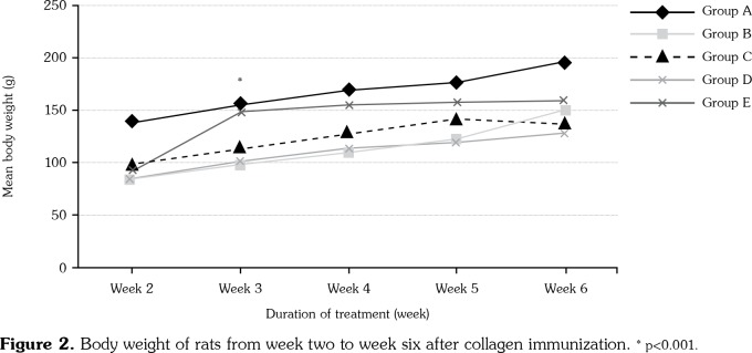
Significant changes were observed in body weight between group A and other groups in the second week before starting the treatment (p<0.001). An insignificant increase was observed in body weight in groups B, C, and D during the course of the treatment. An absolute increment was found in weight of groups A and E during the course of the treatment with no significant difference (p>0.05) (Figure 2). Rats in groups C and D had significant differences in weight compared to group A (p<0.001).
Figure 2. Body weight of rats from week two to week six after collagen immunization. * p<0.001.
A significant elevation in the mean arthritic score was observed in CIA rats (group B) from the second week (3.71±0.48) to the sixth week (3.43±0.53) during the development of RA. Group C showed an insignificant decline in AI during the course of therapy with no significant changes at the end of the experiment compared to positive control group (group B). In contrast, group D and E rats showed a significant decrease in AI during the development of arthritis compared to group B (Table 2).
Table 2. Mean AI scores of rats from week two to week six after collagen immunization.
| Arthritis index | ||||||
| Week 2 | Week 3 | Week 4 | Week 5 | Week 6 | ||
| n | Mean±SE | Mean±SE | Mean±SE | Mean±SE | Mean±SE | |
| Group A | 7 | 00.00±00.00 | 00.00±00.00 | 00.00±00.00 | 00.00±00.0 | 00.00±00.0 |
| Group B | 7 | 3.7±0.9* | 3.7±0.5* | 3.0±0.6* | 3.3±0.5 | 3.4± 0.5 |
| Group C | 7 | 3.3±0.9* | 3.3±0.5* | 2.7±0.5* | 3.0±0.6 | 2.9±0.7 |
| Group D | 7 | 3.7±0.9* | 3.7±0.5* | 3.4±0.5* | 2.4±0.5† | 2.6±1.1† |
| Group E | 7 | 3.9±0.4* | 3.9±0.4* | 2.4±0.4* | 2.0±0.0† | 1.1±0.9† |
| AI: Arthritis index; SE: Standard error; * p<0.05 compared to group A; † p>0.05 compared to group B. | ||||||
Group D had significantly decreased mean serum levels of TNF-α and IL-1β compared to group B (p<0.05) with insignificant changes in the level of IL-6 (p>0.05). On the other hand, low dose of NCL caused a significant difference only in the serum level of IL-1β (p<0.05) compared to group B, with no significant changes in the other two cytokines. A unique result was observed in rats treated with IM injection of diclofenac sodium (group E) for four weeks with a significant reduction in TNF-α, IL-1β, and IL6, with significant differences in the mean of serum level of these parameters compared to group B (Table 3).
Table 3. Effect of NCL on inflammatory biomarkers on collagen induced RA in rats.
| TNF (n=7) | IL-6 (n=7) | IL-1 b (n=7) | |
| Group | Mean±SD | Mean±SD | Mean±SD |
| Group A | 32.4 ±7.1a | 19.8±13.5a | 786.5±453.9a |
| Group B | 56.5±16.5 | 225.1±144.0 | 2206.3±900.3b |
| Group C | 56.7±17.0 | 142.6±131.0 | 1280.0±626.2a |
| Group D | 33.3±8.9a | 131.3±89.7 | 1112.1±540.0a |
| Group E | 30.7±8.5a,b | 101.1±80.2b | 704.8±90.2a,b |
| NCL: Niclosamide; RA: Rheumatoid arthritis; TNF: Tumor necrosis factor; IL: Interleukin; SD: Standard deviation; a p<0.05 comparing to group B. b p<0.05 comparing to baseline before starting treatment. | |||
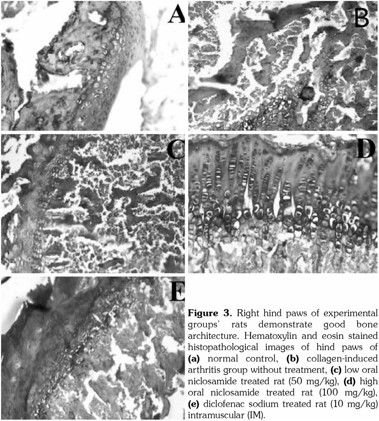
At the end of the experiment, histopathological examination of ankle joints from each group was investigated by a pathologist. Slides from normal rats demonstrated normal cartilage thickness, smooth and continuous lining of the joint surfaces without any bone or cartilage destruction. While light microscope demonstrated the accumulation of a large number of inflammatory cells, a fragment of eroded cartilage and pannus formation was noted in group B. A tissue sample from ankle joints in group C showed the absence of pannus formation; but still, destruction was present in the cartilage with infiltration of inflammatory cells. On the other hand, microscopical examination of the joints from group D showed normal cartilage with inflammatory cell infiltration. Group E did not demonstrate any high-level pathological changes in the joint, having only minor infiltration of inflammatory cells (Figure 3).
Figure 3. Right hind paws of experimental groups’ rats demonstrate good bone architecture. Hematoxylin and eosin stained histopathological images of hind paws of (a) normal control, (b) collagen-induced arthritis group without treatment, (c) low oral niclosamide treated rat (50 mg/kg), (d) high oral niclosamide treated rat (100 mg/kg), (e) diclofenac sodium treated rat (10 mg/kg) intramuscular (IM).
Discussion
Collagen-induced arthritis animal models are important in understanding the mechanism of RA and the development of effective therapy for its optimal management.[11] Several studies have demonstrated that involvement of both humoral and cellular immune responses are needed for the development of CIA.[12-14] NCL is an FDA approved anthelmintic that has a wide safety profile with little side effects, which allow using the drug safely and for long periods in the treatment of chronic diseases.[4]
In 2015, Liang et al.[6] evaluated the anti- inflammatory effect of NCL. They demonstrated the ability of this drug in reducing cytokine expression and release from TNF-α-induced human RA FLS in a dose-dependent manner. Their research also confirmed the anti-inflammatory effect of NCL in vivo by treated CIA mice with an IP injection of NCL (30 mg/kg/day). The pharmacokinetic parameters of NCL have also been studied in rats when administered orally. The drug was rapidly absorbed in less than 30 minutes and had 10% oral bioavailability when given in a dose of 5 mg/kg.[15] Accordingly, this study was designed to evaluate the efficacy of orally administered NCL both in high and low doses in the treatment of arthritis induced in rats and compare their effect with NSAID (diclofenac sodium). Oral treatment of NCL caused a reduction in AI, footpad thickness, and ankle swelling when used in a high dose for 28 days with no significant differences from treatment with diclofenac sodium when given daily as IM injection over the same period. These findings support the anti-inflammatory effect of NCL that has been explored by another research, which demonstrated the ability of NCL to inhibit serum-induced synoviocyte migration and invasion and produced alterations in the filamentous-actin cytoskeletal network in these cells. NCL might exert this effect by decreased TNF-α-stimulated MAP kinase and IKK/NF-κB signaling activity in synoviocytes.[6] The most important pro-inflammatory cytokines involved in the pathogenesis of RA are TNF-α and IL-6. However, IL-1, IL-17, and vascular endothelial growth factor (VEGF) also play an important role in the disease process of RA.[16]
Treatment of arthritis with high-dose NCL (100 mg/kg) in CIA rats caused a significant reduction in TNF-α and IL-1β, with an insignificant decline in IL-6 indicating the powerful anti-inflammatory effects exerted by NCL. This result agrees with another study which showed that NCL potently suppressed the release of pro-inflammatory cytokines (TNF-α, IL-6, and IL-12) in cultured dendritic cells when induced by lipopolysaccharide.[17] In addition, the pathological changes in the ankle joint exerted by collagen type II were also modified by NCL which ameliorated the synovial hyperplasia, infiltration of the inflammatory cell, pannus formation, and cartilage and bone formation. These observations are consistent with another study that found marked reduction in the severity of joint arthritis in mice (induced by collagen type II) when treated with a once-daily IP injection of NCL for 14 days. The drug significantly decreased the clinical scoring, hyperplasia, bone destruction, and pathological changes in the arthritic group compared to normal healthy mice.[6] The therapeutic efficacy of orally administered NCL may be due to the reduction in the inflammatory mediators (TNF-α, IL-1β) suggesting the possibility of using this drug in the treatment of RA.
In RA, angiogenesis is responsible for stabilizing the chronic inflammatory state by transporting inflammatory cells to the site of inflammation and providing nutrients and oxygen to the proliferating inflamed tissue. These new blood vessels are diffused in the synovial membrane and allow the invasion of this tissue supporting the active infiltration of the synovial membrane into cartilage and resulting in erosions and destruction of the cartilage.[18] A recent study showed that NCL effectively inhibits inflammation and angiogenesis through the suppression of VEGF-induced angiogenesis both in vitro and in vivo.[19] Further clinical studies are required to evaluate the efficacy of NCL in the treatment of RA.
In conclusion, to our knowledge, this is the first study to report that orally administered NCL can reduce the severity of arthritis in arthritic animals induced by collagen type II by lowering the inflammatory biomarkers including the TNF-α and IL-1β when used for a 28-day period. Moreover, NCL has the ability to significantly reduce the clinical scores, ankle swelling, synovial hyperplasia, infiltration of the inflammatory cells, and bone and cartilage destruction. However, despite these promising findings, further animal and clinical studies are required to confirm the efficacy and safety of NCL as an anti-rheumatoid drug when used for long periods.
Footnotes
Conflict of Interest: The authors declared no conflicts of interest with respect to the authorship and/or publication of this article.
Financial Disclosure: The authors received no financial support for the research and/or authorship of this article.
References
- 1.Joshi VR. Rheumatology, past, present and future. J Assoc Physicians India. 2012;60:21–24. [PubMed] [Google Scholar]
- 2.Aletaha D, Funovits J, Keystone EC, Smolen JS. Disease activity early in the course of treatment predicts response to therapy after one year in rheumatoid arthritis patients. Arthritis Rheum. 2007;56:3226–3235. doi: 10.1002/art.22943. [DOI] [PubMed] [Google Scholar]
- 3.Verschueren P, Esselens G, Westhovens R. Predictors of remission, normalized physical function, and changes in the working situation during follow-up of patients with early rheumatoid arthritis: an observational study. Scand J Rheumatol. 2009;38:166–172. doi: 10.1080/03009740802484846. [DOI] [PubMed] [Google Scholar]
- 4.Chen W, Mook RA Jr, Premont RT, Wang J. Niclosamide: Beyond an antihelminthic drug. Cell Signal. 2018;41:89–96. doi: 10.1016/j.cellsig.2017.04.001. [DOI] [PMC free article] [PubMed] [Google Scholar]
- 5.Andrews P, Thyssen J, Lorke D. The biology and toxicology of molluscicides, Bayluscide. Pharmacol Ther. 1982;19:245–295. doi: 10.1016/0163-7258(82)90064-x. [DOI] [PubMed] [Google Scholar]
- 6.Liang L, Huang M, Xiao Y, Zen S, Lao M, Zou Y, et al. Inhibitory effects of niclosamide on inflammation and migration of fibroblast-like synoviocytes from patients with rheumatoid arthritis. Inflamm Res. 2015;64:225–233. doi: 10.1007/s00011-015-0801-5. [DOI] [PubMed] [Google Scholar]
- 7.Brand DD, Latham KA, Rosloniec EF. Collagen- induced arthritis. Nat Protoc. 2007;2:1269–1275. doi: 10.1038/nprot.2007.173. [DOI] [PubMed] [Google Scholar]
- 8.Curtis JR, Singh JA. Use of biologics in rheumatoid arthritis: current and emerging paradigms of care. Clin Ther. 2011;33:679–707. doi: 10.1016/j.clinthera.2011.05.044. [DOI] [PMC free article] [PubMed] [Google Scholar]
- 9.Wang Y, Lu Yan, Wang J, Shen Z, Liu H, Ma W, et al. Preparation and analysis of active rat model of rheumatoid arthritis with features of TCM toxic heat-stasis painful obstruction. Journal of Traditional Chinese Medical Sciences. 2015;2:166–172. [Google Scholar]
- 10.Mossiat C, Laroche D, Prati C, Pozzo T, Demougeot C, Marie C. Association between arthritis score at the onset of the disease and long-term locomotor outcome in adjuvant-induced arthritis in rats. Arthritis Res Ther. 2015;17:184–184. doi: 10.1186/s13075-015-0700-8. [DOI] [PMC free article] [PubMed] [Google Scholar]
- 11.Asquith DL, Miller AM, McInnes IB, Liew FY. Animal models of rheumatoid arthritis. Eur J Immunol. 2009;39:2040–2044. doi: 10.1002/eji.200939578. [DOI] [PubMed] [Google Scholar]
- 12.Svensson L, Jirholt J, Holmdahl R, Jansson L. B cell- deficient mice do not develop type II collagen-induced arthritis (CIA) Clin Exp Immunol. 1998;111:521–526. doi: 10.1046/j.1365-2249.1998.00529.x. [DOI] [PMC free article] [PubMed] [Google Scholar]
- 13.Saijo S, Asano M, Horai R, Yamamoto H, Iwakura Y. Suppression of autoimmune arthritis in interleukin-1- deficient mice in which T cell activation is impaired due to low levels of CD40 ligand and OX40 expression on T cells. Arthritis Rheum. 2002;46:533–544. doi: 10.1002/art.10172. [DOI] [PubMed] [Google Scholar]
- 14.Yamaguchi N, Ohshima S, Umeshita-Sasai M, Nishioka K, Kobayashi H, Mima T, et al. Synergistic effect on the attenuation of collagen induced arthritis in tumor necrosis factor receptor I (TNFRI) and interleukin 6 double knockout mice. J Rheumatol. 2003;30:22–27. [PubMed] [Google Scholar]
- 15.Chang Y, Yeh T, Lin K, Chen W, Yao H, Lan S, et al. Pharmacokinetics of anti-SARS-CoV agent niclosamide and its analogs in rats. Journal of Food and Drug Analysis. 2006;14:329–333. [Google Scholar]
- 16.Pickens SR, Volin MV, Mandelin AM, Kolls JK, Pope RM, Shahrara S. IL-17 contributes to angiogenesis in rheumatoid arthritis. J Immunol. 2010;184:3233–3241. doi: 10.4049/jimmunol.0903271. [DOI] [PMC free article] [PubMed] [Google Scholar]
- 17.Wu CS, Li YR, Chen JJ, Chen YC, Chu CL, Pan IH, et al. Antihelminthic niclosamide modulates dendritic cells activation and function. Cell Immunol. 2014;288:15–23. doi: 10.1016/j.cellimm.2013.12.006. [DOI] [PMC free article] [PubMed] [Google Scholar]
- 18.Marrelli A, Cipriani P, Liakouli V, Carubbi F, Perricone C, Perricone R, et al. Angiogenesis in rheumatoid arthritis: a disease specific process or a common response to chronic inflammation. Autoimmun Rev. 2011;10:595–598. doi: 10.1016/j.autrev.2011.04.020. [DOI] [PubMed] [Google Scholar]
- 19.Huang M, Qiu Q, Zeng S, Xiao Y, Shi M, Zou Y, et al. Niclosamide inhibits the inflammatory and angiogenic activation of human umbilical vein endothelial cells. Inflamm Res. 2015;64:1023–1032. doi: 10.1007/s00011-015-0888-8. [DOI] [PubMed] [Google Scholar]


