Abstract
Background
The purpose of this study was to define the relationship of the axillary and radial nerves, particularly how these are affected with changing arm position.
Methods
Twenty cadaveric shoulders were dissected, identifying the axillary and radial nerves. Distances between the latissimus dorsi tendon and these nerves were recorded in different shoulder positions. Positions included adduction/neutral rotation, abduction/neutral rotation for the axillary nerve, adduction/internal rotation, adduction/neutral rotation, adduction/external rotation, and abduction/external rotation for the radial nerve.
Results
Width of the latissimus tendon at its humeral insertion was 29.3 ± 5.7 mm. Mean distance from the latissimus insertion to the axillary nerve in adduction/neutral rotation was 24.2 ± 7.1 mm, the distance increased to 41.1 ± 9.8 mm in abduction/neutral rotation. Mean distance from the latissimus insertion to the radial nerve was 15.3 ± 5.5 mm with adduction/internal rotation, 25.8 ± 6.9 mm in adduction/neutral rotation, and 39.5 ± 6.8 mm in adduction/external rotation. Mean distance increased with abduction/external rotated 51.1 ± 7.4 mm.
Conclusions
Knowing the axillary and radial nerve locations relative to the latissimus dorsi tendon decreases the risk of iatrogenic nerve injury. Understanding the dynamic nature of these nerves related to different shoulder positions is critical to avoid complications.
Keywords: latissimus dorsi, radial nerve, axillary nerve, tendon transfer, revision shoulder arthroplasty
Introduction
The deltopectoral approach provides an internervous and intermuscular plane through which a variety of shoulder procedures can be performed.1 It also allows for distal extension to improve visualization, and access to distal aspects of the humeral shaft, which becomes of particular utility when exposure of the proximal humerus is not sufficient.
Among the procedures which typically necessitate a more extensile approach, revision shoulder arthroplasty is inherently associated with a higher risk of complications than primary shoulder arthroplasty.2,3 Intraoperative periprosthetic humeral fractures occur with significantly greater frequency in revision shoulder arthroplasty procedures, and studies have noted an associated risk of radial nerve palsy.4,5 Similarly, periprosthetic humerus fractures following prior shoulder arthroplasty have been shown to result in radial nerve palsy as high as 28.57%, either as a result of the injury or the subsequent surgical management.5,6
In the setting of massive, nonrepairable rotator cuff tears with teres minor deficiency, latissimus dorsi transfer (with or without teres major) can be used, to restore external rotation.7,8 Boileau et al.9 demonstrated improvement of forward elevation and external rotation following reverse total shoulder arthroplasty combined with latissimus dorsi and teres major transfer for treatment of patients with a massive rotator cuff tear, psuedoparalysis, as well as loss of active external rotation. These findings corroborate those shown in other clinical studies, suggesting this procedure can safely and effectively be performed through a single extended deltopectoral incision.10,11 Similarly, cadaveric latissimus transfer studies have found adequate muscule/tendon excursion for transfer to either greater tuberosity or lesser tuberosity for posterior or anterior cuff deficiency, respectively.12–14
Though not routinely managed with surgery, tears of the latissimus dorsi tendon have been reported in certain patient populations, namely involving the throwing shoulder in baseball pitchers. Tendon repair with suture anchors has been described, highlighting the relevancy of the anatomic relationship of latissimus dorsi tendon to nearby neurologic structures.15
Numerous studies have contributed to our knowledge of the anatomic course as well as clinical relevance of the axillary and radial nerves with the shoulder. The spatial relationship of the radial nerve to various landmarks about the shoulder has been well studied in the literature, particularly relating to the course of the radial nerve as it traverses the spiral groove on the posterior aspect of the humerus.16–21 Similarly, location of the axillary nerve has been of particular clinical interest as it courses from posterior to anterior along the lateral humerus, deep to the deltoid muscle belly.18,22 Many of these studies are limited by static measurement reporting, while true anatomic relationships change in a dynamic fashion based on position of the extremity.
Ultimately, knowledge of the anatomic relationships and course of the axillary and radial nerves in shoulder surgery is critical to avoiding iatrogenic complications. The latissimus dorsi tendon is of surgical importance, particularly in the extensile deltopectoral approach to the shoulder. The purpose of this cadaveric study was to better define distances between the insertion of the latissimus dorsi tendon with the axillary and radial nerves, as well as how these relationships are affected by variations in shoulder position.
Methods
This anatomic study utilized 20 shoulders from 10 fresh-frozen cadaveric torsos with intact bilateral upper extremities from 9 males and 1 female with a mean age of 70.6 years (range = 57–92 years). Mean donor height was 68.5 inches (range = 63–73 inches) and mean donor weight was 148.1 pounds (range =103–188 pounds), representing a mean body mass index (BMI) of 22.2 kg/m2 (range = 16.3–30.2 kg/m2). Of the 20 total shoulders, 10 right-sided and 10 left-sided, none had any evidence of prior surgery, trauma, or obvious gross deformity.
The specimens were dissected through an extended deltopectoral incision. The insertion of the pectoralis major tendon onto the humerus was identified, transected from its humeral insertion, and reflected for better visualization. The same was done with the origin of the conjoined tendon at the coracoid process. The musculotendinous junction of the latissimus dorsi was identified and followed distally to its tendinous insertion onto the humerus. This insertion site was tagged with suture at a point measured to be the middle of the tendon with respect to its superior–inferior width. The axillary artery and brachial plexus was identified, dissected, and the paths of the axillary and radial nerves were followed after bifurcating from the posterior cord of the brachial plexus. The axillary nerve was seen to travel laterally and posteriorly, beneath the glenohumeral joint capsule, remaining superior to the latissimus dorsi tendon. The radial nerve was seen to travel laterally and distally, coursing anterior to the latissimus dorsi tendon before diving posteriorly toward the spiral groove on the posterior aspect of the humeral shaft at the level of the deltoid insertion.
All anatomic relationships were analyzed using silk suture placed in situ and cut to match the length of each respective anatomic measurement. Digital calipers were then used to measure the length of the silk suture, rounded to the nearest millimeter. All measurements were confirmed by a minimum of two observers. The superior–inferior width of the latissimus dorsi tendon at its insertion onto the humeral shaft was also measured in a similar fashion. Distances were recorded from the latissimus dorsi tendon insertion to the axillary nerve in both adduction with neutral rotation and abduction with neutral rotation, taking the shortest linear path. Finally, distance was measured from the latissimus dorsi tendon insertion to the radial nerve as it crossed the midpoint of the latissimus dorsi in different shoulder positions, including adduction internal rotation, adduction neutral rotation, adduction external rotation, and abduction external rotation.
Descriptive statistics were calculated including mean, standard deviations, and range including minimum and maximum values. The data were further analyzed using a Student’s paired t-test for analysis of the axillary nerve and ANOVA for the radial nerve, with statistical significance set at p < 0.05.
Results
In all 20 dissected shoulders, the axillary and radial nerves were found to branch from the posterior cord of the brachial plexus. The course of the axillary nerve consistently remained superior to the latissimus dorsi tendon as it traversed posteriorly beneath the glenohumeral joint capsule, while the course of the radial nerve passed anteriorly and medial over the latissimus dorsi muscle belly before traversing posteriorly to the spiral groove at the level of the deltoid insertion.
The mean width of the latissimus tendon at its humeral insertion was 29.3 ± 5.7 mm (range = 18–41 mm). The mean distance from the latissimus humeral insertion to the axillary nerve in adduction and neutral rotation was 24.2 ± 7.1 mm (range = 8–34 mm), while the mean distance increased to 41.1 ± 9.8 mm (range =22–57 mm) with the shoulder in abduction and neutral rotation (p < 0.0001, mean difference = 16.85 ± 8.0 mm; Table 1).
Table 1.
Distance from latissimus dorsi tendon insertion to axillary nerve in different shoulder positions.
| Arm position | Mean ± SD (mm) | Mean difference ± SD (mm) | p Value |
|---|---|---|---|
| Adduction neutral rotation | 24.2 ± 7.1 | ||
| Abduction neutral rotation | 41.1 ± 9.8 | ||
| Adduction neutral rotation vs. abduction neutral rotation | 16.85 ± 8.0 (95% CI: 13.11–20.59) | <0.0001* |
Note: Paired t-test. SD: standard deviation; CI: confidence interval.
*p < 0.05.
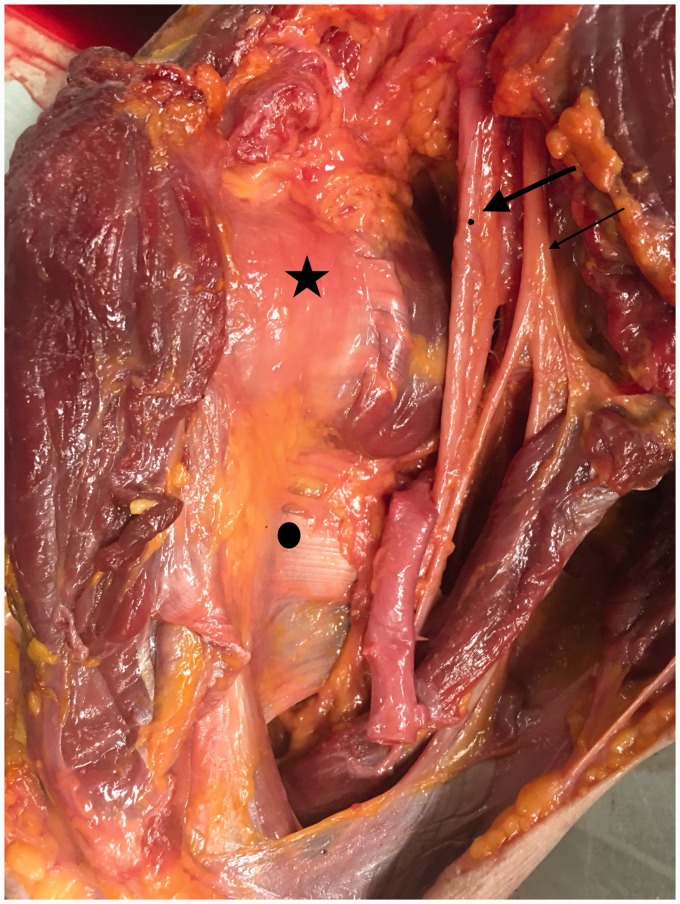
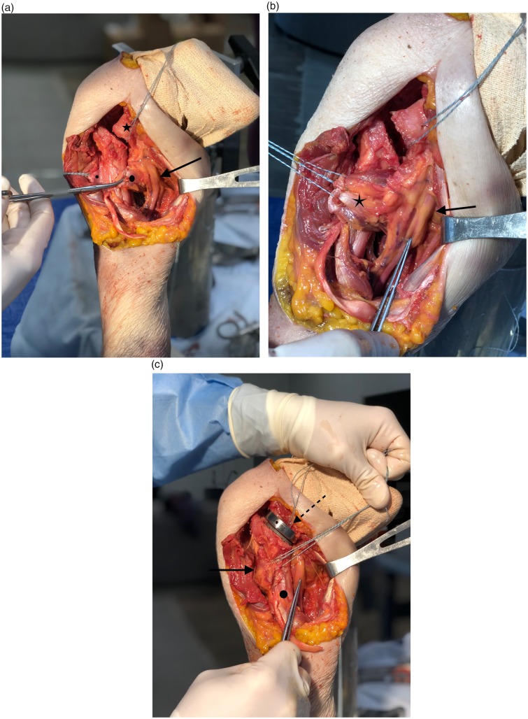
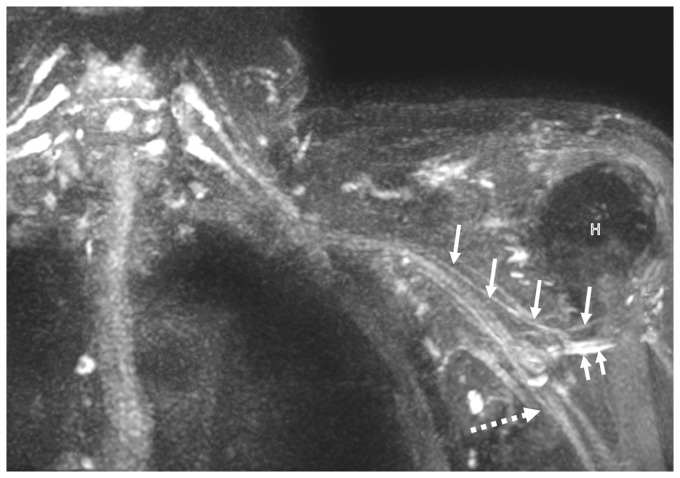
The mean distance from the latissimus humeral insertion to the radial nerve was 15.3 ± 5.5 (range =5–26 mm) in shoulder adduction internal rotation, 25.8 ± 6.9 mm (range = 10–35 mm), adduction neutral rotation, and 39.5 ± 6.8 mm (range = 24–49 mm) in adduction with external rotation. When moved to an abducted and externally rotated position, the mean distance further increased to 51.1 ± 7.4 mm (range = 37–65 mm). Six permutations comparing mean distances of each of the four shoulder positions to one another were all significantly different (p < 0.0001; Table 2). Figure 1 indicates the anatomic relationship of the latissimus dorsi tendon and radial nerve. We also compared these values to evaluate left verses right shoulder side to side differences: The mean width of the latissimus tendon at its humeral insertion was left: 28.5 ± 6.1 mm and right: 30.1 ± 5.5 (p = 0.54). The mean distance from the latissimus humeral insertion to the axillary nerve in adduction and neutral rotation was left: 23.8 ± 5.2 mm and right: 24.6 ± 8.8 mm (p = 0.8), while the mean distance increased to left: 41 ± 10.3 mm and right: 41.1 ± 9.9 (p = 0.98) with the shoulder in abduction and neutral rotation. The mean distance from the latissimus humeral insertion to the radial nerve was left: 16 ± 5.7 mm and right: 14.5 ± 5.6 (p = 5.6) in shoulder adduction internal rotation; left: 27.1 ± 6.8 mm and right: 24.5 ± 7.1 mm (p = 0.41) in adduction neutral rotation; and left: 40.4 ± 7.4 mm and right: 38.5 ± 6.5 (p = 0.55) in adduction with external rotation. When moved to an abducted and externally rotated position, the mean distance further increased left: 50.2 ± 8.7 mm and right: 52 ± 6.2 (p = 0.6). Figure 2(a) to (c) indicates the relationship of the latissimus with the radial nerve and how it is pulled closer to the humeral shaft in the setting of reverse total shoulder arthroplasty and latissimus tendon transfer. Figure 3 demonstrates and MRI image of the anatomic relationship of the course of the axillary and radial nerves.
Table 2.
Distance from latissimus dorsi tendon insertion to radial nerve in different shoulder positions.
| Arm position | Mean ± SD (mm) | ||
|---|---|---|---|
| Adduction internal rotation | 15.3 ± 5.5 | ||
| Adduction neutral rotation | 25.8 ± 6.9 | ||
| Adduction external rotation | 39.5 ± 6.8 | ||
| Abduction external rotation | 51.1 ± 7.4 |
SD: standard deviation; CI: confidence interval; mm: millimeters.
p < 0.05.
Figure 1.
Anatomy of the latissimus dorsi tendon and radial nerve. Star indicates subscapularis, circle indicates latissimus dorsi tendon, thick arrow with dot indicates radial nerve, and thin arrow indicates musculocutaneous nerve. The pectoralis major and conjoint tendon have been cut and reflected medial and inferiorly to better visualize these structures.
Figure 2.
(a) Arrow indicates location of radial nerve, dot latissimus tendon clap at tendon edge sutures in tendon stump at footprint on humerus, star indicates subscapularis. (b) Image demonstrating how pull on the latissimus tendon changes the position of the radial nerve. The latissimus and teres major tendon with sutures pulling across the humerus (star), forceps and arrow indicate the radial nerve. (c) Latissimus transfer with reverse shoulder arthroplasty stem in place. Solid arrow points to the transferred latissimus tendon pulled around the humerus, dashed arrow points to the humeral stem, forceps point to the radial nerve, dot indicates humeral shaft.
Figure 3.
Coronal 3D maximum intensity projection (23 mm slab) magnetic resonance neurography image through the brachial plexus and left shoulder in a 73-year-old woman. Notice the normal left axillary nerve (large arrows) coursing under the axillary pouch of left shoulder along the upper margin of latissimus dorsi and adjacent to the axillary vein (small arrow). H indicates humeral head and dash arrow indicates radial nerve.
Discussion
The latissimus dorsi muscle adducts and internally rotates the humerus at the shoulder joint, and secondarily participates in shoulder extension and scapular retraction.23 The mean width of the latissimus dorsi tendon at its insertion in this series was 29.3 mm, a finding comparable to that of other cadaveric shoulder studies.12,14,24
We found a statistically significant difference in the distance between the latissimus dorsi tendon insertion and the axillary nerve when the shoulder was moved from an adducted to abducted position. The mean distance in shoulder adduction was 24.2 ± 7.1 mm with the smallest distance being 8 mm. With the potential for the axillary nerve to be found less than a centimeter away from the latissimus insertion in shoulder adduction, special care should be taken to avoid surgical maneuvers in this area which are not directly visualized. With a mean distance of 41.1 ± 9.8 mm between the axillary nerve and latissimus dorsi tendon insertion in abduction, we can conclude that shoulder abduction can decrease risk for iatrogenic injury when working near the latissimus insertion.
It should be noted that while our data suggests axillary nerve position changes in relation to the latissimus dorsi tendon insertion, a prior cadaveric study by Simone et al. showed that the distance from the inferior glenohumeral joint to the axillary nerve did not change despite the shoulder being placed into an externally rotated position. In fact, the same study showed that the axillary nerve actually moved closer to the inferior glenohumeral joint with the arm in a position of abduction greater than 45 degrees.25 This example of the dynamic nature of shoulder anatomy predicates the importance of focused spatial awareness and anatomic knowledge intraoperatively.
Similar to results seen for the axillary nerve, statistically significant differences were seen for measured distances between the latissimus dorsi tendon insertion and the radial nerve through sequential shoulder position changes. The mean distances were 15.3 ± 5.5, 25.8 ± 6.9, 39.5 ± 6.8, and 51.1 ± 7.4 mm in shoulder adduction with internal rotation, adduction with neutral rotation, adduction with external rotation, and abduction with external rotation, respectively. Again, we can conclude that the aforementioned progression of shoulder position and rotation acts to protect the radial nerve when working near the latissimus dorsi tendon insertion, providing knowledge that can decrease the risk of iatrogenic injury intraoperatively.
There are several notable limitations of our study. The imbalanced representation of male to female specimens challenges the ability to extrapolate our findings to all patient groups clinically. Similarly, ethnicity was not considered, though some suggest significant differences in anatomic findings between cadavers of different races.26 Other specifications were also ignored for the scope of this study, including how height, weight, and BMI correlate to the distances measured.
While several recent anatomic studies have reported on the relationships of the axillary and radial nerves to defined shoulder landmarks, our study emulates the dynamic range of motion inherent to the shoulder by incorporating measurements at multiple shoulder positions. In addition, because our dissections were performed on shoulders of intact thoraces rather than forequarter specimens, proximal anatomic relationships and muscle origins were not disrupted, mitigating the chance that more distal anatomy would be inappropriately altered.
Conclusion
Knowing the defined measurements of axillary and radial nerve locations relative to the latissimus dorsi tendon insertion can help mitigate the risk of iatrogenic nerve injury. Understanding the dynamic nature of these findings as they relate to different shoulder positions is critical during open anterior shoulder surgery.
Declaration of Conflicting Interests
The author(s) declared no potential conflicts of interest with respect to the research, authorship, and/or publication of this article.
Ethical Review and Patient Consent
Not Applicable to this article.
Funding
The author(s) received no financial support for the research, authorship, and/or publication of this article.
References
- 1.Chalmers PN, Van Thiel GS, Trenhaile SW. Surgical exposures of the shoulder. J Am Acad Orthop Surg 2016; 24: 250–258. [DOI] [PubMed] [Google Scholar]
- 2.Boileau P. Complications and revision of reverse total shoulder arthroplasty. Orthop Traumatol Surg Res 2016; 102: S33–S43. [DOI] [PubMed] [Google Scholar]
- 3.Ingoe HM, Holland P, Cowling P, et al. Intraoperative complications during revision shoulder arthroplasty: a study using the National Joint Registry dataset. Shoulder Elbow 2017; 9: 92–99. [DOI] [PMC free article] [PubMed] [Google Scholar]
- 4.Athwal GS, Sperling JW, Rispoli DM, et al. Periprosthetic humeral fractures during shoulder arthroplasty. J Bone Joint Surg Am 2009; 91: 594–603. [DOI] [PubMed] [Google Scholar]
- 5.Garcia-Fernandez C, Lopiz-Morales Y, Rodriguez A, et al. Periprosthetic humeral fractures associated with reverse total shoulder arthroplasty: incidence and management. Int Orthop 2015; 39: 1965–1969. [DOI] [PubMed] [Google Scholar]
- 6.Mineo GV, Accetta R, Franceschini M, et al. Management of shoulder periprosthetic fractures: our institutional experience and review of the literature. Injury 2013; 44(Suppl 1): S82–S85. [DOI] [PubMed] [Google Scholar]
- 7.Gerber C, Vinh TS, Hertel R, et al. Latissimus dorsi transfer for the treatment of massive tears of the rotator cuff. A preliminary report. Clin Orthop Relat Res 1988; 232: 51–61. [PubMed] [Google Scholar]
- 8.Gerber C. Latissimus dorsi transfer for the treatment of irreparable tears of the rotator cuff. Clin Orthop Relat Res 1992; 275: 152–160. [PubMed] [Google Scholar]
- 9.Boileau P, Rumian AP, Zumstein MA. Reversed shoulder arthroplasty with modified L’Episcopo for combined loss of active elevation and external rotation. J Shoulder Elbow Surg 2010; 19: 20–30. [DOI] [PubMed] [Google Scholar]
- 10.Boileau P, Chuinard C, Roussanne Y, et al. Modified latissimus dorsi and teres major transfer through a single delto-pectoral approach for external rotation deficit of the shoulder: as an isolated procedure or with a reverse arthroplasty. J Shoulder Elbow Surg 2007; 16: 671–682. [DOI] [PubMed] [Google Scholar]
- 11.Zafra M, Carpintero P, Carrasco C. Latissimus dorsi transfer for the treatment of massive tears of the rotator cuff. Int Orthop 2009; 33: 457–462. [DOI] [PMC free article] [PubMed] [Google Scholar]
- 12.Elhassan B, Christensen TJ, Wagner ER. Feasibility of latissimus and teres major transfer to reconstruct irreparable subscapularis tendon tear: an anatomic study. J Shoulder Elbow Surg 2014; 23: 492–499. [DOI] [PubMed] [Google Scholar]
- 13.Henry PD, Dwyer T, McKee MD, et al. Latissimus dorsi tendon transfer for irreparable tears of the rotator cuff: an anatomical study to assess the neurovascular hazards and ways of improving tendon excursion. Bone Joint J 2013; 95B: 517–522. [DOI] [PubMed] [Google Scholar]
- 14.Buijze GA, Keereweer S, Jennings G, et al. Musculotendinous transfer as a treatment option for irreparable posterosuperior rotator cuff tears: teres major or latissimus dorsi? Clin Anat 2007; 20: 919–923. [DOI] [PubMed] [Google Scholar]
- 15.Donohue BF, Lubitz MG, Kremchek TE. Sports injuries to the latissimus dorsi and teres major. Am J Sports Med 2017; 45: 2428–2435. [DOI] [PubMed] [Google Scholar]
- 16.Chaudhry T, Noor S, Maher B, et al. The surgical anatomy of the radial nerve and the triceps aponeurosis. Clin Anat 2010; 23: 222–226. [DOI] [PubMed] [Google Scholar]
- 17.Guse TR, Ostrum RF. The surgical anatomy of the radial nerve around the humerus. Clin Orthop Relat Res 1995; 320: 149–153. [PubMed] [Google Scholar]
- 18.Bono CM, Grossman MG, Hochwald N, et al. Radial and axillary nerves. Anatomic considerations for humeral fixation. Clin Orthop Relat Res 2000; 373: 259–264. [PubMed] [Google Scholar]
- 19.Carlan D, Pratt J, Patterson JM, et al. The radial nerve in the brachium: an anatomic study in human cadavers. J Hand Surg Am 2007; 32: 1177–1182. [DOI] [PubMed] [Google Scholar]
- 20.Gerwin M, Hotchkiss RN, Weiland AJ. Alternative operative exposures of the posterior aspect of the humeral diaphysis with reference to the radial nerve. J Bone Joint Surg Am 1996; 78: 1690–1695. [DOI] [PubMed] [Google Scholar]
- 21.Fu MC, Hendel MD, Chen X, et al. Surgical anatomy of the radial nerve in the deltopectoral approach for revision shoulder arthroplasty and periprosthetic fracture fixation: a cadaveric study. J Shoulder Elbow Surg 2017; 26: 2173–2176. [DOI] [PubMed] [Google Scholar]
- 22.Burkhead WZ, Jr, Scheinberg RR, Box G. Surgical anatomy of the axillary nerve. J Shoulder Elbow Surg 1992; 1: 31–36. [DOI] [PubMed] [Google Scholar]
- 23.Bartlett SP, May JW, Jr, Yaremchuk MJ. The latissimus dorsi muscle: a fresh cadaver study of the primary neurovascular pedicle. Plast Reconstr Surg 1981; 67: 631–636. [PubMed] [Google Scholar]
- 24.Pearle AD, Kelly BT, Voos JE, et al. Surgical technique and anatomic study of latissimus dorsi and teres major transfers. J Bone Joint Surg Am 2006; 88: 1524–1531. [DOI] [PubMed] [Google Scholar]
- 25.Simone JP, Streubel PN, Sanchez-Sotelo J, et al. Change in the distance from the axillary nerve to the glenohumeral joint with shoulder external rotation or abduction position. Hand (N Y) 2017; 12: 395–400. [DOI] [PMC free article] [PubMed] [Google Scholar]
- 26.Chou PH, Shyu JF, Ma HL, et al. Courses of the radial nerve differ between Chinese and Caucasians: clinical applications. Clin Orthop Relat Res 2008; 466: 135–138. [DOI] [PMC free article] [PubMed] [Google Scholar]


