Abstract
Background
Osteosarcoma (OS) is the most common malignant primary bone tumor occurring in children and young adults, which occupies the second important cause of tumor-associated deaths among children and young adults. Recent studies have demonstrated that many microRNAs (miRNAs) have abnormal expression in OS, and can function as prognostic factors of OS patients. However, no previous studies have comprehensively analyzed the relationship between multiple miRNAs and prognosis of OS patients.
Methods
A total of 63 OS patients were retrospectively enrolled. The clinical characteristics were collected, and the expression levels of miRNA-21, miRNA-30c, miRNA-34a, miRNA-101, miRNA-133a, miRNA-214, miRNA-218, miRNA-433 and miRNA-539 in tumor tissues were measured through quantitative real-time polymerasechain reaction. Kaplan–Meier analysis was used to perform univariate survival analysis, and Cox regression model was used to perform multivariate survival analysis which included the variables with P < 0.1 in univariate survival analysis.
Results
The cumulative survival for 1, 2 and 5 years was 90.48%, 68.25% and 38.10%, respectively, and mean survival time was (45.39 ± 3.60) months (95% CI [38.34–52.45]). Kaplan–Meier analysis demonstrated that TNM stage, metastasis or recurrence, miRNA-21, miRNA-214, miRNA-34a, miRNA-133a and miRNA-539 were correlated with cum survival, but gender, age, tumor diameter, differentiation, miRNA-30c, miRNA-433, miRNA-101 and miRNA-218 were not. Multivariate survival analysis demonstrated that miRNA-21 (hazard ratio (HR): 3.457, 95% CI [2.165–11.518]), miRNA (HR: 3.138, 95% CI [2.014–10.259]), miRNA-34a (HR: 0.452, 95% CI [0.202–0.915]), miRNA-133a (HR: 0.307, 95% CI [0.113–0.874]) and miRNA-539 (HR: 0.358, 95% CI [0.155–0.896]) were independent prognostic markers of OS patients after adjusting for TNM stage (HR: 2.893, 95% CI [1.496–8.125]), metastasis or recurrence (HR: 3.628, 95% CI [2.217–12.316]) and miRNA-30c (HR: 0.689, 95% CI [0.445–1.828]).
Conclusions
High expression of miRNA-21 and miRNA-214 and low expression of miRNA-34a, miRNA-133a and miRNA-539 were associated with poor prognosis of OS patients after adjusting for TNM stage, metastasis or recurrence and miRNA-30c.
Keywords: MicroRNAs, Survival, Kaplan–Meier analysis, Multivariate Cox regression analysis
Introduction
Osteosarcoma (OS) is the most common malignant primary bone tumor occurring in children and young adults, which occupies the second important cause of tumor-associated deaths among children and young adults (Mirabello, Troisi & Savage, 2009a, 2009b; Biermann et al., 2013; Yu et al., 2017). It is highly aggressive and occurs mainly in the proximal tibia, proximal humerus, and metaphyseal regions of the distal femur, with an incidence of 4.4 per million people around the world (Zhu et al., 2016). OS responds poorly to chemotherapy and the 5 year survival rate is still very low for OS patients with metastasis or recurrence (Hutanu et al., 2017; Zhou et al., 2016), although its prognosis has been improved gradually over the past 30 years (Rytting et al., 2000; Kunz et al., 2015). Therefore, it is crucial to identify new biomarkers that can exactly evaluate the prognosis of OS.
MicroRNAs (miRNAs) are a group of non-coding RNAs, which consist of 18–25 nucleotides (Ambros, 2004; Chang et al., 2016; Jamieson et al., 2012). They widely exist in animals, plants and even some viruses, and have an important role in post-transcriptional modulation of gene expression and gene silencing (Bartel, 2004; Hayes, Peruzzi & Lawler, 2014; Griffiths-Jones et al., 2008; Liu et al., 2017). Approximately 50% of miRNAs are confirmed to be associated with human tumorigenesis through directly targeting tumor suppressor genes or oncogenes (Li & Rana, 2014; Bracken, Scott & Goodall, 2016). MiRNAs are able to be circulated in body fluid, suggesting their potential as noninvasive markers (Bahrami et al., 2018). In OS, abnormal expression of miRNAs is involved in its occurrence and development. In addition, the expression of some miRNAs is associated with OS chemoresistance. Therfore, miRNAs have been widely applied in prediction of prognosis, detection of patients at early stages, and monitoring of the patients in response to chemotherapy. Studies have demonstrated that many miRNAs can function as prognostic factors of OS patients (Cheng et al., 2017; Zhang et al., 2015). Among them, miRNA-21, miRNA-30c, miRNA-34a, miRNA-101, miRNA-133a, miRNA-214, miRNA-218, miRNA-433 and miRNA-539 have been studied extensively and confirmed a potential association with the prognosis of OS patients. However, no previous studies have comprehensively analyzed the relationship between multiple miRNAs and prognosis of OS patients. There may be interactions among them. In this study, the expression levels of these nine miRNAs in tumor tissues of OS patients were measured through quantitative real-time PCR (qRT-PCR). Kaplan–Meier method was employed to determine the survival rate of OS patients, and long-rank test was employed to compare the survival rates between groups. Multivariate Cox regression analysis was finally performed to identify the independent prognostic factors with adjusting for confounders.
Materials and Methods
Patients
A total of 63 OS patients were retrospectively collected in Heze Municipal Hospital between January 2012 and January 2018. Surgery was performed in all of them, and tumor tissues and adjacent normal bone tissues were sampled. None of them received chemotherapy and radiotherapy before surgery. All tissue samples, obtained during surgery, were frozen immediately in liquid nitrogen and stored at −80 °C. The diagnosis and histological grading were determined with histopathological examination. This study received the approval of the ethic committee of Heze Municipal Hospital (20185261), and was performed according to the Declaration of Helsinki. All patients provided written informed consents.
Quantitative real-time PCR
Total RNA was extract from tumor tissues and adjacent normal bone tissues through miRNeasy kit (Qiagen, Hilden, Germany) in accordance with instructions of the manufacturer. The TaqMan miRNA assey kit (Applied Biosystems, Foster City, CA, USA) was used to quantitate the expression levels of miRNAs. Rotor Gene 6000 Real-Time PCR (Qiagen, Hilden, Germany) was used to perform Real-Time PCR with a TaqMan universal PCR master mix and an invitrogen kit. U6 was chosen as the reference gene, and the 2−ΔΔCt method was used to assess the relative expression levels of miRNAs. The primers of the included miRNAs and U6 were designed and chemosynthesized by Shanghai Jima Biotech Ltd. (Shanghai, China). The primers used were as follows: miRNA-21-3p: 5′-GCCACCACACCAGCTAATTT-3′ (forward) and 5′-CTGAAGTCGCCATGCAGATA-3′ (reverse); miRNA-30c-3p: 5′-GCCCAAGTGGTTCTGTGTTT-3′ (forward) and 5′-TCCATGGCAGAAGGAGTAAA-3′ (reverse); miRNA-34a-5p: 5′-TATGGCAGTGTCTTAGCTGGTTGT-3′ (forward) and 5′-GGCCAACCGCGAGAAGATG-3′ (reverse); miRNA-101-3p: 5′-GCCGAGTACAGTACTGTGA-3′ (forward) and 5′-CTCAACTGGTGTCGTGGA-3′ (reverse); miRNA-133a-5p: 5′-TGCTTTGCTAGAGCTGGTAAAATG-3′ (forward) and 5′-AGCTACAGCTGGTTGAAGGG-3′ (reverse); miRNA-214-3p: 5′-TGCAGTAGTGTCTTAGCTGGAATG-3′ (forward) and 5′-GGCTAACCGCGAGAAGTTT-3′ (reverse); miRNA-218-5p: 5′-GCGCTTGTGCTTGATCTAA-3′ (forward) and 5′-GTGCAGGGTCCGAGGT-3′ (reverse); miRNA-433-3p: 5′-GCTTTAGTGGTTCTGTGTGA-3′ (forward) and 5′-TCCGCGACAGAAGGAGTTTA-3′ (reverse); miRNA-539-3p: 5′-GCTTGTACACCAGCTAGTGC-3′ (forward) and 5′-CTTAGCTCGCCATGCAGAAG-3′ (reverse); and U6: 5′-GATCAAGGATGACACGCAAATTCG-3′ (forward) and 5′-GGCCAACCGCGAGAAGATG-3′ (reverse).
Statistical analysis
Statistical Analysis was conducted using the SPSS version 20.0 for Windows (SPSS Inc., Chicago, IL, USA). Kolmogorov–Smirnov test was used to determine the normality of quantitative data. Normal data were expressed as mean ± standard deviation, and non-normal data were expressed as median (interquartile range). Qualitative data were expressed as percentages or ratios (%). Kaplan–Meier analysis was used to perform univariate survival analysis, and Cox regression model was used to perform multivariate survival analysis which included the variables with P < 0.1 in univariate survival analysis. Significance was set at P < 0.05.
Results
General data
These 63 OS patients included 36 males and 27 females, and the median age of onset for them was 17 years with an interquartile range of 10 years. The other detailed clinical characteristics were demonstrated in Table 1. The follow-up was up to January 2019. The cumulative survival for 1, 2 and 5 years was 90.48%, 68.25% and 38.10%, respectively, and mean survival time was (45.39 ± 3.60) months (95% CI [38.34–52.45]).
Table 1. Clinical characteristics of OS patients.
| Clinical characteristics | No. of patients | Percentages (%) |
|---|---|---|
| Gender | ||
| Male | 36 | 57.14 |
| Female | 27 | 42.86 |
| Age (years) | ||
| ≤25 | 55 | 87.30 |
| >25 | 8 | 12.70 |
| Tumor diameter (cm) | ||
| ≤5 | 37 | 58.73 |
| >5 | 26 | 41.27 |
| TNM stage | ||
| I + II | 25 | 39.68 |
| III + IV | 38 | 60.32 |
| Metastasis or recurrence | ||
| Yes | 37 | 58.73 |
| No | 26 | 41.27 |
| Differentiation | ||
| Well and moderate | 31 | 49.21 |
| Poor | 32 | 50.79 |
Expression levels of miRNAs in tumor tissues and adjacent normal bone tissues
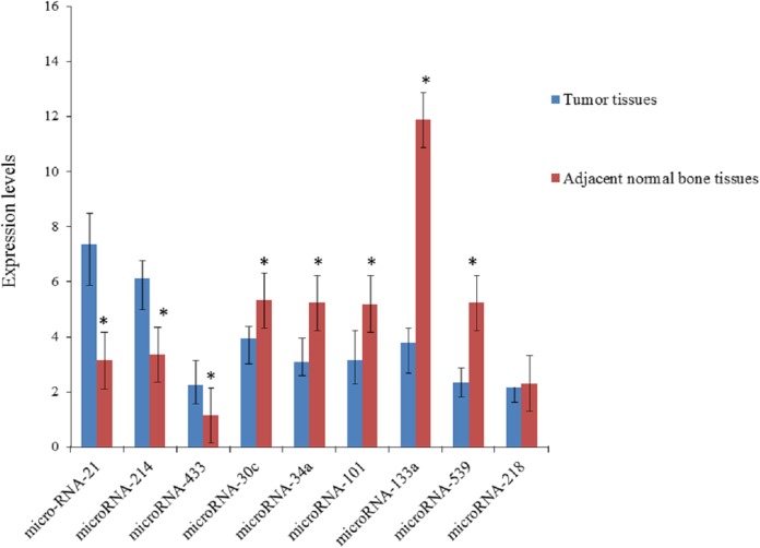
According to the results of quantitative real-time polymerase chain reaction (Table 2; Fig. 1), the expression levels of miRNA-21, miRNA-214 and miRNA-433 were higher in tumor tissues than in adjacent normal bone tissues, and the expression levels of miRNA-30c, miRNA-34a, miRNA-101, miRNA-133a and miRNA-539 was lower in tumor tissues than in adjacent normal bone tissues, and the expression level of miRNA-218 was not statistically different.
Table 2. Expression levels of microRNAs in tumor tissues and adjacent normal bone tissues.
| microRNA-21 | microRNA-214 | microRNA-433 | microRNA-30c | microRNA-34a | microRNA-101 | microRNA-133a | microRNA-539 | microRNA-218 | |
|---|---|---|---|---|---|---|---|---|---|
| Tumor tissues | 7.35 ± 2.96 | 6.12 ± 2.25 | 2.26 ± 1.34 | 3.93 ± 1.77 | 3.09 ± 0.94 | 3.16 ± 1.72 | 3.78 ± 2.17 | 2.35 ± 1.08 | 2.16 ± 1.07 |
| Adjacent normal bone tissues | 3.14 ± 1.58 | 3.37 ± 1.49 | 1.17 ± 0.91 | 5.34 ± 1.32 | 5.24 ± 1.35 | 5.19 ± 2.74 | 11.89 ± 4.16 | 5.23 ± 1.84 | 2.31 ± 1.18 |
| t | 9.959 | 8.088 | 5.341 | −5.069 | −10.374 | −4.981 | −13.719 | −10.714 | −0.747 |
| P | <0.05 | <0.05 | <0.05 | <0.05 | <0.05 | <0.05 | <0.05 | <0.05 | >0.05 |
Figure 1. Expression levels of microRNAs in tumor tissues and adjacent normal bone tissues.
*P < 0.05 vs. tumor tissues.
Univariate survival analysis
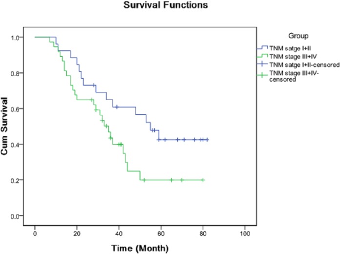
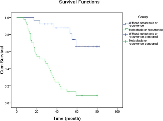
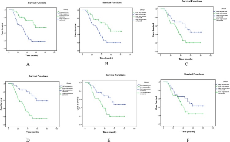
The OS patients were divided into high expression group and low expression group according to the median expression levels of miRNAs. Kaplan–Meier analysis demonstrated that TNM stage (Fig. 2), metastasis or recurrence (Fig. 3), miRNA-21 (Fig. 4A), miRNA-214 (Fig. 4B), miRNA-34a (Fig. 4C), miRNA-133a (Fig. 4D) and miRNA-539 (Fig. 4E) were correlated with cum survival, but gender, age, tumor diameter, differentiation, miRNA-30c (Fig. 4F), miRNA-433, miRNA-101 and miRNA-218 were not. Median time of survival and log rank χ2 were demonstrated in Table 3.
Figure 2. Kaplan–Meier analysis of cumulative survival for TNM stage using Log Rank test.
Figure 3. Kaplan–Meier analysis of cumulative survival for metastasis or recurrence using Log Rank test.
Figure 4. Kaplan–Meier analysis of cumulative survival for microRNAs using Log Rank test.
(A) miRNA-21, (B) miRNA-214, (C) miRNA-34a, (D) miRNA-133a, (E) miRNA-539 and (F) miRNA-30c.
Table 3. Median time of survival and log rank χ2 for the K–M survival plots.
| No. of patients | Median time of survival (months) | Log rank χ2 | P | ||
|---|---|---|---|---|---|
| TNM stage | I + II | 25 | 39.67 ± 4.43 | 4.199 | 0.040 |
| III + IV | 38 | 49.15 ± 5.14 | |||
| Metastasis or recurrence | Yes | 37 | 32.72 ± 3.85 | 28.970 | <0.001 |
| No | 26 | 63.42 ± 6.29 | |||
| microRNA-21 | Low expression | 24 | 61.75 ± 5.60 | 11.847 | 0.001 |
| High expression | 39 | 35.32 ± 4.25 | |||
| microRNA-214 | Low expression | 26 | 58.24 ± 6.17 | 7.338 | 0.007 |
| High expression | 37 | 36.36 ± 4.28 | |||
| microRNA-34a | Low expression | 33 | 35.58 ± 4.22 | 5.372 | 0.020 |
| High expression | 30 | 56.18 ± 5.87 | |||
| microRNA-133a | Low expression | 42 | 36.35 ± 4.38 | 16.258 | <0.001 |
| High expression | 21 | 63.47 ± 5.89 | |||
| microRNA-539 | Low expression | 34 | 35.27 ± 4.13 | 7.390 | 0.007 |
| High expression | 29 | 57.26 ± 6.07 | |||
| microRNA-30c | Low expression | 32 | 42.41 ± 4.72 | 3.378 | 0.066 |
| High expression | 31 | 48.47 ± 5.06 |
Multivariate survival analysis
TNM stage, metastasis or recurrence, miRNA-21, miRNA-214, miRNA-30c, miRNA-34a, miRNA-133a and miRNA-539 were included in Cox proportional hazards model. According to the results of multivariate survival analysis (Table 4), miRNA-21 (hazard ratio (HR): 3.457, 95% CI [2.165–11.518]), miRNA-214 (HR: 3.138, 95% CI [2.014–10.259]), miRNA-34a (HR: 0.452, 95% CI [0.202–0.915]), miRNA-133a (HR: 0.307, 95% CI [0.113–0.874]) and miRNA-539 (HR: 0.358, 95% CI [0.155–0.896]) were independent prognostic markers of OS patients after adjusting for TNM stage (HR: 2.893, 95% CI [1.496–8.125]), metastasis or recurrence (HR: 3.628, 95% CI [2.217–12.316]) and miRNA-30c (HR: 0.689, 95% CI [0.445–1.828]). In other words, high expression of miRNA-21 and miRNA-214 and low expression of miRNA-34a, miRNA-133a and miRNA-539 were associated with poor prognosis of OS patients.
Table 4. Results of Cox proportional hazards model.
| Regression coefficient | Standard error | Wald χ2 | Hazard ratio | 95% Confidence interval | P | |
|---|---|---|---|---|---|---|
| microRNA-21 | 1.107 | 0.465 | 5.923 | 3.457 | 2.165–11.518 | 0.013 |
| microRNA-214 | 1.058 | 0.446 | 5.642 | 3.138 | 2.014–10.259 | 0.017 |
| microRNA-34a | −0.835 | 0.371 | 5.148 | 0.452 | 0.202–0.915 | 0.021 |
| microRNA-133a | −0.946 | 0.382 | 6.137 | 0.307 | 0.113–0.874 | 0.011 |
| microRNA-539 | −0.887 | 0.369 | 5.474 | 0.358 | 0.155–0.896 | 0.018 |
| TNM stage | 0.953 | 0.392 | 5.016 | 2.893 | 1.496–8.125 | 0.024 |
| Metastasis or recurrence | 1.154 | 0.458 | 6.529 | 3.628 | 2.217–12.316 | 0.007 |
| microRNA-30c | −0.738 | 0.426 | 3.045 | 0.689 | 0.445–1.828 | 0.074 |
Discussion
The prognosis of OS patients has been significantly improved with the development of multiple chemotherapy regimens. However, OS patients receiving the same treatment often demonstrate different clinical outcomes, suggesting an urgent need for developing reliable prognostic biomarkers to improve the prognosis of OS patients. MiRNAs modulate protein expression through regulating the degradation and translation of mRNAs at post-transcriptional level (Chang et al., 2016; Jamieson et al., 2012). They play a critical role in various biological processes which are involved in the development and progression of tumors, including proliferation, apoptosis, differentiation and metastasis (Hayes, Peruzzi & Lawler, 2014; Ebert & Sharp, 2012; Rogers & Chen, 2013; Liu et al., 2012).
Additionally, they are very stable and easily detected in the blood and tissues (Gilad et al., 2008). Therefore, plenty of miRNAs are employed as new biomarkers for the diagnosis and prognosis of tumors. Regarding to OS, a variety of miRNAs has been reported to be associated with its prognosis. Kim et al. demonstrated that the pooled HR was 1.40 (95% CI [1.01–1.94]) for OS patients with lower expression miRNAs, and proposed that miRNAs with increased expression should also be investigated for their effects on the prognosis of OS patients. Additionally, the expression of some miRNAs is associated with OS chemoresistance (Xie et al., 2018). In our study, the nine miRNAs, having been studied widely, were chosen as research targets. Our results demonstrated that miRNA-21, miRNA-214, miRNA-34a, miRNA-133a and miRNA-539 were independently associated with the prognosis of OS patients.
MiRNA-21 has been confirmed to act as tumor oncogene in many types of tumors. For OS, it may regulate the proliferation, invasion and metastasis of OS cells through directly targeting PTEN and RECK (Ziyan et al., 2011; Lv, Hao & Tu, 2016). Li et al. (2018) demonstrated that the elevated expression of miRNA-21 might lead to elevated expression of the proteins in the PI3K/AKT signaling pathway and decreased expression of PTEN, which was associated with the increased invasiveness of OS cells. Hu et al. (2018) indicated that inhibition of miRNA-21 might reduce the proliferation of OS cells through modulating the TGF-β1 signaling pathway and targeting PTEN. Additionally, miRNA-21 might decrease the anti-tumor effect of cisplatin through modulating the expression of Bcl-2 (Ziyan & Yang, 2016). Our results demonstrated that high expression of miRNA-21 was independently associated with poor pognosis of OS patients with a HR of 3.457 (95% CI [2.165–11.518]). MiRNA-214 may act as either a tumor suppressor gene or an oncogene. For OS, the elevated expression of miRNA-214 is associated with enhanced invasion and proliferation of OS cells through modulating the expression of LZTS1 (Xu & Wang, 2014). However, Rehei et al. (2018) found that the expression of miRNA-214 was negatively associated with the expression of TRAF3 in OS tissues, and over-expression of miRNA-214 could inhibit the invasion and metastasis of OS cells through targeting TRAF3. Our results demonstrated that high expression of miRNA-214 was independently associated with poor prognosis of OS patients with a HR of 3.138 (95% CI [2.014–10.259]).
MiRNA-34a has various target genes which play important roles in biological function of OS cells, such as Fag1, Wnt, p53 and Notch (Wu et al., 2013; Yan et al., 2012). Gang et al. (2017) demonstrated that miRNA-34a was correlated with the apoptosis, proliferation and adhesion of OS cells, and could function as a new tumor suppressor gene by reducing the expression of DUSP1. Zhang et al. (2018) proved that miRNA-34a was a crucial regulator in the dedifferentiation of OS cells through modulating PAI-1-Sox2 axis. In addition, Wang et al. (2018) showed that down-modulated expression of miRNA-34a was a prognostic biomarker for poor prognosis of OS patients through a meta-analysis. Our results demonstrated that low expression of miRNA-34a was independently associated with poor prognosis of OS patients with a HR of 0.452 (95% CI [0.202–0.915]). MiRNA-133a has been proved to be a crucial modulator for osteogenesis, and have a key role in osteoblast differentiation (Bao et al., 2010). It can act as an antionco-miRNA or a tumor suppressor gene in the development and progression of tumors (Ji et al., 2013). It has been reported to be associated with many cancers, including esophagus cancer, bladder cancer and prostate cancer. The underlying mechanisms of pro-apoptotic function of miRNA-133a may be associated with the inhibition ofMcl-1 and Bcl-xL expression (Wang et al., 2010). Our results confirmed that low expression of miRNA-133a was independently associated with poor prognosis of OS patients with a HR of 0.307 (95% CI [0.113–0.874]). Few reports have investigated the biological functions of miRNA-539. Muthusamy et al. (2014) found that miRNA-539 could inhibit O-GlcNAcase expression. Wang et al. (2014) demonstrated that miRNA-539 was involved in the regulation of apoptosis and mitochondrial activity by means of targeting PHB2. The expression of miRNA-539 is down-regulated in thyroid cancer, and moreover, it has a suppressor role in the invasion and metastasis of thyroid cancer cells through targeting CARMA1 (Gu & Sun, 2015). Our results demonstrated that low expression of miRNA-539 was independently associated with poor prognosis of OS patients with a HR of 0.358 (95% CI [0.155–0.896]).
Conclusions
High expression of miRNA-21 and miRNA-214 and low expression of miRNA-34a, miRNA-133a and miRNA-539 were associated with poor prognosis of OS patients after adjusting for TNM stage, metastasis or recurrence and miRNA-30c.
Supplemental Information
Funding Statement
The authors received no funding for this work.
Additional Information and Declarations
Competing Interests
The authors declare that they have no competing interests.
Author Contributions
Wen Yang performed the experiments, authored or reviewed drafts of the paper, and approved the final draft.
Yu-bin Qi performed the experiments, prepared figures and/or tables, data management, and approved the final draft.
Meng Si performed the experiments, prepared figures and/or tables, and approved the final draft.
Yong Hou performed the experiments, analyzed the data, prepared figures and/or tables, and approved the final draft.
Lin Nie conceived and designed the experiments, authored or reviewed drafts of the paper, and approved the final draft.
Human Ethics
The following information was supplied relating to ethical approvals (i.e., approving body and any reference numbers):
This study received the approval of the ethic committee of Heze Municipal Hospital (20185261).
Data Availability
The following information was supplied regarding data availability:
All raw data are available in the Supplemental Files.
References
- Ambros (2004).Ambros V. The functions of animal microRNAs. Nature. 2004;431(7006):350–355. doi: 10.1038/nature02871. [DOI] [PubMed] [Google Scholar]
- Bahrami et al. (2018).Bahrami A, Aledavood A, Anvari K, Hassanian SM, Maftouh M, Yaghobzade A, Salarzaee O, ShahidSales S, Avan A. The prognostic and therapeutic application of microRNAs in breast cancer: tissue and circulating microRNAs. Journal of Cellular Physiology. 2018;233(2):774–786. doi: 10.1002/jcp.25813. [DOI] [PubMed] [Google Scholar]
- Bao et al. (2010).Bao B, Rodriguez-Melendez R, Wijeratne SSK, Zempleni J. Biotin regulates the expression of holocarboxylase synthetase in the miR-539 pathway in HEK-293 cells. Journal of Nutrition. 2010;140(9):1546–1551. doi: 10.3945/jn.110.126359. [DOI] [PMC free article] [PubMed] [Google Scholar]
- Bartel (2004).Bartel DP. MicroRNAs: genomics, biogenesis, mechanism, and function. Cell. 2004;116(2):281–297. doi: 10.1016/S0092-8674(04)00045-5. [DOI] [PubMed] [Google Scholar]
- Biermann et al. (2013).Biermann JS, Adkins DR, Agulnik M, Benjamin RS, Brigman B, Butrynski JE, Cheong D, Chow W, Curry WT, Frassica DA, Frassica FJ, Hande KR, Hornicek FJ, Jones RL, Mayerson J, McGarry SV, McGrath B, Morris CD, O’Donnell RJ, Randall RL, Santana VM, Satcher RL, Siegel HJ, Von Mehren M, Bergman MA, Sundar H. Bone cancer. Journal of the National Comprehensive Cancer Network. 2013;11(6):688–723. doi: 10.6004/jnccn.2013.0088. [DOI] [PubMed] [Google Scholar]
- Bracken, Scott & Goodall (2016).Bracken CP, Scott HS, Goodall GJ. A network-biology perspective of microRNA function and dysfunction in cancer. Nature Reviews Genetics. 2016;17(12):719–732. doi: 10.1038/nrg.2016.134. [DOI] [PubMed] [Google Scholar]
- Chang et al. (2016).Chang J, Yao M, Li Y, Zhao D, Hu S, Cui X, Liu G, Shi Q, Wang Y, Yang Y. MicroRNAs for osteosarcoma in the mouse: a meta-analysis. Oncotarget. 2016;7(51):85650–85674. doi: 10.18632/oncotarget.13333. [DOI] [PMC free article] [PubMed] [Google Scholar]
- Cheng et al. (2017).Cheng D, Qiu X, Zhuang M, Zhu C, Zou H, Liu Z. MicroRNAs with prognostic significance in osteosarcoma: a systemic review and meta-analysis. Oncotarget. 2017;8(46):81062–81074. doi: 10.18632/oncotarget.19009. [DOI] [PMC free article] [PubMed] [Google Scholar]
- Ebert & Sharp (2012).Ebert MS, Sharp PA. Roles for microRNAs in conferring robustness to biological processes. Cell. 2012;149(3):515–524. doi: 10.1016/j.cell.2012.04.005. [DOI] [PMC free article] [PubMed] [Google Scholar]
- Gang et al. (2017).Gang L, Qun L, Liu WD, Li YS, Xu YZ, Yuan DT. MicroRNA-34a promotes cell cycle arrest and apoptosis and suppresses cell adhesion by targeting DUSP1 in osteosarcoma. American Journal of Translational Research. 2017;9(12):5388–5399. [PMC free article] [PubMed] [Google Scholar] [Retracted]
- Gilad et al. (2008).Gilad S, Meiri E, Yogev Y, Benjamin S, Lebanony D, Yerushalmi N, Benjamin H, Kushnir M, Cholakh H, Melamed N, Bentwich Z, Hod M, Goren Y, Chajut A. Serum microRNAs are promising novel biomarkers. PLOS ONE. 2008;3(9):e3148. doi: 10.1371/journal.pone.0003148. [DOI] [PMC free article] [PubMed] [Google Scholar]
- Griffiths-Jones et al. (2008).Griffiths-Jones S, Saini HK, Van Dongen S, Enright AJ. miRBase: tools for microRNA genomics. Nucleic Acids Research. 2008;36(Database):D154–D158. doi: 10.1093/nar/gkm952. [DOI] [PMC free article] [PubMed] [Google Scholar]
- Gu & Sun (2015).Gu L, Sun W. MiR-539 inhibits thyroid cancer cell migration and invasion by directly targeting CARMA1. Biochemical and Biophysical Research Communications. 2015;464(4):1128–1133. doi: 10.1016/j.bbrc.2015.07.090. [DOI] [PubMed] [Google Scholar]
- Hayes, Peruzzi & Lawler (2014).Hayes J, Peruzzi PP, Lawler S. MicroRNAs in cancer: biomarkers, functions and therapy. Trends in Molecular Medicine. 2014;20(8):460–469. doi: 10.1016/j.molmed.2014.06.005. [DOI] [PubMed] [Google Scholar]
- Hu et al. (2018).Hu X, Li L, Lu Y, Yu X, Chen H, Yin Q, Zhang Y. miRNA‑21 inhibition inhibits osteosarcoma cell proliferation by targeting PTEN and regulating the TGF‑β1 signaling pathway. Oncology Letters. 2018;16(4):4337–4342. doi: 10.3892/ol.2018.9177. [DOI] [PMC free article] [PubMed] [Google Scholar]
- Hutanu et al. (2017).Hutanu D, Popescu R, Stefanescu H, Pirtea L, Candea A, Sarau C, Boruga O, Mehdi L, Ciuca I, Tanasescu S. The molecular genetic expression as a novel biomarker in the evaluation and monitoring of patients with osteosarcoma-subtype bone cancer disease. Biochemical Genetics. 2017;55(4):291–299. doi: 10.1007/s10528-017-9801-1. [DOI] [PubMed] [Google Scholar]
- Jamieson et al. (2012).Jamieson NB, Morran DC, Morton JP, Ali A, Dickson EJ, Carter CR, Sansom OJ, Evans TRJ, McKay CJ, Oien KA. MicroRNA molecular profiles associated with diagnosis, clinicopathologic criteria, and overall survival in patients with resectable pancreatic ductal adenocarcinoma. Clinical Cancer Research. 2012;18(2):534–545. doi: 10.1158/1078-0432.CCR-11-0679. [DOI] [PubMed] [Google Scholar]
- Ji et al. (2013).Ji F, Zhang H, Wang Y, Li M, Xu W, Kang Y, Wang Z, Wang Z, Cheng P, Tong D, Li C, Tang H. MicroRNA-133a, downregulated in osteosarcoma, suppresses proliferation and promotes apoptosis by targeting Bcl-xL and Mcl-1. Bone. 2013;56(1):220–226. doi: 10.1016/j.bone.2013.05.020. [DOI] [PubMed] [Google Scholar]
- Kunz et al. (2015).Kunz P, Fellenberg J, Moskovszky L, Sápi Z, Krenacs T, Machado I, Poeschl J, Lehner B, Szendrõi M, Ruef P, Bohlmann M, Bosch AL, Ewerbeck V, Kinscherf R, Fritzsching B. Improved survival in osteosarcoma patients with atypical low vascularization. Annals of Surgical Oncology. 2015;22(2):489–496. doi: 10.1245/s10434-014-4001-2. [DOI] [PubMed] [Google Scholar]
- Li & Rana (2014).Li Z, Rana TM. Therapeutic targeting of microRNAs: current status and future challenges. Nature Reviews Drug Discovery. 2014;13(8):622–638. doi: 10.1038/nrd4359. [DOI] [PubMed] [Google Scholar]
- Li et al. (2018).Li C, Xu B, Miu X, Deng Z, Liao H, Hao L. Inhibition of miRNA‑21 attenuates the proliferation and metastasis of human osteosarcoma by upregulating PTEN. Experimental and Therapeutic Medicine. 2018;15(1):1036–1040. doi: 10.3892/etm.2017.5477. [DOI] [PMC free article] [PubMed] [Google Scholar]
- Liu et al. (2017).Liu H, Li P, Chen L, Jian C, Li Z, Yu A. MicroRNAs as a novel class of diagnostic biomarkers for the detection of osteosarcoma: a meta-analysis. OncoTargets and Therapy. 2017;10:5229–5236. doi: 10.2147/OTT.S143974. [DOI] [PMC free article] [PubMed] [Google Scholar]
- Liu et al. (2012).Liu Y, Yan W, Zhang W, Chen L, You G, Bao Z, Wang Y, Wang H, Kang C, Jiang T. MiR-218 reverses high invasiveness of glioblastoma cells by targeting the oncogenic transcription factor LEF1. Oncology Reports. 2012;28(3):1013–1021. doi: 10.3892/or.2012.1902. [DOI] [PubMed] [Google Scholar]
- Lv, Hao & Tu (2016).Lv C, Hao Y, Tu G. MicroRNA-21 promotes proliferation, invasion and suppresses apoptosis in human osteosarcoma line MG63 through PTEN/Akt pathway. Tumor Biology. 2016;37(7):9333–9342. doi: 10.1007/s13277-016-4807-6. [DOI] [PubMed] [Google Scholar]
- Mirabello, Troisi & Savage (2009a).Mirabello L, Troisi RJ, Savage SA. Osteosarcoma incidence and survival rates from 1973 to 2004: data from the surveillance, epidemiology, and end results program. Cancer. 2009a;115(7):1531–1543. doi: 10.1002/cncr.24121. [DOI] [PMC free article] [PubMed] [Google Scholar]
- Mirabello, Troisi & Savage (2009b).Mirabello L, Troisi RJ, Savage SA. International osteosarcoma incidence patterns in children and adolescents, middle ages and elderly persons. International Journal of Cancer. 2009b;125(1):229–234. doi: 10.1002/ijc.24320. [DOI] [PMC free article] [PubMed] [Google Scholar]
- Muthusamy et al. (2014).Muthusamy S, DeMartino AM, Watson LJ, Brittian KR, Zafir A, Dassanayaka S, Hong KU, Jones SP. MicroRNA-539 is up-regulated in failing heart, and suppresses O-GlcNAcase expression. Journal of Biological Chemistry. 2014;289(43):29665–29676. doi: 10.1074/jbc.M114.578682. [DOI] [PMC free article] [PubMed] [Google Scholar]
- Rehei et al. (2018).Rehei AL, Zhang L, Fu YX, Mu WB, Yang DS, Liu Y, Zhou SJ, Younusi A. MicroRNA-214 functions as an oncogene in human osteosarcoma by targeting TRAF3. European Review for Medical and Pharmacological Sciences. 2018;22(16):5156–5164. doi: 10.26355/eurrev_201808_15711. [DOI] [PubMed] [Google Scholar]
- Rogers & Chen (2013).Rogers K, Chen X. Biogenesis, turnover, and mode of action of plant microRNAs. Plant Cell. 2013;25(7):2383–2399. doi: 10.1105/tpc.113.113159. [DOI] [PMC free article] [PubMed] [Google Scholar]
- Rytting et al. (2000).Rytting M, Pearson P, Raymond AK, Ayala A, Murray J, Yasko AW, Johnson M, Jaffe N. Osteosarcoma in preadolescent patients. Clinical Orthopaedics and Related Research. 2000;373:39–50. doi: 10.1097/00003086-200004000-00007. [DOI] [PubMed] [Google Scholar]
- Wang et al. (2018).Wang W, Hu S, Chang J, Ruan H, Zhi W, Wang X, Shi Q, Wang Y, Yang Y. Down-regulated microRNA-34a expression as a prognostic marker for poor osteosarcoma in mice: a systematic review and meta-analysis. Journal of Cancer. 2018;9(22):4179–4186. doi: 10.7150/jca.27483. [DOI] [PMC free article] [PubMed] [Google Scholar]
- Wang et al. (2014).Wang K, Long B, Zhou L-Y, Liu F, Zhou Q-Y, Liu C-Y, Fan Y-Y, Li P-F. CARL lncRNA inhibits anoxia-induced mitochondrial fission and apoptosis in cardiomyocytes by impairing miR-539-dependent PHB2 downregulation. Nature Communications. 2014;5(1):3596. doi: 10.1038/ncomms4596. [DOI] [PubMed] [Google Scholar]
- Wang et al. (2010).Wang Z-X, Yang J-S, Pan X, Wang J-R, Li J, Yin Y-M, De W. Functional and biological analysis of Bcl-xL expression in human osteosarcoma. Bone. 2010;47(2):445–454. doi: 10.1016/j.bone.2010.05.027. [DOI] [PubMed] [Google Scholar]
- Wu et al. (2013).Wu X, Zhong D, Gao Q, Zhai W, Ding Z, Wu J. MicroRNA-34a inhibits human osteosarcoma proliferation by downregulating ether à go-go 1 expression. International Journal of Medical Sciences. 2013;10(6):676–682. doi: 10.7150/ijms.5528. [DOI] [PMC free article] [PubMed] [Google Scholar]
- Xie et al. (2018).Xie B, Li Y, Zhao R, Xu Y, Wu Y, Wang J, Xia D, Han W, Chen D. Identification of key genes and miRNAs in osteosarcoma patients with chemoresistance by bioinformatics analysis. BioMed Research International. 2018;2018(4):1–10. doi: 10.1155/2018/4761064. [DOI] [PMC free article] [PubMed] [Google Scholar]
- Xu & Wang (2014).Xu Z, Wang T. miR-214 promotes the proliferation and invasion of osteosarcoma cells through direct suppression of LZTS1. Biochemical and Biophysical Research Communications. 2014;449(2):190–195. doi: 10.1016/j.bbrc.2014.04.140. [DOI] [PubMed] [Google Scholar]
- Yan et al. (2012).Yan K, Gao J, Yang T, Ma Q, Qiu X, Fan Q, Ma B. MicroRNA-34a inhibits the proliferation and metastasis of osteosarcoma cells both in vitro and in vivo. PLOS ONE. 2012;7(3):e33778. doi: 10.1371/journal.pone.0033778. [DOI] [PMC free article] [PubMed] [Google Scholar]
- Yu et al. (2017).Yu W, Zhu J, Wang Y, Wang J, Fang W, Xia K, Shao J, Wu M, Liu B, Liang C, Ye C, Tao H. A review and outlook in the treatment of osteosarcoma and other deep tumors with photodynamic therapy: from basic to deep. Oncotarget. 2017;8(24):39833–39848. doi: 10.18632/oncotarget.16243. [DOI] [PMC free article] [PubMed] [Google Scholar]
- Zhang et al. (2018).Zhang Y, Pan Y, Xie C, Zhang Y. miR-34a exerts as a key regulator in the dedifferentiation of osteosarcoma via PAI-1-Sox2 axis. Cell Death & Disease. 2018;9(7):777. doi: 10.1038/s41419-018-0778-4. [DOI] [PMC free article] [PubMed] [Google Scholar]
- Zhang et al. (2015).Zhang J, Yan Y-G, Wang C, Zhang S-J, Yu X-H, Wang W-J. MicroRNAs in osteosarcoma. Clinica Chimica Acta. 2015;444:9–17. doi: 10.1016/j.cca.2015.01.025. [DOI] [PubMed] [Google Scholar]
- Zhou et al. (2016).Zhou H, Zhang M, Yuan H, Zheng W, Meng C, Zhao D. MicroRNA-154 functions as a tumor suppressor in osteosarcoma by targeting Wnt5a. Oncology Reports. 2016;35(3):1851–1858. doi: 10.3892/or.2015.4495. [DOI] [PubMed] [Google Scholar]
- Zhu et al. (2016).Zhu K, Liu L, Zhang J, Wang Y, Liang H, Fan G, Jiang Z, Zhang C-Y, Chen X, Zhou G. MiR-29b suppresses the proliferation and migration of osteosarcoma cells by targeting CDK6. Protein & Cell. 2016;7(6):434–444. doi: 10.1007/s13238-016-0277-2. [DOI] [PMC free article] [PubMed] [Google Scholar]
- Ziyan et al. (2011).Ziyan W, Shuhua Y, Xiufang W, Xiaoyun L. MicroRNA-21 is involved in osteosarcoma cell invasion and migration. Medical Oncology. 2011;28(4):1469–1474. doi: 10.1007/s12032-010-9563-7. [DOI] [PubMed] [Google Scholar]
- Ziyan & Yang (2016).Ziyan W, Yang L. MicroRNA-21 regulates the sensitivity to cisplatin in a human osteosarcoma cell line. Irish Journal of Medical Science. 2016;185(1):85–91. doi: 10.1007/s11845-014-1225-x. [DOI] [PubMed] [Google Scholar]
Associated Data
This section collects any data citations, data availability statements, or supplementary materials included in this article.
Supplementary Materials
Data Availability Statement
The following information was supplied regarding data availability:
All raw data are available in the Supplemental Files.


