Abstract
A circumflex retroesophageal left aortic arch with a right-sided ductus is an extremely rare cause of a complete vascular ring, which may result in severe tracheobronchial compression, leading to respiratory compromise, especially in children. We present a case of a 6-month-old female child with stridor and feeding difficulties since birth with interspersed self-resolving episodes of cyanosis and apnea, secondary to the presence of the above-mentioned vascular ring.
Keywords: Circumflex retroesophageal aortic arch, complete vascular ring, computed tomography angiography, Kommerell's diverticulum
CLINICAL SUMMARY
A 6-month-old female child presented to the outpatient department with a history of “noisy” and “rapid” breathing and feeding difficulties since birth with few interspersed self-resolving episodes of cyanosis and apnea, following bouts of crying. During the outpatient visit, the patient developed an episode of sudden-onset cyanosis, apnea, and bradycardia and became unresponsive. Following cardiopulmonary resuscitation, the child underwent endotracheal and nasogastric intubation and was referred for a computed tomography angiography (CTA) to evaluate for the presence of any vascular ring.
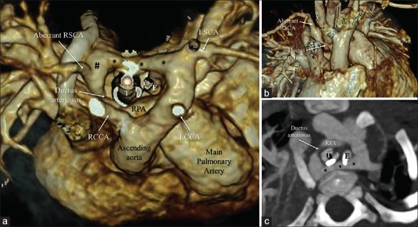
CTA demonstrated the presence of a left-sided aortic arch crossing the midline (*) posterior to the trachea (T) and the esophagus (E) in the upper mediastinum and further descending on the right side of spine. A Kommerell's diverticulum (#) arising from the anterosuperior aspect of aorta, on the right side, gave rise to an aberrant right subclavian artery (RSCA). A ductus arteriosus was seen connecting the descending aorta to the right pulmonary artery completing the vascular ring [Figure 1a-c].
Figure 1.
Volume-rendered images (a and b) and oblique axial maximum intensity projection image (c) of computed tomography angiography shows left-sided aortic arch crossing the midline (indicated by *) posterior to the trachea (T) and the esophagus (E). A Kommerell's diverticulum (indicated by #) giving rise to an aberrant RSCA is seen with a ductus arteriosus connecting the descending aorta to the RPA also noted. RCCA: Right common carotid artery, LCCA: Left common carotid artery, RPA: Right pulmonary artery, RSCA: Right subclavian artery
DISCUSSION
A circumflex retroesophageal left aortic arch with a right-sided ductus is an extremely rare cause of a complete vascular ring and results from regression of the right fourth arch between the right common carotid artery and RSCA, with persistence of right-sided sixth arch component forming the ductus arteriosus, along with a right-sided descending aorta. The definitive distal aortic arch is formed by the distal left dorsal aorta, which passes posterior to the trachea and esophagus to a right-sided descending aorta.[1] The vascular ring is completed by a segment of aortic arch on the left side along with a retroesophageal segment of aortic arch, ductus on the right, and pulmonary artery on the anterior aspect.[2] This uncommon anomaly may result in severe tracheobronchial compression, leading to respiratory compromise, especially in children, necessitating surgery. The compression may be relieved by division of the ductus as was done in the current case. Patients may also require aortic uncrossing procedure, where the distal aortic arch is transected; the retroesophageal segment mobilized and brought anterior to trachea followed by its anastomosis with the ascending aorta.[3]
Declaration of patient consent
The authors certify that they have obtained all appropriate patient consent forms. In the form, the patient's parents have given their consent for his images and other clinical information to be reported in the journal. The patient's parents understand that his names and initials will not be published and due efforts will be made to conceal their identity, but anonymity cannot be guaranteed.
Financial support and sponsorship
Nil.
Conflicts of interest
There are no conflicts of interest.
REFERENCES
- 1.Hanneman K, Newman B, Chan F. Congenital variants and anomalies of the aortic arch. Radiographics. 2017;37:32–51. doi: 10.1148/rg.2017160033. [DOI] [PubMed] [Google Scholar]
- 2.Priya S, Thomas R, Nagpal P, Sharma A, Steigner M. Congenital anomalies of the aortic arch. Cardiovasc Diagn Ther. 2018;8:S26–44. doi: 10.21037/cdt.2017.10.15. [DOI] [PMC free article] [PubMed] [Google Scholar]
- 3.Russell HM, Rastatter JC, Backer CL. The aortic uncrossing procedure for circumflex aorta. Oper Techn Thoracic Cardiovasc Surg. 2013;18:15–31. [Google Scholar]


