Abstract
Ultrasound is commonly the first-line imaging modality for assessing the pediatric abdomen. An abnormal size of the liver, spleen, or kidneys may indicate disease, but the evaluation is challenging because the normal size changes with age. In addition, published normal value charts for children may vary by population and methods. In this systematic review, we summarized published data on the normal size of the pediatric liver, spleen, and kidneys as measured by ultrasound in which we found similar values across different populations, ages, and sexes.
Keywords: kidney, liver, normal size, pediatric, spleen, ultrasound
Ultrasound (US) is an important imaging modality in children because it is safe, quick, and portable. It is an established diagnostic and screening tool to assess a variety of clinical concerns.1 Ultrasound is used in everyday practice for emergency, inpatient, and outpatient care. Measurement of abdominal organ dimensions in children of all ages is performed in the monitoring of abdominal organ growth patterns, diagnosis, and follow-up of patients with a variety of diseases.2
Organ size is crucial to the image interpretation of disease: for example, diseases of the liver, spleen, and kidneys can affect organ size and development, but a physical examination is not enough accurate to detect small increases in organ size.3 For example, the spleen may be palpable in 15% to 17% of healthy neonates4 and 10% of healthy children, but in most children, it must be 2 to 3 times its normal size before it is palpable.1 Ultrasound may therefore first detect organ size abnormalities that indicate disease. Normative data for organ size are challenging in the pediatric population because of changes that occur with growth and development and the effects of the body habitus, including height and weight.
In contrast to adults, for whom there are established normal ranges of organ size, organ size in children relies on growth and development.5–8 Studies exist that define the normal ranges of organ size in healthy children.1,3,4,7–23 For example hepatomegaly is a frequent clinical finding in children that may be caused by intrinsic liver diseases or by other diseases with liver involvement.16 With the growing epidemic of childhood obesity, early detection of hepatic steatosis is critical to avoiding premature liver failure.24 In children with kidney disease, the renal size may be increased or decreased.15 Measurements of renal length and volume reportedly correlate with height and weight, but the exact measurements are not homogeneous in the available literature.3,9,21 This systematic review was conducted to establish available literature published in indexed journals of US measurements of the liver, spleen, and kidneys in healthy children.
Methods
This study was approved by our Institutional Review Board, as well was registered on the PROSPERO registry, with number CRD42018094714, and performed according to the Preferred Reporting Items for Systematic Reviews and Meta-analyses and the Meta-analysis of Observational Studies in Epidemiology guidelines.25
Literature Search and Selection of Studies
A PICO strategy for searching in the databases was used, with P (population): healthy children from 0 to 18 years old; I (intervention): measurement of the spleen, liver, and kidney using US; C (comparison): not applicable; and O (outcome): determination of abdominal organ sizes. The Institutional Review Board at Children’s Hospital of Philadelphia approved the study, and a waiver of informed consent was obtained.
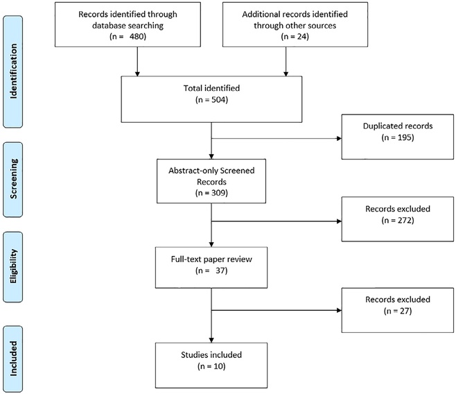
A systematic review was performed of all cross-sectional, case-control, and cohort studies, clinical trials, and randomized clinical trials related to the abdominal organ size of the liver, spleen, and kidneys in children published in the English language. A bibliographic search was performed using the following databases: Embase, MEDLINE, PubMed, SciELO, CINAHL, and Lilacs, using medical subject heading key words (spleen, liver, kidney, ultrasonography, child, pediatrics), with search dates up to May 1, 2018. The search terms were entered as shown in the following example used for PubMed: (ultrasonography and organ size) or (ultrasonography and spleen) or (ultrasonography and liver) or (ultrasonography and kidneys) and (children or pediatric). In addition, the reference lists of the retrieved articles were screened for further material for inclusion. In addition, we searched using the same search terms on Google Scholar. For extracting those articles suitable for our research, we performed a number of exclusion steps, as highlighted in the flow diagram (Figure 1). The first exclusion step was removing duplicated studies (using My EndNote Web; Clarivate Analytics, Philadelphia, PA); the second step involved exclusion of studies performed in patients with previous disease in the target organ, studies of contrast-enhanced US, studies using imaging modalities other than US, studies in animals and phantoms, and those studies that included adults who had escaped the age-filtering process of the search.
Figure 1.
Systematic review flowchart: organ size study selection process.
Assessment of Methodological Quality
The qualitative assessment of articles selected for retrieval was based on methodological validity before inclusion in the review, using standardized critical appraisal instruments from the Joanna Briggs Institute Qualitative Assessment and Review Instrument (Appendix 1). A cutoff score of 70% was used in this review.26
Data Extraction
The information extracted from each study included study setting, population demographics and baseline characteristics, US technique, study methods, outcomes, and assessment of the risk of bias. Information was organized in a general table (Appendixes 1 and 2) and in 4 subtables by each organ.
Data Synthesis
The data analysis was conducted from the findings of the included studies, structured according to the type of intervention, the target population characteristics and outcome measures. It was anticipated that there would be a small number of studies on this topic.
Results
We selected from the 504 studies only those suitable for our research as summarized in the flow diagram (Figure 1). The first exclusion step was for duplicated studies (195), resulting in 309 abstracts for review. A total of 272 studies in patients with prior disease in the target organ, studies performed with contrast-enhanced US, studies using imaging modalities other than US, studies in animals and phantoms, and those studies that included adults who had escaped the age-filtering process of the search were excluded. This resulted in 37 full-text studies remaining for complete review. From these, 27 were further excluded for 1 of 3 reasons: the organ size was reported by volume instead of a linear measure; the study focused on preterm neonates and fetuses; or the study assessed patients with a previous organ disease. The 10 studies remaining for evaluation in this review were published between 1991 and 2018 (Figure 1). Five studies included data of the liver,7,8,17,27,28 6 of the spleen,1,7,8,22,28,29 and 4 of both kidneys.7,28,30,31
The studies included patients from 0 to 20 years of age; however, only data from those aged 0 to 18 years are included here because those 19 years and older are considered young adults. Two studies included patients from 0 to 18 years1,7; 1 study included infants from 0 to 1 year30; and the remainder included groups between 0 and 16 years.7,8,17,27–29,31
Most were prospective studies (6 or 10), and none were multicenter. Application of the Joanna Briggs Institute Qualitative Assessment and Review Instrument tool for assessment of study quality showed that 8 of 10 studies had at least 80% of the required characteristics for a good-quality research study; the other 2 had scores of 70% and 75%, respectively (Appendixes 1 and 2).
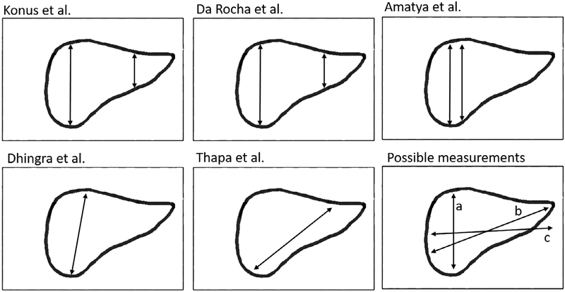
In general, similar organ sizes were reported between the included studies. The liver was measured in the sagittal plane longitudinally in all 5 studies included for liver measurement and ranged from 7 cm7 in younger patients, to a maximum of 12.1 cm7 in older patients (Table 1), although not all of them used the same technique to measure the liver size (Figure 2), and the craniocaudal measurement was the most commonly used.32,33 The spleen in all studies was measured in the sagittal plane longitudinally and ranged from 5.2 cm18,22 in younger patients, to a maximum of 12.5 cm7 in older patients (Table 2). The right kidney in all studies was measured in the sagittal plane longitudinally, and the reported measurements ranged from 4.5 cm23 in younger patients, to a maximum of 10.7 cm23 in older patients (Table 3). The left kidney size ranged from 4.5 cm23 in younger patients to a maximum of 10.7 cm7 in older patients (Table 4).
Table 1.
Included Studies Measuring Liver Size in Healthy Children
| Age | Turkey, Konuş et al, 1998 | Brazil, da Rocha et al, 2009 | India, Dhingra, 2010 | India, Amatya et al, 2014 | Nepal, Thapa et al, 2016 | ||||||||||||||||||||
|---|---|---|---|---|---|---|---|---|---|---|---|---|---|---|---|---|---|---|---|---|---|---|---|---|---|
| n | Mean | SD | 5th | 95th | n | Mean | SD | 5th | 95th | n | Mean | SD | 3th | 97th | n | Mean | SD | 5th | 95th | n | Mean | SD | 5th | 95th | |
| 0–<3 mo | 53 | 61.0 | 10.4 | 48.0 | 90.0 | 32 | 66.0 | 5.7 | 47.0 | 75.0 | 21 | 63.5 | 9.3 | 48.5 | 80.5 | 45 | 53.0 | 5.2 | 44.0 | 57.0 | 36 | 68.0 | 7.0 | 54.0 | 80.0 |
| 3–<6 mo | 40 | 73.0 | 10.8 | 53.0 | 86.0 | 34 | 76.0 | 7.2 | 58.0 | 90.0 | 35 | 71.5 | 8.6 | 56.0 | 84.5 | 45 | 64.0 | 9.2 | 49.0 | 83.0 | |||||
| 6–<9 mo | 20 | 79.0 | 8.0 | 70.0 | 90.0 | 72 | 84.0 | 9.1 | 62.0 | 101.0 | 51 | 77.0 | 9.0 | 62.0 | 95.5 | 35 | 76.0 | 9.3 | 47.0 | 75.0 | |||||
| 9–<12 mo | 36 | 84.0 | 6.9 | 65.0 | 96.0 | ||||||||||||||||||||
| l–<2 y | 18 | 85.0 | 10.0 | 68.0 | 98.0 | 102 | 92.3 | 7.5 | 77.3 | 110.3 | 77 | 85.5 | 11.8 | 67.0 | 106.5 | 45 | 89 | 8.6 | 71 | 103 | 62 | 84.0 | 7.4 | 69.5 | 94.5 |
| 2–<4 y | 27 | 86.0 | 11.8 | 63.0 | 105.0 | 48 | 99.0 | 6.7 | 78.0 | 11.0 | 132 | 89.5 | 11.4 | 70.5 | 116.0 | 43 | 87.3 | 8.9 | 73.4 | 105.2 | |||||
| 4–<6 y | 30 | 100.0 | 13.6 | 77.0 | 124.0 | 181 | 104.0 | 8.2 | 86.5 | 126.0 | 115 | 100.5 | 12.6 | 69.0 | 140.0 | 45 | 92.0 | 9.1 | 80.0 | 108.0 | 41 | 92.2 | 9.1 | 75.3 | 106.6 |
| 6–<8 y | 38 | 105.0 | 10.6 | 90.0 | 123.0 | 109 | 109.0 | 8.7 | 91.0 | 133.0 | 51 | 108.5 | 11.2 | 86.0 | 128.0 | 25 | 98.7 | 8.7 | 84.6 | 117.6 | |||||
| 8–<10 y | 30 | 105.0 | 12.5 | 83.0 | 128.0 | 62 | 118.0 | 11.0 | 97.0 | 140.5 | |||||||||||||||
| 10–<12 y | 16 | 115.0 | 14.0 | 95.0 | 136.0 | 53 | 132.5 | 12.5 | 103.5 | 153.5 | 45 | 107 | 11.1 | 91 | 129 | 19 | 106.3 | 10.7 | 92.0 | 127.0 | |||||
| 12–<14 y | 23 | 118.0 | 14.6 | 94.0 | 136.0 | 11 | 116.1 | 8.8 | 104.0 | 130.0 | |||||||||||||||
| 14–<18 y | 12 | 121.0 | 11.7 | 104.0 | 139.0 | ||||||||||||||||||||
Measurements are reported in millimeters
Figure 2.
Custom representation of the linear measurements performed in the longitudinal plane in 5 of the main studies as well as a diagram of possible methods, with a being the ventrodorsal dimension (depth), b the maximum dimension, and c the craniocaudal dimension.
Table 2.
Included Studies Measuring Spleen Size in Healthy Children
| Age | USA, Rosenberg et al, 1991 | Turkey, Konus et al, 1998 | USA, Megremis et al, 2004 | India, Dhingra, 2010 | Nepal, Thapa et al, 2016 | Turkey, Özdikici et al, 2018 | |||||||||||||||||||||||
|---|---|---|---|---|---|---|---|---|---|---|---|---|---|---|---|---|---|---|---|---|---|---|---|---|---|---|---|---|---|
| n | Median | 10th | 90th | n | Mean | SD | 5th | 95th | n | Mean | SD | Min | Max | n | Mean | SD | 3th | 97th | n | Mean | SD | 5th | 95th | n | Mean | SD | 5th | 95th | |
| 0–<3 mo | 28 | 45.0 | 33.0 | 58.0 | 53 | 53.0 | 7.8 | 40.0 | 65.0 | 57 | 45.0 | 7.1 | 30.0 | 61.5 | 21 | 47.0 | 9.7 | 34.5 | 69.5 | 36 | 50.0 | 7.9 | 36.1 | 62.4 | 21 | 46 | 10 | 33 | 61 |
| 3–<6 mo | 13 | 53.0 | 49.0 | 64.0 | 40 | 59.0 | 6.3 | 47.0 | 67.0 | 16 | 55.0 | 5.6 | 47.0 | 63.0 | 35 | 54.5 | 5.1 | 45.5 | 65.5 | 24 | 54 | 5 | 47 | 62 | |||||
| 6–<9 mo | 17 | 62.0 | 52.0 | 68.0 | 20 | 63.0 | 7.6 | 53.0 | 74.0 | 27 | 63.5 | 7.3 | 52.5 | 74.5 | 51 | 58.0 | 7.4 | 45.5 | 77.5 | 35 | 60.0 | 7.9 | 47.2 | 75.8 | 24 | 62 | 7 | 54 | 74 |
| 9–<12 mo | |||||||||||||||||||||||||||||
| l–<2 y | 12 | 69.0 | 54.0 | 75.0 | 18 | 70.0 | 9.6 | 55.0 | 85.0 | 35 | 65.5 | 7.1 | 53.5 | 82.5 | 77 | 62.5 | 8.8 | 46.0 | 87.0 | 62 | 61.0 | 8.2 | 48.5 | 76.5 | 30 | 70 | 8 | 57 | 80 |
| 2–<4 y | 24 | 74.0 | 64.0 | 86.0 | 27 | 75.0 | 8.4 | 61.0 | 88.0 | 46 | 75.5 | 9.5 | 58.0 | 94.0 | 132 | 68.0 | 8.8 | 47.0 | 88.0 | 43 | 68.1 | 9.2 | 54.2 | 81.6 | 27 | 75 | 11 | 62 | 89 |
| 4–<6 y | 39 | 78.0 | 69.0 | 88.0 | 30 | 84.0 | 9.0 | 70.0 | 100.0 | 54 | 80.5 | 8.8 | 65.5 | 97.0 | 115 | 72.5 | 9.5 | 51.0 | 101.0 | 28 | 69.3 | 7.0 | 55.4 | 80.6 | 27 | 79 | 10 | 67 | 94 |
| 6–<8 y | 21 | 82.0 | 70.0 | 96.0 | 38 | 85.0 | 10.5 | 69.0 | 100.0 | 51 | 85.5 | 9.5 | 70.0 | 102.5 | 51 | 77.5 | 9.7 | 59.0 | 96.0 | 38 | 95.5 | 9.3 | 79.0 | 110.5 | 31 | 86 | 11 | 74 | 101 |
| 8–<10 y | 16 | 92.0 | 79.0 | 105.0 | 30 | 86.0 | 10.7 | 70.0 | 100.0 | 41 | 88.5 | 9.7 | 69.0 | 108.5 | 62 | 82.0 | 10.2 | 66.5 | 103.5 | 26 | 92 | 12 | 76 | 107 | |||||
| 10–<12 y | 17 | 99.0 | 86.0 | 109.0 | 16 | 97.0 | 9.7 | 81.0 | 108.0 | 53 | 94.5 | 10.7 | 70.5 | 113.5 | 53 | 87.0 | 15.2 | 65.0 | 115.0 | 19 | 106.3 | 10.7 | 92.0 | 127.0 | 27 | 97 | 14 | 73 | 111 |
| 12–<14 y | 26 | 101.0 | 87.0 | 114.0 | 23 | 101.0 | 11.7 | 85.0 | 118.0 | 48 | 100.0 | 9.2 | 82.0 | 116.5 | 11 | 116.1 | 8.8 | 104.0 | 130.0 | 38 | 100 | 10 | 85 | 114 | |||||
| 14–<18 y | 17 | 101.0 | 95.5 | 121.5 | 12 | 101.0 | 10.3 | 88.0 | 115.0 | 26 | 130.0 | 8.0 | 91.0 | 117.5 | 35 | 104 | 9 | 94 | 118 | ||||||||||
Measurements are reported in millimeters.
Table 3.
Included Studies Measuring Right Kidney Size in Healthy Children
| Age | Turkey, Konuş et al, 1998 | Serbia, Vujic et al, 2007a | India, Otiv et al, 2012a | Nepal, Thapa et al, 2016a | ||||||||||||||
|---|---|---|---|---|---|---|---|---|---|---|---|---|---|---|---|---|---|---|
| n | Mean | SD | 5th | 95th | n | SD | Mean | 3th | 97th | n | Mean | SD | n | Mean | SD | 5th | 95th | |
| 0–<3 mo | 50 | 50.0 | 5.8 | 40.0 | 58.0 | 582 | 5.6 | 48.0 | 38.0 | 60.0 | 71 | 43.0 | 6.0 | 36 | 48.0 | 4.8 | 40.0 | 60.0 |
| 3–<6 mo | 39 | 53.0 | 5.3 | 50.0 | 64.0 | 448 | 4 | 57.0 | 50.0 | 65.0 | 49 | 47.0 | 7.0 | |||||
| 6–<9 mo | 17 | 59.0 | 5.2 | 52.0 | 66.0 | 496.0 | 4.1 | 58.0 | 51.0 | 67.0 | 61 | 55.0 | 7.0 | 35 | 54.0 | 6.4 | 44.0 | 68.0 |
| 9–<12 mo | 458 | 5 | 61.0 | 50.0 | 69.0 | 81 | 56.0 | 6.0 | ||||||||||
| 1–<2 y | 18 | 61.0 | 3.4 | 55.0 | 65.0 | 122 | 57.0 | 4.0 | 62 | 56.0 | 5.5 | 49.1 | 66.5 | |||||
| 2–<4 y | 22 | 67.0 | 5.1 | 59.0 | 75.0 | 133 | 64.0 | 6.5 | 43 | 63.0 | 6.8 | 52.0 | 74.0 | |||||
| 4–<6 y | 26 | 74.0 | 5.5 | 65.0 | 83.0 | 129 | 67.5 | 6.0 | 28 | 69.8 | 5.6 | 60.0 | 80.1 | |||||
| 6–<8 y | 32 | 80.0 | 6.6 | 70.0 | 91.0 | 102 | 69.5 | 5.0 | 38 | 74.5 | 6.2 | 62.5 | 84.5 | |||||
| 8–<10 y | 27 | 80.0 | 7.0 | 69.0 | 89.0 | 115 | 78.0 | 6.5 | ||||||||||
| 10–<12 y | 15 | 89.0 | 6.2 | 82.0 | 100.0 | 75 | 82.5 | 7.5 | 19 | 83.0 | 7.2 | 68.0 | 95.0 | |||||
| 12–<14 y | 22 | 94.0 | 5.9 | 85.0 | 102.0 | 62 | 86.0 | 8.0 | 11 | 91.0 | 5.3 | 82.0 | 97.0 | |||||
| 14–<18 y | 11 | 92.0 | 7.0 | 83.0 | 102.0 | |||||||||||||
Measurements are reported in millimeters.
Study did not divide by side.
Table 4.
Included Studies Measuring Left Kidney Size in Healthy Children
| Age | Turkey, Konuş et al, 1998 | Serbia, Vujic et al, 2007a | India, Otiv et al, 2012a | Nepal, Thapa et al, 2016a | ||||||||||||||
|---|---|---|---|---|---|---|---|---|---|---|---|---|---|---|---|---|---|---|
| n | Mean | SD | 5th | 95th | n | Mean | SD | 3th | 97th | n | Mean | SD | n | Mean | SD | 5th | 95th | |
| 0–<3 mo | 50 | 50.0 | 5.5 | 42.0 | 59.0 | 582 | 48.0 | 5.6 | 38.0 | 60.0 | 71 | 43.0 | 6.0 | 36 | 50.3 | 5.0 | 41.7 | 59.1 |
| 3–<6 mo | 39 | 56.0 | 5.5 | 47.0 | 64.0 | 448 | 57.0 | 4 | 50.0 | 65.0 | 49 | 47.0 | 7.0 | |||||
| 6–<9 mo | 17 | 61.0 | 4.6 | 54.0 | 68.0 | 496.0 | 58.0 | 4.1 | 51.0 | 67.0 | 61 | 55.0 | 7.0 | 35 | 55.8 | 5.2 | 48.6 | 67.2 |
| 9–<12 mo | 458 | 61.0 | 5 | 50.0 | 69.0 | 81 | 56.0 | 6.0 | ||||||||||
| 1–<2 y | 18 | 66.0 | 5.3 | 57.0 | 72.0 | 122 | 570 | 4.0 | 62 | 59.2 | 5.4 | 51.0 | 67.5 | |||||
| 2–<4 y | 22 | 71.0 | 4.5 | 610 | 76.0 | 133 | 64.0 | 6.5 | 43 | 66.4 | 8.0 | 53.8 | 81.4 | |||||
| 4–<6 y | 26 | 79.0 | 5.9 | 70.0 | 87.0 | 129 | 67.5 | 6.0 | 28 | 71.2 | 5.8 | 63.0 | 83.1 | |||||
| 6–<8 y | 32 | 84.0 | 6.6 | 73.0 | 93.0 | 102 | 69.5 | 5.0 | 38 | 77.1 | 6.4 | 64.8 | 88.5 | |||||
| 8–<10 y | 27 | 84.0 | 7.4 | 75.0 | 97.0 | 115 | 78.0 | 6.5 | ||||||||||
| 10–<12 y | 15 | 91.0 | 8.4 | 77.0 | 102.0 | 75 | 82.5 | 7.5 | 19 | 85.6 | 6.2 | 74.0 | 98.0 | |||||
| 12–<14 y | 22 | 96.0 | 8.9 | 84.0 | 110.0 | 62 | 86.0 | 8.0 | 11 | 94.0 | 6.2 | 86.0 | 105.0 | |||||
| 14–<18 y | 11 | 99.0 | 7.5 | 90.0 | 110.0 | |||||||||||||
Measurements are reported in millimeters.
Study did not divide by side.
Discussion
Normative liver, spleen, and kidney sizes as measured by US change with the child’s age, which is expected because of the normal growth and development in a healthy child. Currently published evidence demonstrates that there is no difference in abdominal organ size between boys and girls.1,4,7,9–11,14,15,18–20,22,23,27,31,34–36 However, a correlation was demonstrated between the organ length and the child’s weight, height, body mass index, and body surface area,12,27,28,37 in which patients with a higher weight, height, body mass index, and body surface area tended to have larger organs.1,13,21,22,27,30,31,38
In clinical practice, differences of just a centimeter may make a difference between a read of “normal” versus “hepatomegaly,” with the latter prompting further testing and even biopsy. Standard practice in our institution and many others is to only measure the craniocaudal dimension.39 Radiologists and their ordering clinicians need valid measurements that account for variability in the organ measurement, patient size, and statistical analysis. Although we cannot be sure of the technical differences in transducer placement and the technique descriptions provided, taken as a true representation, we have provided an image (Figure 2).
This review had several strengths: there were no restrictions regarding the year of publication; quality criteria were based on the available evidence and were agreed on independent of the reviewers; and the use of a quality score form allowed an objective rather than empirical assessment of quality for determining the risk of bias.
However, we recognize several limitations: the data were entirely from observational studies; different inclusion and exclusion criteria were used in the studies, including different patient populations and ethnicities; and only English-language studies were included for practical reasons. Additionally, the tool that was used to rate the quality of each study was developed more recently than some of the article publication dates.
One limitation of US is the user-dependent image acquisition, resulting in interobserver variability.5,6,40–46 Novel problems include improved visualization of different anatomic layers resulting from more advanced US scanners, which can lead to erroneous placement of measurement markers.2,45 Biometric studies in children by means of US have been reported; however, some of them used different measurement methods.9,13,14,19
In conclusion, the size of the liver, spleen, and kidneys increases consistently as the patient grows, and according to the data from the included studies, dimensions of these organs have a similar growth pattern. A true meta-analysis of available data from around the globe would be of international value.
Abbreviation
- US
ultrasound
Appendix 1.
Critical Appraisal Results for Included Studies Using the Joanna Briggs Institute Qualitative Assessment and Review Instrument Critical Appraisal Checklist
| Author | Q1 | Q2 | Q3 | Q4 | Q5 | Q6 | Q7 | Q8 | Q9 | Q10 | Total % |
|---|---|---|---|---|---|---|---|---|---|---|---|
| Amatya et al | Y | Y | Y | Y | N | N | Y | Y | U | U | 75 |
| da Rocha et al | Y | Y | Y | Y | Y | Y | Y | NA | NA | Y | 80 |
| Dhingra | Y | Y | Y | Y | Y | Y | Y | NA | NA | Y | 80 |
| Konuş, et al | Y | Y | Y | Y | Y | Y | Y | Y | NA | Y | 90 |
| Megremis et al | Y | Y | Y | Y | Y | Y | Y | NA | NA | Y | 80 |
| Otiv et al | Y | Y | Y | Y | Y | Y | Y | NA | NA | N | 70 |
| Özdikici | Y | Y | Y | Y | N | Y | Y | Y | U | U | 88 |
| Rosenberg et al | Y | Y | Y | Y | Y | Y | Y | Y | NA | Y | 90 |
| Thapa et al | Y | Y | Y | Y | Y | Y | Y | Y | U | U | 100 |
| Vujic et al | Y | Y | Y | Y | Y | Y | Y | NA | NA | Y | 80 |
| Average score | 83 |
N indicates no; NA, not applicable; U, unclear; and Y, yes.
Appendix 2.
Included Studies List
| Year | Journal | 1st Author | Country | Age Range, y | Organ | n | Modality |
|---|---|---|---|---|---|---|---|
| 2018 | J Ultrasound | Özdikici | Turkey | 0–16 | Spleen | 310 | Retrospective |
| 2016 | Kathmandu Univ Med J | Thapa | Nepal | 0–15 | Left kidney, right kidney, liver, spleen | 272 | Prospective |
| 2014 | Indian J Pediatr | Amatya | India | 0–15 | Liver | 500 | Cross-sectional |
| 2012 | Indian Pediatr | Otiv | India | 0–12 | Left kidney, right kidney | 1000 | Cross-sectional |
| 2009 | Radiol Brasil | da Rocha | Brazil | 0–7 | Right kidney, | 584 | Prospective |
| 2010 | Indian Pediatr | Dhingra | India | 1–12 | Liver, spleen | 597 | Cross-sectional |
| 2007 | Pediatr Nephrol | Vujic | Serbia | 0–1 | Left kidney, right kidney | 992 | Prospective |
| 2004 | Radiology | Megremis | USA | 0–18 | Spleen | 512 | Prospective |
| 1998 | AJR Am J Roentgenol | Konuş | Turkey | 0–16 | Left kidney, right kidney, liver, spleen | 307 | Prospective |
| 1991 | AJR Am J Roentgenol | Rosenberg | USA | 0–20 | Spleen | 230 | Prospective |
Footnotes
All of the authors of this article have reported no disclosures.
References
- 1.Rosenberg HK, Markowitz RI, Kolberg H, Park C, Hubbard A, Bellah RD. Normal splenic size in infants and children: sonographic measurements. AJR Am J Roentgenol 1991; 157:119–121. [DOI] [PubMed] [Google Scholar]
- 2.Sippel S, Muruganandan K, Levine A, Shah S. Review article: use of ultrasound in the developing world. Int J Emerg Med 2011; 4:72. [DOI] [PMC free article] [PubMed] [Google Scholar]
- 3.Safak AA, Simsek E, Bahcebasi T. Sonographic assessment of the of liver, spleen, and kidney dimensions. J Ultrasound Med 2005; 24:1359–1364. [DOI] [PubMed] [Google Scholar]
- 4.Zhang B, Lewis SM. A study of the reliability of clinical palpation of the spleen. Clin Lab Haematol 1989; 11:7–10. [DOI] [PubMed] [Google Scholar]
- 5.Renfrew DL, Franken EA, Berbaum KS, Weigelt FH, Abu-Yousef MM. Error in radiology: classification and lessons in 182 cases presented at a problem case conference. Radiology 1992; 183:145–150. [DOI] [PubMed] [Google Scholar]
- 6.Fitzgerald R. Error in radiology. Clin Radiol 2001; 56:938–946. [DOI] [PubMed] [Google Scholar]
- 7.Konuş OL, Ozdemir A, Akkaya A, Erbaş G, Celik H, Işik S. Normal liver, spleen, and kidney dimensions in neonates, infants, and children: evaluation with sonography. AJR Am J Roentgenol 1998; 171:1693–1698. [DOI] [PubMed] [Google Scholar]
- 8.Dhingra B. Normal values of liver and spleen size by ultrasonography in Indian children. Indian Pediatr 2010; 47:475–476. [DOI] [PubMed] [Google Scholar]
- 9.Dittrich M, Milde S, Dinkel E, Baumann W, Weitzel D. Sonographic biometry of liver and spleen size in childhood. Pediatr Radiol 1983; 13:206–211. [DOI] [PubMed] [Google Scholar]
- 10.Kaplan SL, Edgar JC, Ford EG, et al. Size of testes, ovaries, uterus and breast buds by ultrasound in healthy full-term neonates ages 0–3 days. Pediatr Radiol 2016; 46:1837–1847. [DOI] [PMC free article] [PubMed] [Google Scholar]
- 11.Middleton W, McAlister. Kidney dimensions in ultrasound compared to somatometric parameters in normal children. Pediatr Radiol 1986; 16:387–391. [DOI] [PubMed] [Google Scholar]
- 12.Umeh E, Adeniji-Sofoluwe AT, Adekanmi AJ, Atalabi OM. Normal sonographic dimensions for liver, spleen and kidneys in healthy southwest Nigerian children: a pilot study. West Afr J Ultrasound 2015; 16:1–7. [Google Scholar]
- 13.Rylance GW, Moreland TA, Cowan MD, Clark DC. Liver volume estimation using ultrasound scanning. Arch Dis Child 1982; 57: 283–286. [DOI] [PMC free article] [PubMed] [Google Scholar]
- 14.Holder LE, Strife J, Padikal TN, Perkins PJ, Kereiakes JG. Liver size determination in pediatrics using sonographic and scintigraphic techniques. Radiology 2014; 117:349–353. [DOI] [PubMed] [Google Scholar]
- 15.Kim JH, Kim MJ, Lim SH, Kim J, Lee MJ. Length and volume of morphologically normal kidneys in Korean children: ultrasound measurement and estimation using body size. Korean J Radiol 2013; 14:677–682. [DOI] [PMC free article] [PubMed] [Google Scholar]
- 16.Walker WA, Mathis RK. Hepatomegaly: an approach to differential diagnosis. Pediatr Clin North Am 1975; 22:929–942. [PubMed] [Google Scholar]
- 17.da Rocha SMS, Ferrer APS, de Oliveira IRS, et al. Determinação do tamanho do fígado de crianças normais, entre 0 e 7 anos, por ultrassonografia. Radiol Bras 2009; 42:7–13. [Google Scholar]
- 18.Megremis S, Alegakis A, Koropouli M. Ultrasonographic spleen dimensions in preterm infants during the first 3 months of life. J Ultrasound Med 2007; 26:329–335. [DOI] [PubMed] [Google Scholar]
- 19.Friis H, Ndhlovu P, Mduluza T, et al. Ultrasonographic organometry: liver and spleen dimensions among children in Zimbabwe. Trop Med Int Health 1996; 1:183–190. [DOI] [PubMed] [Google Scholar]
- 20.Wladimiroff JW, Sekeris A. Ultrasonic assessment of liver size in the newborn. J Clin Ultrasound 1977; 5:316–320. [DOI] [PubMed] [Google Scholar]
- 21.Pantoja Zuzuárregui JR, Mallios R, Murphy J. The effect of obesity on kidney length in a healthy pediatric population. Pediatr Nephrol 2009; 24:2023–2027. [DOI] [PubMed] [Google Scholar]
- 22.Megremis SD, Vlachonikolis IG, Tsilimigaki AM. Spleen length in childhood with US: normal values based on age, sex, and somatometric parameters. Radiology 2004; 231:129–134. [DOI] [PubMed] [Google Scholar]
- 23.Han BK, Babcock DS. Sonographic measurements and appearance of normal kidneys in children. AJR Am J Roentgenol 1985; 145:611–616. [DOI] [PubMed] [Google Scholar]
- 24.Shelim R, Mohsin F, Begum T, Baki M, Mahbuba S, Islam R. Hepatic steatosis among obese children and adolescents. J Bangladesh Coll Physicians Surg 2017; 34:57–63. [Google Scholar]
- 25.Moher D, Liberati A, Tetzlaff J, et al. Preferred reporting items for systematic reviews and meta-analyses: the PRISMA statement. PLoS Med 2009; 6:e10000097. [DOI] [PMC free article] [PubMed] [Google Scholar]
- 26.Joanna Briggs Institute. JBI QARI data extraction form for interpretive & critical research Joanna Briggs Institute website; 2014. http://joannabriggs.org/assets/docs/jbc/operations/dataExtractionForms/JBC_Form_DataE_IntCrit.pdf.
- 27.Amatya P, Shah D, Gupta N, Bhatta NK. Clinical and ultrasonographic measurement of liver size in normal children. Indian J Pediatr 2014; 81:441–445. [DOI] [PubMed] [Google Scholar]
- 28.Thapa NB, Shah S, Pradhan A, Rijal K, Pradhan A, Basnet S. Sonographic assessment of the normal dimensions of liver, spleen, and kidney in healthy children at tertiary care hospital. Kathmandu Univ Med J 2016; 13:286–291. [DOI] [PubMed] [Google Scholar]
- 29.Özdikici M. The relationship between splenic length in healthy children from the Eastern Anatolia region and sex, age, body height and weight. J Ultrason 2018; 18:5–8. [DOI] [PMC free article] [PubMed] [Google Scholar]
- 30.Vujic A, Kosutic J, Bogdanovic R, Prijic S, Milicic B, Igrutinovic Z. Sonographic assessment of normal kidney dimensions in the first year of life-A study of 992 healthy infants. Pediatr Nephrol 2007; 22:1143–1150. [DOI] [PubMed] [Google Scholar]
- 31.Otiv A, Mehta K, Ali U, Nadkarni M. Sonographic measurement of renal size in normal Indian children. Indian Pediatr 2012; 49:533–536. [DOI] [PubMed] [Google Scholar]
- 32.Niederau C, Sonnenberg A, Müller JE, Erckenbrecht JF, Scholten T, Fritsch WP. Sonographic measurements of the normal liver, spleen, pancreas, and portal vein. Radiology 1983; 149:537–540. [DOI] [PubMed] [Google Scholar]
- 33.Gosink BB, Leymaster CE. Ultrasonic determination of hepatomegaly. J Clin Ultrasound 1981; 9:37–41. [DOI] [PubMed] [Google Scholar]
- 34.Ayede AI, Agunloye AM, Gram L, Omokhodion SI. Normal ultrasonographic dimensions of the liver in Neonates in southwest Nigeria. West Afr J Med 2014; 33:183–188. [PubMed] [Google Scholar]
- 35.Phuapradit P, Assadamongkol K, Udompanich O, Varavithya W. Liver size and serum alkaline phosphatase in normal preschool Thai children. J Med Assoc Thai 1986; 69(suppl 2):69–76. [PubMed] [Google Scholar]
- 36.Bacopoulos C, Papahatzi-Kalmadi M, Karpathios T, Thomaidis T, Matsaniotis N. Renal-vertebral index in normal children. Arch Dis Child 1981; 56:390–391. [DOI] [PMC free article] [PubMed] [Google Scholar]
- 37.Paul L, Talhar S, Sontakke B, Shende M, Jwalant W. Renal length and its relationship with the height of an individual: a review. J Pharm Biol Sci 2016; 11:36–40. [Google Scholar]
- 38.Rousan LA, Fataftah J, Al-Omari MH, Hayajneh WA, Miqdady M, Khader YS. Sonographic assessment of liver and spleen size based on age, height, and weight: evaluation of Jordanian children. Minerva Pediatr 2019; 71:28–33. [DOI] [PubMed] [Google Scholar]
- 39.Havill DA, Wilmot DM. Diagnostic ultrasound. In: Pediatric Imaging for the Technologist. Philadelphia, PA: Elsevier/Mosby; 2011:137–147. [Google Scholar]
- 40.Lee CS, Nagy PG, Weaver SJ, Newman-Toker DE. Cognitive and system factors contributing to diagnostic errors in radiology. AJR Am J Roentgenol 2013; 201:611–617. [DOI] [PubMed] [Google Scholar]
- 41.Reason J. Human error: models and management. West J Med 2000; 172:393–396. [DOI] [PMC free article] [PubMed] [Google Scholar]
- 42.FitzGerald R. Radiological error: analysis, standard setting, targeted instruction and teamworking. Eur Radiol 2005; 15:1760–1767. [DOI] [PubMed] [Google Scholar]
- 43.Kim YW, Mansfield LT. Fool me twice: delayed diagnoses in radiology with emphasis on perpetuated errors. AJR Am J Roentgenol 2014; 202:465–470. [DOI] [PubMed] [Google Scholar]
- 44.Berlin L, Berlin JW. Malpractice and radiologists in Cook County, IL: trends in 20 years of litigation. AJR Am J Roentgenol 1995; 165:781–788. [DOI] [PubMed] [Google Scholar]
- 45.Brady AP. Error and discrepancy in radiology: inevitable or avoidable? Insights Imaging 2017; 8:171–182. [DOI] [PMC free article] [PubMed] [Google Scholar]
- 46.Bruno MA, Walker EA, Abujudeh HH. Understanding and confronting our mistakes: the epidemiology of error in radiology and strategies for error reduction. Radiographics 2015; 35:1668–1676. [DOI] [PubMed] [Google Scholar]


