FIG. 10.
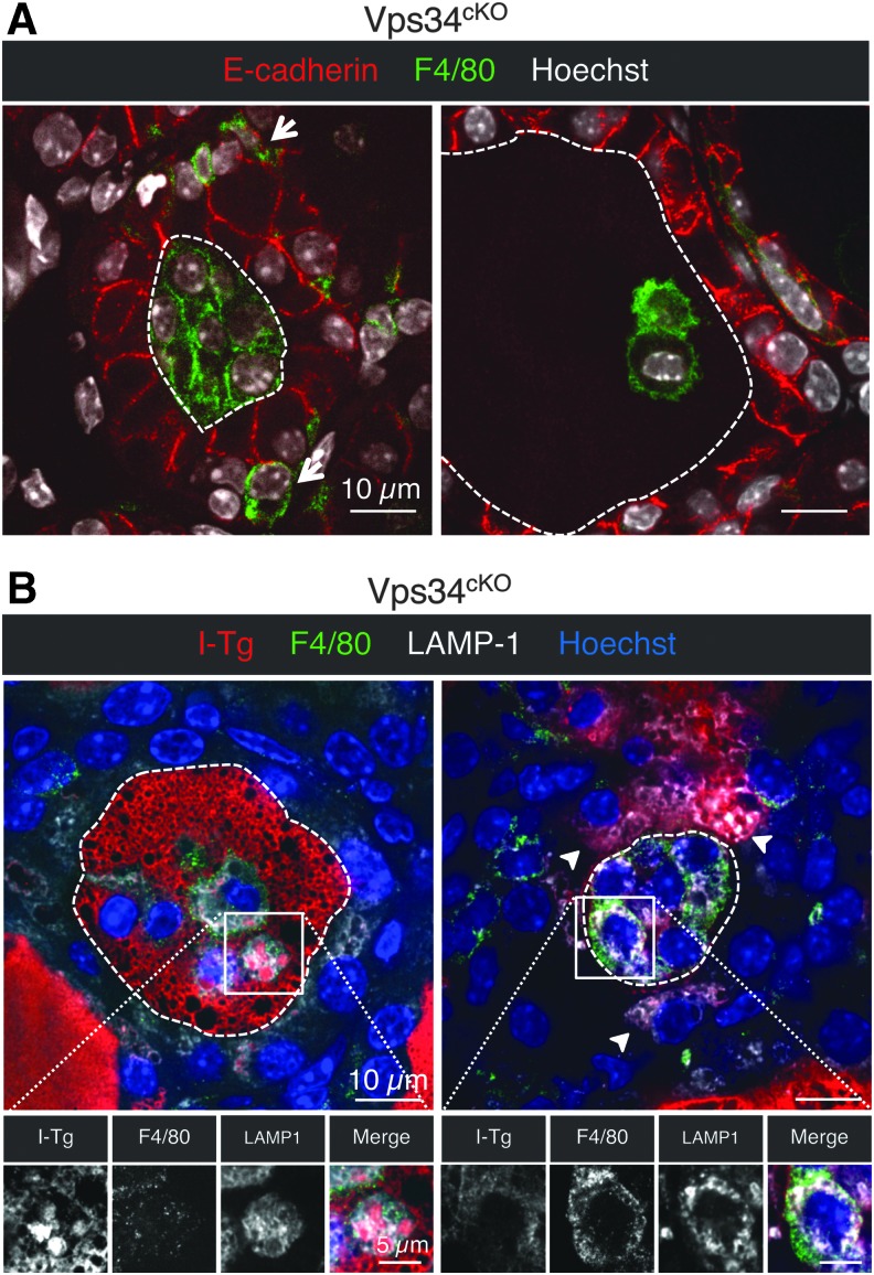
Luminal cells in Vps34cKO thyroid are macrophages. (A) Immunolabeling for E-cadherin (red), and for the conventional macrophage marker (F4/80, green); nuclei are visualized with the Hoechst stain (shown in white). In Vps34cKO thyroid, F4/80 signal can be found in the interstitium (arrows), but also in the colloidal space, thus identifying luminal cells as infiltrating macrophages. (B) Luminal cells are taking up the colloid. Immunolabeling for I-Tg (red), F4/80 (green), and LAMP-1 (white). Nuclei are visualized with the Hoechst stain (shown in blue). Luminal cells in Vps34cKO have abundant LAMP-1 structures that surround I-Tg droplets. Arrowheads show additional I-Tg in thyrocytes. Broken lines indicate luminal contours.

