
Figure 3.
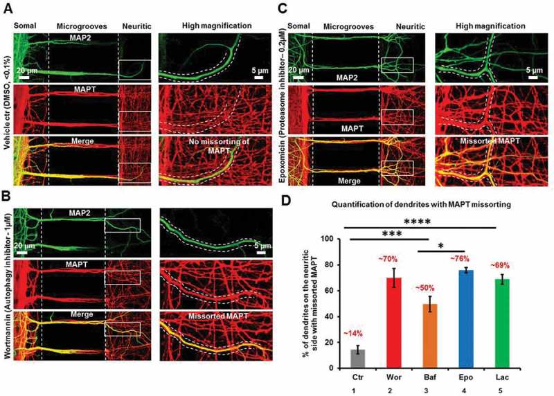
Inhibition of protein degradation (autophagy, proteasome) leads to MAPT missorting. Rat hippocampal neurons (DIV 21–25) cultured in microfluidic chambers were treated on the neuritic side for 24 h either with DMSO (control, a) or with the autophagy inhibitor (wortmannin [Wor], b) or the proteasomal inhibitor (epoxomicin [Epo], c). Dendrites were stained with MAP2 antibody (green) and total MAPT with K9JA antibody (red). Magnified images of the insets are shown on the right with a pair of eye-guiding dotted lines highlighting a dendrite with or without MAPT. (a) In the vehicle-treated control (DMSO, < 0.1%), MAPT is predominantly localized to the axons (see merged images at the bottom in a). Only a small fraction of dendrites colocalizes with MAPT. (b and c) In cultures treated with wortmannin (b, 1 µM, 24 h) or with epoxomicin (c, 0.2 µM, 24 h), the fraction of dendrites with MAPT increases strongly (see quantification in D) where a clear colocalization of MAPT with MAP2 (merged images at the bottom in b and c) becomes visible. Scale bars in all overview images on left: 20 µm; in all magnifications of insets on right: 5 µm. (d) Quantification of dendrites on the neuritic side showing colocalization of MAPT with MAP2 following treatment with DMSO (ctr, bar 1) or with the autophagy (bars 2 and 3) or proteasomal (bars 4 and 5) inhibitors. (n > 180 dendrites/dendritic branches from 3–6 experiments; one-way ANOVA with Tukey’s post hoc test; F [4,17] = 30.09; *p < 0.05; ***p < 0.001; ****p < 0.0001).