

Abstract
Objective
Cup-cage reconstruction has emerged as a possible solution for managing massive acetabular defects with a few existing studies reporting encouraging results at mid-term follow-up. We present our experience with this unitised construct.
Method
Six patients (7 hips) with a mean age of 76 years (73–81) were revised due to catastrophic aseptic failure of a primary cup implanted 10–19 years previously, having a Paprosky type 3B acetabular defect.
Results
At a mean follow-up of 72 months (63–140) no cases have required re-revision. Oxford Hip Scores improved from an average of 8 (1–17) preoperatively to an average of 36 (18–45) at the last follow-up. WOMAC scores preoperatively averaged 76 (49–96) and postoperatively averaged 26.5 points (0–69) at the last follow-up. SF-12 scores improved in both components. One patient showed non-progressive osteolysis around the ischial flange and one had less than 5 mm migration of the construct. One patient died of unrelated causes.
Conclusion
Our study presents one of the longest follow-up of cup-cage construct and supports the previously reported good results; it encourages the use of this construct in reconstruction of massive acetabular defect, with or without pelvic discontinuity.
Keywords: Cup-cage construct, Massive acetabular defect, Revision hip arthroplasty
1. Introduction
Revision total hip arthroplasty (THA) is an increasingly common procedure.10 The goals of revision THA are to remove existing components as indicated, manage bone defects and provide stable fixation of new component(s).
In the vast majority of cases, revision of the acetabular component can be achieved with a cementless, porous-coated hemispherical cup inserted with screws.9,21 However, massive bone loss and pelvic discontinuity caused by osteolysis, infection or occasionally fracture present a greater challenge to the revision surgeon.
Current options for the management of massive bone loss or osteolytic pelvic discontinuity in revision THA include: the use of porous metal augments with a porous metal acetabular component,26 standard ilioischial cage reconstruction,29 a cup-cage construct with a porous metal acetabular component and inset cage fixed proximally and distally,1 bulk acetabular allograft with plating31 and custom triflange components.33
Regarding cage reconstruction, a standard ilioischial cage does not have the potential for biological integration and therefore failure over time is inevitable. The cup-cage construct, first described by Hanssen and Lewallen in 200515, was developed in order to promote biological integration in association with an ilioischial cage. The ilioischial cage provides support for a porous or highly porous cup that would not be stable in isolation. The cage is placed into the cup and stabilised with screws as necessary and a liner is then cemented into this construct. This unitised construct allows potential osseointegration of the cup and, if this occurs, provides a decrease in stresses applied to the cage, protecting it from long-term failure.15 This technique has few outcomes reported in the literature. Our unit has been utilising the cup-cage technique for the treatment of large acetabular defects since 2005 and present in this paper a technical case-series report with mid to long-term follow-up.
2. Patients and methods
3–4% of patients presenting to our revision unit present with massive acetabular defects. The cup-cage construct was used in 7 revision hip arthroplasties in 6 patients. All procedures were performed by the senior author (MS). Our cohort included 5 females and 1 male with a mean age of 76 years (range 73–81). All patients had a Paprosky 3B acetabular defect.27 Indication for revision in all cases was catastrophic aseptic failure of a primary uncemented cup implanted 10–19 years prior to revision. Preoperative digital templating and computed tomography were performed in all cases to assess acetabular bone stock and potential component sizing in order to restore hip centre and offset.
The posterior approach was used in all cases. The femoral component was revised in 2 of 7 procedures. Where the femoral component was retained appropriate exposure of the acetabulum was achieved by careful dissection and retraction. The sciatic nerve was carefully protected throughout the procedure. Following exposure and removal of the failed component the acetabular bone loss was evaluated, confirming the preoperative radiological findings of massive bone loss and Paprosky 3B defect. The acetabular rim deficiency was located postero-laterally, with deficiency present also medially. Pelvic discontinuity was present in one case.
Thorough debridement was performed followed by gentle hemispherical reaming of the acetabulum until bleeding host bone was evident. Using trial components, stability of hemispherical cup was assessed. In the 7 procedures described the hemispherical trial cup was unstable. Therefore, the surgeon proceeded with cup-cage reconstruction.
In four hips trabecular metal acetabular revision system (TMARS) (Zimmer, Warsaw, Indiana) was used and in 3 hips a multihole porous cup was used in conjunction with a standard ilioischial cage. The average size of the hemispherical cup used was 60 mm (range 56–68 mm). All the cups were fixed with screws. The acetabular defect was filled with morsellised bone allograft which was compacted using large round pushers and reverse reaming. Appropriate exposure of the ilium and ischium was achieved to allow fitting the appropriately sized cage into the cup. Ischium was identified by palpating down the posterior rim of the acetabulum and a small osteotome was used to create a slot in the ischium. Once the starter slot was created, the ischial flange of the cage was inserted to complete the slot. The ischial flange was inserted first and then the cage was impacted, seating the superior flange on the ilium. Two or three 6.5 mm fully threaded cancellous screws were used to secure the superior flange to the bone (Fig. 1). Careful contouring of the cage was done prior to insertion in order to provide a good fit against host bone. Repeated manipulation was avoided in order to prevent potential weakening of the cage. Four Zimmer cages Protasul-Ti (Zimmer, Indiana) and three Burch-Schneider cage (Protek, Berne, Switzerland) were used. Face-changing polyethylene liners (3 with 20° and 4 with 10°) were prepared by roughening the back surface with a high-speed burr and then cemented into the cage in the correct version and inclination, independent of the version/orientation of the cage. Five 32 mm, one 36 mm and one 28 mm metallic heads were used. After washout the wound was closed in layers over one deep drain.
Fig. 1.

Intra-operative picture of the Cup-cage Construct.
Post-operative care consisted of assisted mobilisation in bed until the patient regained control of the hip musculature. Weight-bearing as tolerated was then allowed with a frame or crutches, under the supervision of physiotherapists.
All patients were reviewed prospectively in a dedicated revision hip clinic. The patients were assessed using Oxford Hip Score (OHS), WOMAC score and SF-12 scores before surgery and at six weeks, 6 months, 12 months postoperatively after which they were reviewed annually. At each follow-up appointment radiographs were performed, including anteroposterior views of the pelvis and a horizontal beam lateral view of the operated hip. The radiographs were reviewed by an independent assessor for evidence of migration or loosening of the components. Acetabular migration was evaluated according to Massin, Schmidt and Engh's criteria23 with more than 5 mm considered as loosening.
Loosening of the cage was determined according to the criteria of Gill, Sledge and Muller,12 with the modification added by Kosashvili et al..20 This modification considers the presence of a non-progressive radiolucency around the tip of the ischial flange a common finding, likely due to micro movement until biological stabilization and not a sign of “definite loosening”.
3. Results
Mean follow-up in this prospective study was 72 months (range 63–140 months). The mean Oxford Hip Score (OHS) improved from 8 (range 1–17) preoperatively to 36 at the last follow-up (range 18–45). The mean preoperative WOMAC score improved from 76 points (range 49–96) to 26.5 points (range 0–69) at the last follow-up. SF-12 scores, as a reflection of overall health-related-quality of life, improved in both components: the physical component score (PCS) improved from a mean 24.78 points (range 21–29.7) before operation to 40.15 points (range 29.2–46.8) at the last follow-up; the mental component score (MCS) improved from mean 35.5 points (range 28.6–41.9) to 46.64 points (range 34.2–56.9) at the last follow-up. Visual Analogue Score (VAS) for pain at the last follow-up was 0 in 3 patients and 2 in two cases. One patient died of unrelated causes.
Radiologically all patients showed signs of graft incorporation and cup osseointegration (Fig. 2, Fig. 3, Fig. 4, Fig. 5). One patient had less than 5 mm superior migration of the construct at the last follow-up, 4 years after surgery, but was clinically pain-free and mobilising well. No osteolysis around the screws or breaking of the cage screws was seen. One hip showed osteolysis around the ischial flange but this was not progressive and was not interpreted as a sign of loosening. One patient had a Trendelenburg gait but remained satisfied with the results of surgery.
Fig. 2.
Case example showing pre-operative radiograph.
Fig. 3.

Cup-Cage radiograph at 7 years follow-up.
In this case a Trident Tritanium Cup (Stryker, Michigan) and a Burch-Schneider Cage was used.
Fig. 4.

Another Case example showing pre-operative radiograph.
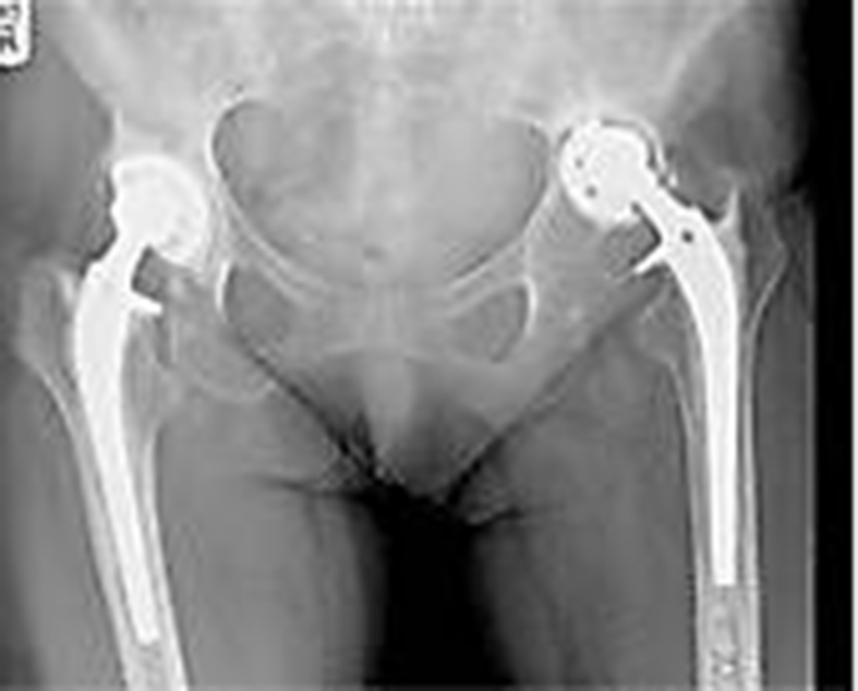
Fig. 5.

Cup-Cage radiograph at 9 years follow-up.
In this case TMARS System was used.
4. Discussion
Reconstruction of the failed acetabular component of a THA aims to obtain immediate and log-term implant stability, preserve or augment the bone stock, restore the centre of rotation and offset of the hip and to avoid significant leg-length discrepancy.
The choice of reconstruction depends on the severity of bone loss.24 Several different classification systems have been developed for femoral and acetabular bone deficiencies in an attempt to facilitate communications between orthopaedic surgeons and provide an algorithm to guide treatment.6,27,30 The Paprosky classification system is most commonly used.27 In type 3B defects the acetabular rim is non-supportive. In these cases, where there is usually less than 50% host bone contact, a conventional hemispherical cup alone is usually unsuccessful.1, 2, 11 Trabecular metal implants have demonstrated encouraging short-term results.7,19,32 The highly porous configuration of trabecular metal enables rapid and extensive bone ingrowth5 making this product very useful even in massive bone defects. Trabecular metal augments can be used to deal with bone defects and achieve better primary stability of the shell.2,17
Other techniques have been employed to deal with massive acetabular bone defects. Antiprotrusio cages have been used to protect the graft whilst remodelling takes place, providing a scaffold at the correct anatomic level and a bed into which the cup is cemented. The major disadvantage of cages is their non-biological fixation, which makes them prone to mechanical failure within seven to ten years of implantation.14,17 Use of bulk structural allografts in association with cementless acetabular components for massive bone defects has shown a failure rate of 18% at 31-month follow-up25 and 64% at 6 years follow-up.28 Impaction bone grafting, while reconstructing the bone stock, remains technically demanding and carries a risk of acetabular fracture, graft resorbtion, implant migration and disease transmission.17,34 The custom triflange cup has been designed as a salvage implant for acetabular reconstruction in the presence of significant bone loss with favourable results at mid-term follow-up.33 However, previous reports showed a high dislocation rate and underline the fact that it may take up to six weeks to generate the implant from a computer tomographic scan of the acetabulum.8,16
The cup-cage construct was first described in 200515 as an alternative for the treatment of massive bone loss. Few reports exist in the literature regarding this type of reconstruction in revision hip surgery (Table 1). A literature review revealed only seven papers, four of which originate from the same Lower Extremity Reconstruction Unit.1,3,4,18,20,22 In all but one, the cup-cage construct was used in the treatment of pelvic discontinuity. In our series cases without discontinuity predominate.
Table 1.
Reported results with Cup-Cage Construct in Revision Hip Arthroplasty.
| Reported Results with Cup-Cage Construct | ||||
|---|---|---|---|---|
| Author | Year | No of hips | Mean Follow up | Success rate |
| Kosashvili et al. | 2009 | 26 | 44 mths | 88.5% |
| Kellet et al. | 2010 | 14 | 27 mths | 82% |
| Alfaro et al. | 2010 | 5 | 26 mths | 100% |
| Malhotra et al. | 2012 | 1 | 30 mths | 100% |
| Abolghasemian et al. | 2014 | 26 | 82 mths | 87% |
| Amenabar et al. | 2016 | 67 | 74 mths | 93% |
| Sculco et al. | 2017 | 57 | 60 mths | 89% |
| Sood et al. (our series) | 2018 | 7 | 72 mths | 100% |
Kosashvili Y et al. reviewed a consecutive series of 26 cases (24 patients) with pelvic discontinuity treated using cup-cage technique at a mean follow-up of 44.6 months (range 24–68) and had a success rate of 88.5% with significant improvement in Harris Hip Score.20 Abolghasemian M et al. reviewed the same series at a mean follow-up of 82 months (12–113) and compared the results of cup-cage reconstruction with conventional cages used in 19 patients with a mean follow-up of 69 months (1–170).1 The seven-year survivorship was 87.2% for the cup-cage group and 49.9% for the cage-alone group. There were 3 revisions of the cup-cage due to loosening and/or migration with a mean time to failure of 46.7 months (12–85). Complications of the cup-cage group consisted of two dislocations, one deep infection and one sciatic nerve injury, which partially recovered. This is the only study comparing cup-cage with another implant. The use of the cup-cage construct in the treatment of pelvic discontinuity is also supported by a recent biomechanical study that has shown that the stiffness of fixation is similar to a combined cage and posterior-column plate.13
Ballester Alfaro et al. published their results with trabecular metal buttress augments and trabecular metal cup-cage construct in revision hip arthroplasty for severe acetabular bone loss.4 Amongst their 19 participants, five had pelvic discontinuity. In these the cup-cage construct was used. At a mean follow-up of 26 months (range 18–43) no mechanical failure had occurred and all patients had radiographically stable cups.
Kellett CF et al. examined early results in 14 patients with pelvic discontinuity treated with Cup-cage construct at a mean follow-up of 27 months (range 1–39). Outcomes were excellent or good in 82% of cases, with improvements in Oxford Hip Scores, WOMAC scores and SF-36 scores. Radiographically, at the last follow-up, all implants were stable and none had migrated.18
Malhotra R at al presented a case of failed hip reconstruction managed with TMARS cup-cage construct. Follow-up radiographs at 30 months showed no signs of failure and the patient was walking unaided and had a Harris Hip Score of 9022.
The study of Amenabar T et al. presented the outcome of sixty-seven cup-cage reconstructions for a mean of 74 months (range 24–135). Sixty-one percent of patients had pelvic discontinuity. The five and ten-year survival rate were 93% and 85%, respectively when failure was defined as revision for any cause. Complications included deep infection in three patients, dislocation in three and sciatic nerve injury in two patients.3
The most recent publication about cup-cage construct came from the originators of the technique.35 Fifty-seven patients were retrospectively reviewed at a mean follow-up of 4.6 years (range 1.4–10.6 years). There were 30 full cup-cage patients and in 27 half cup-cage (inferior half was removed) patients. The survivorship free from re-revision for any cause was 89% (83% and 96% for full and half cup-cage cohorts, respectively. Postoperative complications included sciatic nerve injury (2 cases-3.5%) and hip dislocation (4 cases-7%).
We present our results with cup-cage construct in 7 cases at a mean 72 months follow-up (range 63–140 months). All patients had massive bone loss (Paprosky type 3B) and one had a pelvic discontinuity. No cases have required re-revision, with only one patient showing 5 mm migration of the construct at 4- year radiographic follow-up. This does not appear to have clinically significant consequences.
This case-series is, to our knowledge, the third longest follow-up available in the literature, the longest from a non-designer centre. Our “independent” modest experience support the use of cup-cage construct as a reliable technique in addressing massive acetabular bone loss encountered by revision hip arthroplasty surgeon in both pelvic discontinuity and non-discontinuity cases.
We acknowledge the limitation of our study due to the small number of cases. Hence, a meaningful statistical analysis was not possible. We agree that longer follow-up is needed to determine whether this combination will prevent mechanical failures seen with ilioischial cages.
Funding
This research did not receive any specific grant from funding agencies in the public, commercial, or not-for-profit sectors.
Footnotes
The study was carried out at Bedford Hospital, UK.
Contributor Information
Dan Arvinte, Email: dan_arvinte@hotmail.com.
Manish Kiran, Email: drmanishkiran@gmail.com.
Manoj Sood, Email: manojsood@yahoo.com.
References
- 1.Della Valle C.J., Rosenberg A.G. Revision Total Hip and Knee Arthroplasty. Lippincot Williams & Wilkins; 2012. Cementless acetabular reconstruction in revision total hip arthroplasty; pp. 155–162. [Google Scholar]
- 2.Della Valle C.J., Berger R.A., Rosenberg A.G. Cementless acetabular reconstruction in revision total hip arthroplasty. Clin Orthop Relat Res. 2004;420:96–100. doi: 10.1097/00003086-200403000-00013. [DOI] [PubMed] [Google Scholar]
- 3.Leopold S.S., Rosenberg A.G., Bhatt R.D. Cementless acetabular revision. Evaluation at an average of 10.5 years. Clin Orthop. 1999;369:179–186. [PubMed] [Google Scholar]
- 4.Paprosky W.G., O'Rourke M., Sporer S.M. The treatment of acetabular bone defects with an associated pelvic discontinuity. Clin Orthop Relat Res. 2005;441:216–220. doi: 10.1097/01.blo.0000194311.20901.f9. [DOI] [PubMed] [Google Scholar]
- 5.Perka C., Ludwig R. Reconstruction of segmental defects during revision procedures of the acetabulum with the Burch-Schneider anti-protrusion cage. J Arthroplast. 2001;16(5):568–574. doi: 10.1054/arth.2001.23919. [DOI] [PubMed] [Google Scholar]
- 6.Abolghasemian M., Tangsaraporn S., Drexler M. The challenge of pelvic discontinuity. Cup-cage reconstruction does better than conventional cages in mid-term. Bone Joint Lett J. 2014;96-B:195–200. doi: 10.1302/0301-620X.96B2.31907. [DOI] [PubMed] [Google Scholar]
- 7.Stiehl J.B., Saluja R., Diener T. Reconstruction of major column defects and pelvic discontinuity in revision total hip arthroplasty. J Arthroplast. 2000;15:849–857. doi: 10.1054/arth.2000.9320. [DOI] [PubMed] [Google Scholar]
- 8.Taunton M.J., Fehring T.K., Edwards P.E. Pelvic discontinuity treated with custom triflange component. Clin Orthop Relat Res. 2012;470:428–434. doi: 10.1007/s11999-011-2126-1. [DOI] [PMC free article] [PubMed] [Google Scholar]
- 9.Hanssen A.D., Lewallen D.G. Modular acetabular augments: composite void fillers. Orthopedics. 2005;28:971–972. doi: 10.3928/0147-7447-20050901-29. [DOI] [PubMed] [Google Scholar]
- 10.Paproski W.G., Perona P.G., Lawrence J.M. Acetabular defect classification and surgical reconstruction in revision arthroplasty: a 6 year follow-up evaluation. J Arthroplast. 1994;9:33–44. doi: 10.1016/0883-5403(94)90135-x. [DOI] [PubMed] [Google Scholar]
- 11.a Massin P., Schmidt L., Engh C.A. Evaluation of cementless acetabular component migration: an experimental study. J Arthroplasty. 1989;4:245–251. doi: 10.1016/s0883-5403(89)80020-8. [DOI] [PubMed] [Google Scholar]; b Abolghasemian M., Tangsataporn S., Sternheim A. Combined trabecular metal acetabular shell and augment for acetabular revision with substantial bone loss. Bone Joint J. 2013;95-B:166–172. doi: 10.1302/0301-620X.95B2.30608. [DOI] [PubMed] [Google Scholar]
- 12.Gill T.J., Sledge J.B., Muller M.E. The Burch-Sneider anti-protrusion cage in revision total hip arthroplasty: indications, principles and long term results. J Bone Joints Surg (Br) 1998;80-B:946–953. doi: 10.1302/0301-620x.80b6.8658. [DOI] [PubMed] [Google Scholar]
- 13.Kosashvili Y., Backstein D., Safir O., Lakstein D., Gross A.E. Acetabular revision surgery using an anti-protrusion (ilio-ischial) cage and trabecular metal acetabular component for severe acetabular bone loss associated with pelvic discontinuity. J Bone Jt Surg. 2009;91-B:870–876. doi: 10.1302/0301-620X.91B7.22181. [DOI] [PubMed] [Google Scholar]
- 14.O'Brien D.A., Rorabeck C.H. Managing bone loss in revision total hip arthroplasty: the acetabulum. Instr Course Lect. 2006;55:263–277. [PubMed] [Google Scholar]
- 15.D'Antonio J.A., Capello W.N., Borden L.S. Classification and management of acetabular abnormalities in total hip arthroplasty. Clin Orthop. 1989;243:126–137. [PubMed] [Google Scholar]
- 16.Saleh K.J., Holtzman J., Gafni A. Development, test reliability and validation of a classification for revision hip arthroplasty. J Orthop Res. 2001;19:50–56. doi: 10.1016/S0736-0266(00)00021-8. [DOI] [PubMed] [Google Scholar]
- 17.Garcia-Cimbrela E. Porous-coated cementless acetabular cups in revision surgery : a 6- to 11-year follow-up study. J Arthroplast. 1999;14(4):397–406. doi: 10.1016/s0883-5403(99)90094-3. [DOI] [PubMed] [Google Scholar]
- 18.Kim W.Y., Greidanus N.V., Duncan C.P., Masri B.A., Garbuz D.S. Porous tantalum uncemented acetabular shells in revision total hip replacement: two to four year clinical and radiographic results. Hip Int. 2008;18(1):17–22. doi: 10.1177/112070000801800104. [DOI] [PubMed] [Google Scholar]
- 19.Kim W.Y., Greidanus N.V., Duncan C.P., Masri B.A., Garbuz D.S. Porous tantalum uncemented acetabular shells in revision total hip replacement: two to four year clinical and radiographic results. Hip Int. 2008;18(1):17–22. doi: 10.1177/112070000801800104. [DOI] [PubMed] [Google Scholar]
- 20.Skytta E.T., Eskelinen A., Paavolainen P.O., Remes V.M. Early results of 827 trabecular metal revision shells in acetabular revision. J Arthroplast. 2011;26(3):342–345. doi: 10.1016/j.arth.2010.01.106. [DOI] [PubMed] [Google Scholar]
- 21.Bobyn J.D., Stackpool G.J., Hacking S.A., Tanzer m, Krygier J.J. Characteristics of bone ingrowth and interface mechanics of a new porous tantalum biomaterial. J Bone Jt Surg. 1999;81-B:907–914. doi: 10.1302/0301-620x.81b5.9283. [DOI] [PubMed] [Google Scholar]
- 22.Abolghasemian M., Tangsataporn S., Sternheim A. Combined trabecular metal acetabular shell and augment for acetabular revision with substantial bone loss. Bone Joint Lett J. 2013;95-B:166–172. doi: 10.1302/0301-620X.95B2.30608. [DOI] [PubMed] [Google Scholar]
- 23.Issack P.S. Current concepts review. Use of porous tantalum for acetabular reconstruction in revision hip arthroplasty. J Bone Joint Surg Am. 2013;95:1981–1987. doi: 10.2106/JBJS.L.01313. [DOI] [PubMed] [Google Scholar]
- 24.Goodman S., Saastamoinen H., Sasha N., Gross A. Complications of ilioischial reconstruction rings in revision total hip arthroplasty. J Arthroplast. 2004;19:436–446. doi: 10.1016/j.arth.2003.11.015. [DOI] [PubMed] [Google Scholar]
- 25.Paprosky W.G., Bradford M.S., Jablonsky W.S. Acetabular reconstruction with massive acetabular allografts. Instr Course Lect. 1996;45:149–159. [PubMed] [Google Scholar]
- 26.Paprosky W.G., Sekundiak T.D. Total acetabular allografts. Instr Course Lect. 1999;48:67–76. [PubMed] [Google Scholar]
- 27.Van Egmond N., De Kam D.C., Gardeniers J.W., Schreurs B.W. Revision of extensive acetabular defects with impaction grafting and a cement cup. Clin Orthop Relat Res. 2011;469(2):562–573. doi: 10.1007/s11999-010-1618-8. [DOI] [PMC free article] [PubMed] [Google Scholar]
- 28.DeBoer D.K., Christie M.J., Brinson M.F., Morrison J.C. Revision total hip arthroplasty for pelvic discontinuity. J Bone Joint Surg Am. 2007;89(4):835–840. doi: 10.2106/JBJS.F.00313. [DOI] [PubMed] [Google Scholar]
- 29.Holt G.E., Dennis D.A. Use of custom triflanged acetabular components in revision total hip arthroplasty. Clin Orthop Relat Res. 2004;429:209–214. doi: 10.1097/01.blo.0000150252.19780.74. [DOI] [PubMed] [Google Scholar]
- 30.Kellett C.F., Gross A.E., Backstein D., Safir O. Massive acetabular bone loss: the cup-cage solution. Semin Arthroplasty. 2010;21:57–61. [Google Scholar]
- 31.Ballester Alfaro J.J., Fernandez J.S. Trabecular metal buttress augment and the Trabecular Metal cup-cage construct in revision hip arthroplasty for severe acetabular bone loss and pelvic discontinuity. Hip Int. 2010;20(Suppl 7):119–127. doi: 10.1177/11207000100200s720. [DOI] [PubMed] [Google Scholar]
- 32.Malhotra R., Ramprasad K., Kumar V., Soral A. Trabecular metal acetabular revision system (cup-cage construct) to address the massive acetabular defects in revision arthroplasty. Indian J Orthop. 2012;46(4):483–486. doi: 10.4103/0019-5413.97264. [DOI] [PMC free article] [PubMed] [Google Scholar]
- 33.Amenabar T., Rahman W.A., Hetaimish B.M. Promising Mid-Term results with Cup-Cage construct for large acetabular defects and pelvic discontinuity. Clin Orthop Relat Res. 2016;474(2):408–414. doi: 10.1007/s11999-015-4210-4. [DOI] [PMC free article] [PubMed] [Google Scholar]
- 34.Gililland J.M., Anderson L.A., Henninger H.B., Kubiak E.N., Peters C.L. Biomechanical analysis of acetabular revision constructs: is pelvic discontinuity best treated with bicolumnar or traditional unicolumnar fixation? J Artroplasty. 2013;28:178–186. doi: 10.1016/j.arth.2012.04.031. [DOI] [PubMed] [Google Scholar]
- 35.Sculco P.K., Ledford C.K., Hanssen A.D., Abdel M.P., Lewallen D.G. The evolution of the Cu-cage technique for major acetabular defects. J Bone Joint Surg Am. 2017;99:1104–1110. doi: 10.2106/JBJS.16.00821. [DOI] [PubMed] [Google Scholar]