Abstract
Nail coloration has many causes and may reflect systemic disease. White nails (leukonychia) are common; rubronychia is rare, whereas green (chloronychia) is occasionally evident. Chloronychia, the Fox–Goldman syndrome, is caused by infection of an often damaged nail plate by Pseudomonas aeruginosa. P. aeruginosa is an opportunistic pathogen known for localized and systemic infections. It can spread cryptically in a variety of ways, whether from an infected nail to a wound either autologously or to a patient as a surgical site infection, and many represent a threat to elderly, neonatal, or immunocompromised patients who are at increased risk of disseminated pseudomonas infection. We will review the Goldman–Fox syndrome as an occupational disorder of homemakers, nurses, plumbers, and others often with wet hands. At a time when hand washing is being stressed, especially in healthcare settings, examination of nails should be emphasized too, recalling the possibility of surgical site infection even with a properly washed and gloved medical care provider. Pseudomonas may be a community-acquired infection or a hospital or medical care setting-acquired one, a difference with therapeutic implications. Since healthcare workers represent a threat of nosocomial infections, possible guidelines are suggested.
KEY WORDS: Chloronychia, chromonychia, green, nails, pseudomonas
Introduction
Changes in nail appearance can be indicative of an underlying systemic disease.[1,2,3,4] Persistent greenish pigmentation of the nail plate was originally described in 1944 by Goldman and Fox,[4] who linked it with nail plate involvement by Pseudomonas aeruginosa; hence, its designation as the Goldman–Fox syndrome (GFS) honoring the legendary professor of dermatology and medical laser pioneer at the University of Cincinnati, Leon Goldman MD. They described two patients, each with a secondary P. aeruginosa infection followed by greenish nail pigmentation of the entire nail plate. They postulated that an associated paronychia led to nail matrix spread with pigment diffusion through the nail plate to produce a bright green color that ultimately transitioned to dull green but persisted for 6 months after the infection had been successfully treated.
The goal of this review is to explore green nails as a warning sign.[5,6,7,8,9,10,11,12] GFS is basically a nontender paronychial infection that appears most often in persons whose hands are constantly exposed to water, soaps and detergents, sometimes from occupational exposure, or have damaged nails by trauma, psoriasis, onychomycosis, or other disorders. An outbreak of sternal surgical site infections due to P. aeruginosa was traced to a scrub nurse with onychomycosis, illuminating its importance in an era of rigorous hand washing.[5]
Clinical Features
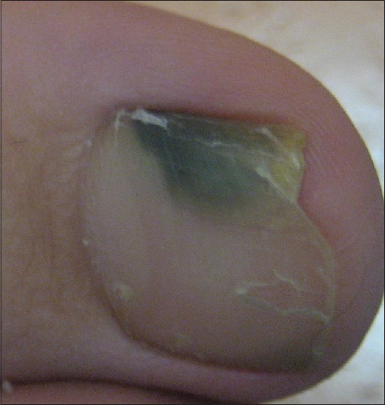
The GFS has been described as a triad of green discoloration of the nail plate, proximal paronychia, and distal onycholysis.[4,5,6,7,8] It is typically seen in patients with nail diseases, such as onychotillomania, psoriasis, or paronychia, particularly in those whose abnormal nails have been exposed to a chronic moist environment. The green discoloration can vary from blue-green, green-brown to green-yellow [Figure 1]. This condition can involve the fingernails or toenails and is often confined to only one or two nails with partial or complete involvement of the nail plate. The nail is usually painless; however, the skin around the nail may be erythematous or tender. Horizontal green-striped fingernails may result from a P. aeruginosa paronychia with the periodicity of the linear coloration corresponding to clinical episodes of infection.[10] Growth of the causative organism from the green portion of the nail plate was documented on suitable bacteriological media. The largest series was probably that of 40 patients described by Chernosky and Dukes.[7] Of these, 34 had occupational exposure, 11 had nails damaged by trauma, and 3 had concurrent onychomycosis. The infection might be acquired when a patient was gardening or dishwashing or at work in a healthcare setting, despite the wearing of gloves when caring for patients.
Figure 1.

Green nail in otherwise health individual
Dissemination has not been emphasized. An outbreak of sternal surgical site P. aeruginosa infections in 16 of 185 patients traced to a gloved scrub nurse with onychomycosis emphasizes its importance.[5] In another situation, GFS of the second fingernail of someone employed on a postoperative intensive care unit resulted in many patients developing P. aeruginosa infection or colonization. Less than one-third of 168 nurses responded in a questionnaire that they knew the significance of a green fingernail.[12] GFS also has the potential of autologous spread in an infected individual who scratches or rubs his or her own skin, especially if the cutaneous surface is damaged. Although intact skin is usually impervious, this bacterium may more efficiently colonize wounds, ulcers, and burns.[6] Some patients have coexistent mycological or bacterial infections with organisms such as Staphylococcus aureus and Klebsiella. [12,13] A nail dystrophy assay now including the “Green Nail Syndrome Test” may be of value in confirming that the P. aeruginosa nail infection has been cured to reassure an affected nurse or surgeon.[14] Rarely, Aspergillus flavus nail infection, green dyes, and certain chemicals have also been linked with green nails.[15,16]
Pathogenesis
P. aeruginosa is a Gram-negative, aerobic coccobacilli that is ubiquitous in nature.[4,5,6,7] This pathogen secretes the blue-green pigments pyocyanin and pyoverdin which result in the characteristic green discoloration of the nail and sometimes detectable green fluorescence caused by the fluorescent siderophore pyoverdin. The color may simply diffuse into the nail from the surrounding diseased tissue.[13] In humans, P. aeruginosa is an opportunistic pathogen known to cause pneumonia, otitis externa, urinary tract infections, osteomyelitis, and sepsis.
Although ubiquitous in nature, P. aeruginosa, also known as “the water bug,” is not part of the normal flora in healthy human skin. Because the dermis is relatively dry, this pathogen is not able to survive and therefore cutaneous infections in intact human skin are rare. When exposed to moist environments, cutaneous infections such as P. aeruginosa of the nail have the potential to spread in an infected patient who scratches his or her own skin. One should be concerned about an enhanced risk of serious and life-threatening P. aeruginosa infections, such as necrotizing fasciitis and ecthyma gangrenosum, that may result in immunocompromised patients.
One bacteriological study of 26 patients with green discoloration of one or more nails documented 23 of them with P. aeruginosa in pure or mixed culture.[7] Many of them had occupational exposure to water, soap. and detergents, some showing evidence of mechanical trauma or concurrent onychomycosis. Nonpigmented onychopathic or normal nails from 50 patients were cultured with only one patient yielding P. aeruginosa. Histologically, Gram-negative bacilli consistent with P. aeruginosa were demonstrated within the nail plate. P. aeruginosa invasion of the nail plate may be facilitated by local trauma, nail deformities. or coinfections. The large number of other low-grade pathogens suggests P. aeruginosa may be a secondary invader whether as colonizer or as an actual infection.[13] Infection or colonization of an intact nail by P. aeruginosa is also unusual. Affected patients typically have a history of prior nail problems such as onycholysis, paronychia, psoriasis, or trauma. Separation of the nail bed allows entry of bacteria and therefore infection or colonization by this pathogen.
Treatment
There are, to our knowledge, no controlled double-blind clinical trials for treating the GFS. In addition, treatment to our knowledge has not separated health care facility and community-acquired infections, a concept of considerable importance with the former possibly exhibiting enhanced antibiotic resistance. Treatment may consist of removing and cleaning the onycholytic portion of the nail, and keeping the nails dry to prevent P. aeruginosa persistence. Application of compresses of 1% acetic acid can be effective.[4] Topical silver sulfadiazine, gentamicin, ciprofloxacin, bacitracin, and polymyxin B are also often beneficial. Topical nadifloxacin has been employed with success in a few HIV-positive patients[17] and others, including an intensive care nurse with psoriasis involving nails.[8] She had an infection with both fluoroquinolone-sensitive P. aeruginosa and Klebsiella pneumonia which, after 2 weeks of topical nadifloxacin, had the nail color returned to normal. Another approach consists of cutting of the detached nail plate and brushing the nail bed with a 2% sodium hypochlorite solution twice daily.[18]
There are many favorable anecdotal reports. Tobramycin eye drops may also be a good choice.[19] Another is topical 1% silver sulfasalazine together with 2% miconazole.[20] Amikacin may be injected daily for ten days into the nail matrix of an the infected nail if it has not responded to oral and topical antibiotics and antifungal agents.[21] Home remedies have been employed too, including chlorine bleach diluted 1:4 with water and various concentrations of vinegar (acetic acid). When topical therapies fail, the oral antibiotic ciprofloxacin has been effective in many cases. This oral quinolone has been recommended, particularly in aged patients, utilizing a regimen of ciprofloxacin 500 mg/day for 3 weeks.[9] Any coexistent onychomycosis can be treated, with a regime such as itraconazole 200 mg/day 14 days per month for 3 months. Coexistent C. albicans and P. aeruginosa nail infections can be treated with oral fluconazole 200 mg per week, topical ciprofloxacin drops, and topical ciclopirox.[22] Another topical approach is soaking the affected nails twice a day for 10 minutes in 0.1% octenidine dihydrochloride solution for 6 weeks, an option curative for 12 of 15 patients.[23] However, despite the various treatments options, removal of the whole nail is occasionally necessary. Careful evaluation of the nail employing dermatoscopy may be of value in diagnosis and treatment follow-up for nail mixed infections caused by P. aeruginosa and other organisms.[22]
Conclusion
The GFS is caused by a superficial and localized infection of the nail by P. aeruginosa, which is unusual in intact nails and often seen in patients with predisposing conditions that favor the growth of this pathogen. After all, Pseudomonas is a common and often overlooked cause of nail infection easily recognized by its green coloration. It may be viewed as both an occupational disease of homemakers, nurses, and plumbers and an opportunistic infection of nails damaged by trauma, disease, or fungal infection. It may spread in autologous manner. It is of considerable concern in the health care setting. Healthcare workers, even after hand washing and wearing of gloves, may spread it to others, with a special focus in surgical operative settings.
Further research should explore the guidelines to prevent transmission of pseudomonas from healthcare workers with the GFS as well as diminishing their enhanced risk of acquiring GFS with mandated frequent hand-washing. Suggestions are noted in Table 1.
Table 1.
Goldman-Fox Green Nail Syndrome: Patient Protection Suggestions
| Examine fingernails of healthcare workers for green coloration |
| Discourage painted nails in healthcare workers, which may hide nail color changes |
| Investigate surgical gloves used to assess protection against Pseudomonas aeruginosa penetration |
| Educate healthcare workers about Pseudomonas aeruginosa, the “water bug,” and methods to promptly dry hands after mandated frequent washing |
| Evaluate closely coworkers of infected healthcare workers |
| Consider whether or not infected healthcare workers should be allowed patient contact while still infected |
| Encourage randomized double-blind control studies on the most effective treatment |
| Consider separating iatrogenic, medical care provider, and community-acquired infections with regard to treatment for culture and sensitivity testing and for clinical responses to therapy |
| Educating medical staff on the risk of frequent hand-washing, including the acquisition of the Goldman-Fox syndrome |
Financial support and sponsorship
Nil.
Conflicts of interest
There are no conflicts of interest.
References
- 1.Witkowski AM, Jasterzbski TJ, Schwartz RA. Terry's nails: A sign of systemic disease. Indian J Dermatol. 2017;62:309–11. doi: 10.4103/ijd.IJD_98_17. [DOI] [PMC free article] [PubMed] [Google Scholar]
- 2.Almohssen AA, Schwartz RA. Rubronychia: A rose by any other name. J Eur Acad Dermatol Venereol. 2019;33:e103–4. doi: 10.1111/jdv.15265. [DOI] [PubMed] [Google Scholar]
- 3.Schwartz RA, Barnett CR. Muehrcke Lines of the Fingernails Medscape Reference Updated May 23, 2019. [Last accessed on 2019 Aug 19]. Available from: http://emedicinemedscapecom/article/1106423-overview .
- 4.Goldman L, Fox H. Greenish pigmentation of the nail plates from bacillus pyocyaneus infection. AMA Arch Dermatol Syphilol. 1944;68:136–7. [Google Scholar]
- 5.McNeil SA, Nordstrom-Lerner L, Malani PN, Zervos M, Kauffman CA. Outbreak of sternal surgical site infections due to Pseudomonas aeruginosa traced to a scrub nurse with onychomycosis. Clin Infect Dis. 2001;33:317–23. doi: 10.1086/321890. [DOI] [PubMed] [Google Scholar]
- 6.Vergilis I, Goldberg LH, Landau J, Maltz A. Transmission of Pseudomonas aeruginosa from nail to wound infection. Dermatol Surg. 2011;37:105–6. doi: 10.1111/j.1524-4725.2010.01827.x. [DOI] [PubMed] [Google Scholar]
- 7.Chernosky ME, Dukes CD. Green nails. Importance of Pseudomonas aeruginosa in onychia. Arch Dermatol. 1963;88:548–53. doi: 10.1001/archderm.1963.01590230056008. [DOI] [PubMed] [Google Scholar]
- 8.Hengge UR, Bardeli V. Images in clinical medicine. Green nails. N Engl J Med. 2009;360:1125. doi: 10.1056/NEJMicm0706497. [DOI] [PubMed] [Google Scholar]
- 9.Chiriac A, Brzezinski P, Foia L, Marincu I. Chloronychia: Green nail syndrome caused by Pseudomonas aeruginosa in elderly persons. Clin Interv Aging. 2015;10:265–7. doi: 10.2147/CIA.S75525. [DOI] [PMC free article] [PubMed] [Google Scholar]
- 10.Shellow WV, Koplon BS. Green striped nails: Chromonychia due to Pseudomonas aeruginosa. Arch Dermatol. 1968;97:149–53. [PubMed] [Google Scholar]
- 11.Ratka P, Słoboda T. Green nails. Przegl Dermatol. 1986;73:212–5. [PubMed] [Google Scholar]
- 12.Greenberg JH. Green fingernails: A possible pathway of nosocomial pseudomonas infection. Mil Med. 1975;140:356–7. [PubMed] [Google Scholar]
- 13.Stone OJ, Mullins JF. The role of Pseudomonas aeruginosa in nail disease. J Invest Dermatol. 1963;41:25–6. [PubMed] [Google Scholar]
- 14.Nail Dystrophy Assay Includes Green Nail Syndrome Test. [Last accessed on 2019 Jan 14]. Available from: https://bakodx.com/green-nail-syndrome/
- 15.Bereston ES, Keil H. Onychomycosis due to Aspergilus flavus. Arch Derm Syphilol. 1941;44:420–5. [Google Scholar]
- 16.Leung LK, Harding J. A chemical mixer with dark-green nails. BMJ Case Rep. 2015;2015 doi: 10.1136/bcr-2014-209203. pii: bcr2014209203. [DOI] [PMC free article] [PubMed] [Google Scholar]
- 17.Rallis E, Paparizos V, Flemetakis A, Katsambas A. Pseudomonas fingernail infection successfully treated with topical nadifloxacin in HIV-positive patients: Report of two cases. AIDS. 2010;24:1087–8. doi: 10.1097/QAD.0b013e32833819aa. [DOI] [PubMed] [Google Scholar]
- 18.Maes M, Richert B, de la Brassinne M. Green nail syndrome or chloronychia. Rev Med Liege. 2002;57:233–5. [PubMed] [Google Scholar]
- 19.Bae Y, Lee GM, Sim JH, Lee S, Lee SY, Park YL. Green nail syndrome treated with the application of tobramycin eye drop. Ann Dermatol. 2014;26:514–6. doi: 10.5021/ad.2014.26.4.514. [DOI] [PMC free article] [PubMed] [Google Scholar]
- 20.de Almeida HL, Jr, Duquia RP, de Castro LA, Rocha NM. Scanning electron microscopy of the green nail. Int J Dermatol. 2010;49:962–3. doi: 10.1111/j.1365-4632.2009.04251.x. [DOI] [PubMed] [Google Scholar]
- 21.Aksakal AB, Adisen EM, Gurer MA. Green nail syndrome. Gazi Tip Dergisi. 2006;17:216–7. [Google Scholar]
- 22.Romaszkiewicz A, Sławińska M, Sobjanek M, Nowicki RJ. Nail dermoscopy (onychoscopy) is useful in diagnosis and treatment follow-up of the nail mixed infection caused by Pseudomonas aeruginosa and Candida albicans. Postepy Dermatol Alergol. 2018;35:327–9. doi: 10.5114/ada.2018.76232. [DOI] [PMC free article] [PubMed] [Google Scholar]
- 23.Rigopoulos D, Rallis E, Gregoriou S, Larios G, Belyayeva Y, Gkouvi K, et al. Treatment of pseudomonas nail infections with 0.1% octenidine dihydrochloride solution. Dermatology. 2009;218:67–8. doi: 10.1159/000171816. [DOI] [PubMed] [Google Scholar]


