Abstract
Electrophysiological studies can provide objective and quantifiable assessments of movement disorders. They are useful in the diagnosis of hyperkinetic movement disorders, particularly tremors and myoclonus. The most commonly used measures are surface electromyography (sEMG), electroencephalography (EEG) and accelerometry. Frequency and coherence analyses of sEMG signals may reveal the nature of tremors and the source of the tremors. The effects of voluntary tapping, ballistic movements and weighting of the limbs can help to distinguish between organic and functional tremors. The presence of Bereitschafts-potentials and beta-band desynchronization recorded by EEG before movement onset provide strong evidence for functional movement disorders. EMG burst durations, distributions and muscle recruitment orders may identify and classify myoclonus to cortical, subcortical or spinal origins and help in the diagnosis of functional myoclonus. Organic and functional cervical dystonia can potentially be distinguished by EMG power spectral analysis. Several reflex circuits, such as the long latency reflex, blink reflex and startle reflex, can be elicited with different types of external stimuli and are useful in the assessment of myoclonus, excessive startle and stiff person syndrome. However, limitations of the tests should be recognized, and the results should be interpreted together with clinical observations.
Keywords: Accelerometry, Dystonia, Electroencephalography, Electromyography, Electrophysiology, Myoclonus, Psychomotor disorders, Tremor
Electrophysiological assessments are valuable in diagnosing patients with movement disorders. Together with clinical information, the results help identify the correct diagnosis and can be used to evaluate treatment efficacy. Electrophysiological testing is an important tool in the assessment of tremor and myoclonus and in differentiating functional disorders from organic disorders. Surface electromyography (sEMG), electroencephalography (EEG) and accelerometry are the main electrophysiological measurements used. In this review, we will outline the basic settings and techniques used in electrophysiological studies for movement disorders, data analysis techniques and the typical response patterns corresponding to several movement disorders.
BASIC ASSESSMENT METHODS
sEMG
In sEMG studies, muscle activity is recorded noninvasively over the skin. Usually, active and reference electrodes are attached to the target muscle in a tendon-belly arrangement. For larger muscles, both the active and reference electrodes can be placed on the muscle belly with a distance of approximately 2 cm between the electrodes. The following amplifier settings are recommended: at least a 1,000 Hz sampling rate and bandpass filters between 20 and 500 Hz. A limitation of sEMG is the potential for crosstalk from other adjacent muscles and the inability to record the activity of deep muscles. For some deep muscles, such as the diaphragm, needle EMG is needed to obtain high quality recordings (Supplementary Figure 1 in the online-only Data Supplement) [1]. The analysis usually includes the amplitude, frequency and burst durations of the sEMG signals. A fast Fourier transformation of the rectified EMG signal is performed to obtain the peak frequencies in the power spectrum distribution and to estimate the coherence between different sEMG signals. Coherence represents a linear correlation between the power of two oscillatory signals and ranges from 0 (no coherence) to 1 (perfect coherence) [2]. High coherence between two signals suggests that they originated from the same source.
Accelerometry
Accelerometers detect motion and provide acceleration information, which is the second derivative of displacement. Accelerometers are usually mounted on rigid and non-movable surfaces, such as bony surfaces, to detect motion. However, if there are severe tremors or other movements in the contralateral limb, accelerometer signals can be contaminated by transmitted body movements, and the results need to be interpreted with caution. A high-pass filter > 2 Hz is suggested to remove the direct current components from the accelerometer signal. The sampling rate for accelerometer and sEMG recordings should be identical if coherence analyses between these two signals are to be performed.
EEG
For the investigation of movement disorders, EEG channels at Cz, FCz, C3/C4, Cp3/Cp4 (with the international 10-20 system) are sufficient in most clinical conditions.
Several EEG measurements may be recorded: 1) Somatosensory evoked potentials (SEPs) are averaged responses from sensory stimulation. Stimulation of a mixed peripheral nerve, such as the median nerve at the wrist, is commonly used. The median nerve stimulation intensity is usually set at the motor threshold, which is the intensity that produces a small twitch in the abductor pollicis brevis (APB) muscle. The stimulation rates may affect different portions of the SEP response. Stimulation rates from 2 to 4 Hz have been suggested to be the optimal frequency in the upper limb to study cortical potentials with peak latencies later than 25 ms [3]. Response amplitudes are depressed, and latencies are changed if the stimulation frequency is higher than 8 Hz [4]. Five hundred sweeps are usually sufficient to assess SEPs. Reproducibility can be assessed by the superimposition of recordings from two separate runs. 2) For the evaluation of jerks, EEG backaveraging with recording epochs time-locked to EMG onset is a useful way to characterize the nature of the jerks. EEG potentials that occur 20 to 40 ms before EMG onset in upper limb muscles after back-averaging is performed indicate that the jerks are of cortical origin and likely represent cortical myoclonus [5]. 3) Another EEG signal often used in the assessment of muscle jerks is the premovement potential, which is also called the Bereitschaftspotential (BP). BP is a negative, slow rising potential that begins 2,500 to 1,500 ms before voluntary movement onset (Supplementary Figure 2 in the online-only Data Supplement) [5] and is considered to reflect movement preparation. The early component is generated from the supplementary motor area and the later component is generated from the primary motor cortex. BPs can be recorded from the C3, C4 and Cz electrodes, but they are usually most prominent in the Cz electrode, which is located near the supplementary motor area. BPs are typically observed in volitional movements, and their presence provides strong support for the diagnosis of a functional movement disorder (FMD). However, the absence of a BP does not rule out a FMD [6]. 4) Event-related desynchronization (ERD) of the beta (13–30 Hz) or mu rhythms (8–12 Hz) from the sensorimotor area (C3/C4) is an alternative marker for voluntary movements. Suppression of the beta and mu rhythms in the motor cortical area begins approximately 1.5 to 2 seconds before EMG onset during the preparation for a volitional movement [7]. Time-frequency transformations, such as wavelet or short time Fourier transformations, are used to measure ERD. If the movement utilizes the voluntary motor system, the beta power can be reduced up to 20–30% compared to baseline in the movement preparation stage [8]. The baseline power can be defined by a 0.5 to 1 second time window at least 1.5 seconds before EMG onset [8,9]. A previous study found that beta ERD combined with BP measurements resulted in 53% additional diagnostic gain compared to BP measurements alone in the detection of functional jerks [10].
External stimulation
Electrical stimulation for the long latency reflex
A bar electrode with a cathode on the proximal side to stimulate a specific nerve (e.g., median nerve) or ring electrodes with a cathode at the proximal phalanx and an anode at the distal phalanx can be used to generate sensory stimuli. The long latency reflex (LLR) test is a common test that uses electrical stimulation. An LLR is generally considered a cortical reflex. An LLR test protocol involves recording the sEMG signal from the APB muscle during a background contraction of 20 to 30% of the maximum contraction. Recordings of at least 250 trials usually lead to clear results after averaging. There are different nomenclatures for an LLR depending on the exact protocol used. The responses to a typical protocol with a stimulus delivered at 3 Hz with a 200 μs pulse width at the motor threshold intensity for mixed nerves are termed LLRs and are categorized as LLR I, II or III according to the response latencies. Another protocol involves the stimulation of a pure sensory nerve (e.g., radial superficial nerve) at 2.5 times the sensory threshold, and the responses are termed cutaneous LLRs (cLLRs) and are categorized as cLLR I, II or III according to the response latencies. If the stimulation site is at the digital nerves, the response is called a cutaneo-muscular reflex (CMR). Although the terms are different, cLLRs and CMRs probably represent the same response. LLR II has an onset time of approximately 50 ms after median nerve stimulation and is elicited by the stimulation of Ia fibers, which is transmitted through the dorsal column to the motor cortex and descends through the corticospinal tract to activate spinal motor neurons [11]. However, it is unclear which pathways mediate LLR I and LLR III. LLR II is the only wave that is always present in normal subjects. The CMR test protocol involves digit stimulation with ring electrodes often with trains of 5 pulses at 500 Hz with a 1 to 1.5 second inter-train interval at 2.5 times the sensory threshold intensity. The CMR results include short latency excitation (E1, onset 40 ms after stimulation), short latency inhibition (I1, about 50 ms after stimulation) and long latency excitation (E2, about 60 ms after stimulation) components [12,13]. E1 and I1 are due to spinal reflexes, but I1 is also affected by the descending input. E2 likely originates from the cortex, with afferent impulses that transmit through the dorsal column to the sensorimotor cortex and descend through the corticospinal tract [14]. Both LLR II and E2 correspond to the C (cortical)-reflex.
Transcranial magnetic stimulation
Transcranial magnetic stimulation (TMS) is a noninvasive way to stimulate the motor cortex to produce motor evoked potentials. With single or paired pulse TMS, measures such as the cortical silent period or short interval intracortical inhibition can be tested. Different movement disorders have different patterns of motor cortex excitability.
TREMOR ASSESSMENT
General suggestions for tremor measurements
For limb tremors, we suggest recording at least 30 seconds at baseline before performing task evaluations. During tremor evaluations, the participant should have rest periods, maintain specific postures and perform specific movement tasks if necessary. For each test, it is recommended that the recordings last for at least 20 seconds and are repeated at least once. sEMG electrodes are attached to the muscles that contribute the most to the tremor. To record the tremor pattern, we suggest recording EMG signals from both agonist and antagonist muscles and including distal and proximal muscle groups if complex tremors are observed. Accelerometers should be attached to the distal part of the limb to record limb movements. An optical device can also evaluate tremor amplitude, frequency and acceleration in three dimensions without devices being attached to fingers and is a potential method to measure tremors [15].
For the tremor analysis, the burst duration, amplitude and frequency of the tremors are the basic measurements obtained from sEMG recordings. The sEMG signal amplitude alone cannot represent the tremor amplitude accurately. Double integrals of the accelerometer data can serve as an estimate of the distal limb displacement. sEMG signals recorded from the agonist and antagonist muscles can reflect synchronized or alternating patterns of tremor activities. The regularity and frequency of EMG bursts can be ascertained by visual inspection of the data and by Fourier transformation in a power spectral analysis.
General interpretations of the tremor measurements
Although it is often difficult to diagnose specific diseases by the peak frequency alone due to overlap in tremor frequencies, it still provides useful information. For example, tremor associated with parkinsonism usually occurs at 4–7 Hz, the frequency of primary orthostatic tremor (OT) typically ranges from 13 to 18 Hz, and intentional tremor and Holmes tremor usually occur at frequencies less than 5 Hz. According to the recent consensus on the classification of tremors, essential tremor syndrome is classified as either essential tremor or essential tremor plus [16]. The essential tremor frequency is usually between 7 and 11 Hz [17,18]. EMG patterns can be useful in some cases. For instance, the pattern of Parkinson’s disease resting tremor is usually alternating. The postural tremor of essential tremor (ET) is most commonly synchronized but is alternating in a minority of patients, and that of functional tremor is usually alternating [19]. For the sEMG burst characteristics of the resting tremor, a synchronous sEMG firing pattern with a long burst duration and low tremor amplitude favors an ET diagnosis. Parkinson’s disease (PD) resting tremor showed alternating agonist-antagonist firing, with high EMG amplitudes and short burst durations [20]. These tremor characteristics may also be used to differentiate drug-induced resting tremor from PD resting tremor [21]. In addition, transient resting tremor suppression during movement onset is observed in 90% of PD patients with resting tremor but only in 6.5% of ET patients with resting tremor [22].
Tremor generators are often classified as central or peripheral in origin. Loading of the limb with a weight is a good way to differentiate a centrally generated versus a peripherally generated tremor. Decreased tremor frequency with the addition of weight to the arm has been noted in individuals with enhanced physiological tremor, which implies that peripheral mechanical or reflex components are partially involved in generating the tremor. Tremors produced by central generators alone should be associated with changes in the peak frequency of less than 1 Hz with the weighting of the limb [23]. High coherence between the EMG signals in two muscles involved in tremor suggests that the tremor arises from the same central generator.
Electrophysiological testing is particularly useful in the diagnosis of OT and functional tremor. These conditions are discussed in more detail below.
Orthostatic tremor
In individuals with OT, there are high frequency tremors in both legs upon standing. OT can be associated with postural arm tremor [24]. OT leads to unsteadiness, shaking and a tendency to fall when patients stand, but the symptoms improve when patients lean on objects or walk. High frequency oscillations in bilateral thigh muscles may be palpated by the examiner when OT patients stand. Upon standing, the sEMG signals from the quadriceps (Quad), tibialis anterior (TA) and gastrocnemius (GN) muscles show regularly synchronized EMG bursts at 13 to 18 Hz, which is the classic frequency range for OT (Figure 1A). Frequencies greater than 20 Hz have also been reported. When OT patients lean on a chair or table and support weight with their arms, the sEMG burst amplitude in the Quad, TA and GN muscles decrease, and synchronized tremor bursts at the same frequency occur in upper limb muscles such as the triceps, with high coherence between the upper and lower limb muscles at the peak tremor frequency (Figure 1B) [25]. This finding indicates that upper and lower limb tremors share the same central oscillatory generator in weight-bearing conditions in individuals with OT. Slow OT with a frequency of less than 10 Hz has also been reported.
Figure 1.
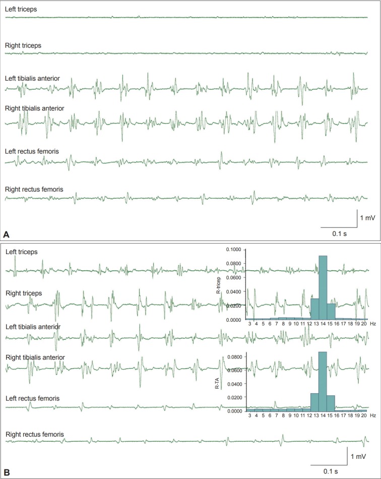
Example of orthostatic tremor. A: Surface electromyography (sEMG) signals recorded from a patient with orthostatic tremor. EMG bursts in the bilateral rectus femoris and tibialis anterior muscles reveal regular firing at a frequency of 14 Hz. B: The patient leaned forward and partially supported his weight with both arms by pressing on a table. Tremors at 14 Hz are now evident in both triceps muscles that are used for support. The inset histograms show the same power spectrum peak at 14 Hz in both the triceps and the tibialis anterior muscles. R-TA: right tibialis anterior muscle.
These frequencies are usually associated with cerebellar ataxia, paraneoplastic syndrome, Graves’ disease or Parkinson’s disease [26]. Slow OT usually has a longer EMG burst duration and less synchronization between the proximal (Quad) and distal muscles (GN) in the lower limb compared to classic OT. In individuals with slow OT, the tremor is not always transmitted to the upper limbs when weight is supported by the arms [24,26].
Functional tremor
Electrophysiological studies provide useful information to differentiate functional and organic tremors. However, functional tremors may coexist with organic disorders. Separation of the movements that are functional versus organic can provide important information for diagnostic and treatment decisions. The tests are based on the principle that although functional movements are considered involuntary by the patient, they utilize the voluntary motor system. It is difficult to maintain different movement frequencies on the right and left sides at the same time. Several assessments may be used to differentiate organic versus functional tremors.
Entrainment or suppression of tremors during rhythmic tapping
An accelerometer is placed on the distal part of the limb, and sEMG is recorded from muscles with tremors. The patient is asked to perform finger or foot tapping with the contralateral limb. The tapping rate can be paced by a metronome at low (1 Hz), medium (3 Hz), and high (5 Hz) frequencies. In patients with functional tremors, the tremors may be suppressed, or the frequency of the tremors may shift from the original frequency to the tapping frequency (entrainment). The results are regarded as abnormal if the tremors cease or the peak frequency shifts by more than 19%, 26.9% and 25.7% during tapping of the contralateral limb at 1, 3, and 5 Hz, respectively [27]. Generally, normal subjects are able to perform finger tapping at frequencies up to 5 Hz. Poor tapping performance is also an indicator of functional tremor [27,28].
High sEMG coherence when performing rhythmic tapping
Functional tremor patients may show significant coherence (over 99% confidence limit) at the tapping frequency between the limb with tremors and the tapping limb. This is not observed in organic tremors [29].
Distractibility and ballistic movement-related pause of tremors
In addition to finger tapping, other tasks that may take the patients’ attention away from the tremors, such as mental subtraction, precise motor tasks or ballistic movements, may also interrupt functional tremors (Supplementary Figure 3 in the online-only Data Supplement). When patients are performing a ballistic movement with the contralateral arm (e.g., touching or grasping an object in front of the body as quickly as possible upon command), functional tremors may transiently pause [30] or have a greater than 50% decrease in amplitude in more than 7 out of 10 trials (Figure 2) [27]. Transient pauses in tremors has high test specificity. However, a reduction in resting tremor amplitude occurred in some Parkinson’s disease patients while they performed contralateral ballistic movements [31].
Figure 2.
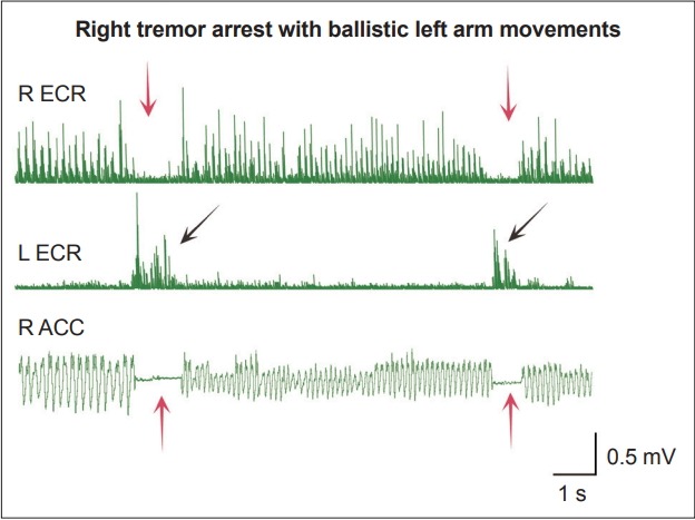
Contralateral ballistic movement induced a transient pause of functional tremor. A case of functional right arm postural tremor. EMG signals were recorded from the bilateral extensor carpi radialis muscles (ECR), and an accelerometer (ACC) was attached to the right middle finger. Transient pauses of the tremors in the right ECR muscle and in the accelerometer recording (red arrows) were observed when the left hand performed voluntary ballistic movements (black arrows).
Tonic-coactivation sign
Tonic coactivation in both agonist and antagonist muscles approximately 300 ms before the onset of a tremor is considered a feature of functional tremor (Supplementary Figure 4 in the online-only Data Supplement). This is because coactivation of agonist-antagonist muscles can lead to a clonus state, which contributes to the subsequent limb tremors [32]. There are no tonic-coactivation signs before tremor onset in organic tremor patients.
Loading test
When a 500 g (approximately 1 pound) weight is attached to the affected wrist, the tremor amplitude is usually reduced or unchanged in individuals with organic tremor. In contrast, the tremor amplitude may increase in individuals with functional tremor. However, the characteristic of changing amplitude after loading in individuals with functional tremor had high specificity (92%) but low sensitivity (22%) [33]. The tremor frequency is usually not changed in individuals with organic or functional tremor during the loading test [32], but an increase in tremor frequency is suggestive of functional tremor. With mass loading, an increase in tremor peak frequency of more than 130% is considered suggestive of functional tremor [27]. However, this may also be observed in some patients with essential tremor or Parkinson’s disease tremor [34].
It is often the case that only some but not all of the features described above are observed in individuals with functional tremor. Therefore, a battery of tests is usually needed for the diagnosis. The above tests have been incorporated into a laboratory-supported functional tremor scoring system, and scores of more than 3 out of 10 are considered highly suggestive functional tremor [35]. It should also be noted that the demonstration of functional tremors does not exclude an organic neurological disorder because functional and organic disorders may coexist in the same patient.
MYOCLONUS ASSESSMENT
Myoclonus can be classified according to its distribution as focal, segmental or generalized myoclonus. It can also be categorized by its physiological origin as cortical, subcortical or spinal myoclonus. Cortical and brainstem myoclonus are generally associated with EMG burst durations of less than 100 ms, and the durations are typically less than 70 ms [36]. Spinal myoclonus may be associated with longer EMG burst durations. For example, EMG bursts in individuals with propriospinal myoclonus (PSM) can last longer than 200 ms and may last for multiple seconds. EMG bursts from volitional jerks can occasionally only last for 50 to 80 ms. Therefore, to distinguish organic versus functional myoclonus, other electrophysiological methods, such as BP assessments or distraction maneuvers, are often necessary. Another form of myoclonus is negative myoclonus, which refers to the transient interruption of a muscle contraction with a silent period of 50 to 120 ms in the EMG signal [37]. Most cases of negative myoclonus are of epileptic cortical origin, but they can also be subcortical in origin, as it is in individuals with hepatic encephalopathy-associated asterixis [38].
Cortical myoclonus
Cortical myoclonus is usually most prominent in the distal arm and face, which is the area that has the largest representation in the homunculus in the motor cortex. Cortical myoclonus symptoms may increase while the patient performs voluntary actions compared to a resting state. There are many causes of cortical myoclonus, including hereditary diseases, such as progressive myoclonic epilepsies, and neurodegenerative disorders, such as Lewy body dementia and cortico-basal syndrome. The EMG burst durations of cortical myoclonus are usually approximately 30 to 40 ms. The morphology of the burst can comprise a single or several compound muscle action potentials (Supplementary Figure 5 in the online-only Data Supplement).
In some cases, EEG back-averaging may reveal time-locked discharges from the contralateral primary motor cortex that precede the EMG discharge or EMG silence in cases of negative myoclonus by 20 to 40 ms.
Cortical myoclonus can also be stimulus sensitive, indicating increased excitability of the sensorimotor cortex. In some cases, this abnormality can be detected by increased SEP amplitudes. The first postcentral cortical component of median nerve SEPs is a negative wave with a latency about 20 ms (N20), which represents arrival of the sensory stimulus at the primary somatosensory cortex, and it is usually normal in individuals with cortical myoclonus. The subsequent components P25 and N35 may have increased amplitudes. If the amplitude from P25 (trough) to N35 (peak) is larger than 10 μV, it is termed a giant SEP and is highly suggestive of cortical myoclonus (Supplementary Figure 6 in the online-only Data Supplement). Paired stimulation with interstimulus intervals shorter than 100 ms normally decreases the cortical SEP response. In patients with cortical myoclonus not displaying a giant SEP by single median nerve stimulation, decreased attenuation of the SEP response by paired stimulation of the SEP is an alternative way to demonstrate a cortical origin of myoclonus [39]. However, not all patients with cortical myoclonus have giant SEPs. For example, patients with benign forms of juvenile myoclonic epilepsies, Creutzfeldt-Jakob disease and post-hypoxic myoclonus frequently do not show giant SEPs or only present them in the late stages of the disease [39]. It should be noted that even if there is evidence of a cortical origin of myoclonus with giant SEPs or time-locked discharges in the EEG signals, the main disease pathology may involve distant brain structures, such as the cerebellum [40].
Stimulus sensitive myoclonus can be assessed with LLRs and CMRs. LLR or CMR responses occurring at rest are definitely abnormal. Patients with cortical reflex myoclonus may show enlarged LLR I (Figure 3), with or without giant SEPs. The absence of an LLR response may be due to inconsistent muscle contractions or negative myoclonus. For CMRs, the magnitude of the E2 response is exaggerated with multiple system atrophy, and cortico-basal syndrome is associated with an increased amplitude and a shortened latency of the E2 response [41].
Figure 3.
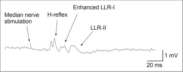
Enhanced long-latency reflex (LLR) in a patient with cortical myoclonus. The long latency reflex showed normal H-reflex and LLR-II responses and enhanced an LLR-I response in a patient with myoclonus. EMG signals were recorded from the abductor pollicis brevis muscle with 20% background activation. Three Hz median nerve stimulations at the motor threshold stimulation intensity were delivered, and a total of 250 trials were recorded.
Subcortical myoclonus
Subcortical myoclonus can arise from structures between the cortex and the spinal cord. A classic form of subcortical myoclonus is reticular myoclonus (RM), which originates from the brainstem. RM usually presents as generalized jerks, mostly affecting proximal limb flexor muscles. It can be stimulus sensitive and tends to be periodic. A characteristic of RM is the sequence of muscle recruitment. A jerk typically starts at the trapezius or sternocleidomastoid muscle, in which brainstem nuclei are closest to the reticular formation, and spreads rostrally to the orbicularis oculi muscle and caudally to limb muscles (Figure 4) [42]. Abnormal LLR can also be observed in individuals with stimulus-sensitive RM [43]. Myoclonus dystonia (MD) is another example of subcortical myoclonus. Features of cortical myoclonus, including jerk-locked EEG cortical spikes preceding EMG signal onset, giant SEPs, and enlarged LLRs, have not been observed in individuals with MD. In addition, the intracortical facilitation and inhibition circuits measured by TMS in MD patients were normal. These results suggest a subcortical origin of myoclonus in individuals with MD [44].
Figure 4.
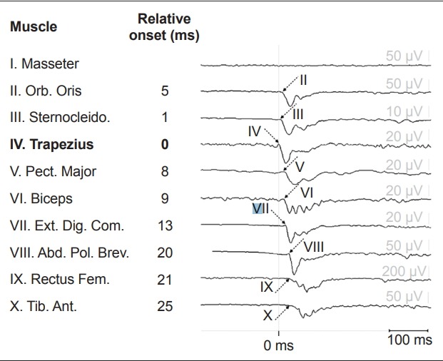
Example of reticular reflex myoclonus due to left medulla compression by the vertebral artery. The myoclonus started in the trapezius muscle with subsequent muscle activation rostrally to the orbicularis oris and caudally to the tibialis anterior muscles. The corresponding onset latency relative to the first contracted trapezius muscle (bold) was shown in ms. The myoclonus has short EMG burst durations. Adapted from Beudel et al.[42] Orb. Oris: orbicularis oris muscle, Sternocleido.: sternocleidomastoid muscle, Pect. Major: pectoralis major muscle, Ext. Dig. Com.: extensor digitorum communis muscle, Abd. Pol. Brev.: Abductor pollicis brevis muscle, Rectus Fem.: rectus femoris muscle, Tib. Ant.: tibialis anterior muscle.
Startle reflex and hyperekplexia
The startle reflex (SR) is a normal brainstem response induced by sudden stimuli, such as tapping on the forehead, mental space or sternum or an unexpected loud sound. The response latencies are different depending on the location and nature of the stimulation, but the muscle recruitment order is identical irrespective of the absolute latency. The first response is a bilateral orbicularis oculi blink response (onset latency 9–20 ms with taps on the nose or jaw, ≤ 30 ms with auditory stimuli), which is not considered part of the SR because it does not habituate, unlike other muscle responses [45]. The first EMG response of the SR is in the sternocleidomastoid muscle, followed by the masseter, trunk and limb muscles. The EMG burst durations usually last between 150 and 400 ms [46]. This rostral-caudal muscle recruitment order implies that the SR originates from the brainstem. While both the SR and RM arise from the brainstem, a difference between the SR and RM is the much longer latencies from cranial muscles to intrinsic hand and foot muscles with the SR. In addition, EMG burst durations are also longer with the SR than with RM. In normal subjects, the SR habituates after several stimuli. In patients with hyperekplexia with an exaggerated startle response due to a mutation in the glycine receptor, patients present with a startle response that does not habituate to repeated stimuli and may have tonic contractions lasting several seconds after stimulation. However, the muscle recruitment pattern in individuals with hyperekplexia is the same as that in individuals with normal SR [46]. Symptomatic hyperekplexia is rare but can be observed in individuals with brainstem encephalopathy [47]. Longer EMG burst durations, relatively long latencies in distal limb muscles and a lack of spontaneous jerks distinguish symptomatic hyperekplexia from RM (Figure 5).
Figure 5.
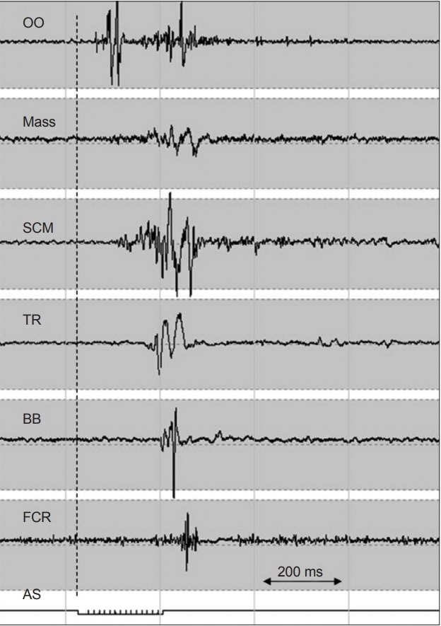
Symptomatic hyperekplexia caused by brainstem encephalopathy. EMG recording of the startle response from an unexpected acoustic stimulus (AS, bottom line). The vertical dashed line indicates the beginning of the AS. After the initial blink response, the first muscle response to the AS was from the sternocleidomastoid muscle, with both rostral and caudal spreading. Relatively late EMG responses in the limb muscle and generally long EMG burst durations were the characteristics of the startle reflex. Adapted from van de Warrenburg et al.[47] OO: orbicularis oculi, Mass: master, SCM: sternocleidomastoid, TR: trapezius, BB: biceps brachii, FCR: flexor carpi radialis.
Spinal myoclonus
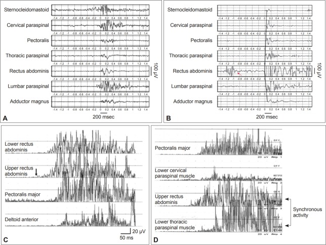
There are two forms of spinal myoclonus: spinal segmental myoclonus and PSM. Spinal segmental myoclonus is often associated with focal spinal lesions. It is usually confined to a few contiguous myotomes and is not affected by supraspinal influences, such as sleep [38]. It can be irregular or semirhythmic. The EMG burst duration may be longer than 1,000 ms. PSM usually presents with flexion jerks in the truncal axial muscles, hips and knees in arrhythmic patterns. It may or may not be stimulus sensitive and often becomes more frequent when the individual is lying in a supine position. In individuals with PSM, the EMG burst durations are usually longer than 200 ms. An abnormal impulse is presumed to spread along the propriospinal pathway, which refers to white matter tracts that connect different segments of the spinal cord. The typical EMG recruitment pattern begins in trunk muscles at a certain myotome level and spreads rostrally to neck muscles and caudally to lower limb muscles (Supplementary Figure 7 in the online-only Data Supplement) [48]. PSM does not affect cranial muscles. Some reports have suggested that synchronous activities of the truncal flexor and extensor muscles are a feature of PSM [49], but this was not observed in other studies [50,51]. However, several studies have shown that patients with axial jerks that appeared consistent with PSM may have functional axial jerks [48]. A FMD presenting as PSM was first reported in 2008 [52]. Since then, many PSM cases have been diagnosed as FMDs on the basis of the presence of a BP, combined facial movements or vocalization, a history of somatization and distractibility in neurological examinations. In a literature review, 104 out of 179 (58%) PSM cases reported as FMD cases, 29 (16%) were idiopathic PSM, and 46 (26%) were due to secondary etiologies, such as myelopathy, infection or drug abuse [53,54]. Although PSM symptoms can be mimicked by healthy subjects [55], with a similar propagation sequence but synchronous activity of the truncal flexor and extensor muscles, less consistent bursts, longer EMG burst durations lasting more than 1,000 ms and higher conduction velocities (> 16 m/s) may be useful to differentiate functional PSM from organic PSM [56]. Moreover, eye blinks occurring before axial muscle contractions are suggestive of functional jerks rather than PSM [57]. Figure 6 shows several functional PSM EMG presentations. Combining EMG with recordings of BPs or beta ERD is a useful way to distinguish PSM from functional axial jerks. The diagnosis of functional jerks should be considered in patients presenting with PSM symptoms.
Figure 6.
Functional propriospinal myoclonus (PSM) and PSM mimicked by healthy subjects. Functional PSM can sometimes be differentiated from idiopathic or symptomatic PSM by (A) the absence of a typical rostral and caudal recruitment order, (B) a burst duration longer than 1,000 ms and isolated muscle activity in the rectus abdominis muscle (red arrow). PSM symptoms can be mimicked. Healthy subjects can mimic (C) typical PSM propagation patterns starting from the upper rectus abdominus muscle (black arrow) and (D) synchronous activation of the truncal flexors and extensors. Adapted from Erro et al.[56] and Kang and Sohn[55].
Functional myoclonus
Functional myoclonus in limb muscles can be differentiated from organic myoclonus by the EMG burst durations, muscle recruitment order and tests of distractibility. A variable order of muscle recruitment, a normal LLR, the absence of short latency cortical discharge with EEG back averaging and the presence of BP or beta ERD before EMG onset are strong indicators of functional jerks. However, there are limitations to BP and ERD tests. Functional jerks occurring excessively frequently with less than approximately 2 seconds between jerks make BP or ERD assessments difficult due to the absence of a stable baseline period. On the other hand, jerks that are excessively infrequent are difficult to assess due to the low number of trials available for averaging. Moreover, BP and ERD are useful for the assessment of spontaneous jerks but not action-induced jerks.
DYSTONIA ASSESSMENT
Co-contractions of agonist and antagonist muscles with an overflow of contractions in muscles not relevant to the task are considered electrophysiological characteristics of dystonia [58]. However, co-contractions are not always present in individuals with organic dystonia. It can be produced volitionally or can be observed in individuals with other conditions, such as stiff person syndrome.
Organic dystonia
Although there is no established measurement to diagnose organic dystonia in an individual patient, some measures can still provide valuable information. In individuals with cervical dystonia, a study found that the sEMG power spectrum in the affected neck muscles showed an absence of a 12 Hz peak, which was present in normal subjects. In addition, agonist-antagonist muscle pairs, such as left splenius capitis and right sternocleidomastoid muscles, revealed prominent coherence at low frequencies up to 7 Hz in these patients compared to normal subjects who exhibited coherence at 10–20 Hz [59]. However, a subsequent study revealed that only the absence of spectral power at approximately 10 Hz in a dystonic muscle is a robust finding in individuals with cervical dystonia [60,61]. However, additional studies are needed to determine whether this method has sufficient sensitivity and specificity to identify dystonic muscles in individual patients. The extent of co-contractions can be assessed by asking the patient activate agonist and antagonist muscles in an alternating pattern, such as turning the neck from side to side or wrist flexion-extension movements. sEMG signals may show simultaneous active firing of both the agonist and antagonist muscles. In addition, with the assessment of a cross-correlogram from sEMG recordings, the extent of a peak at time zero may be used to quantify the extent of dystonia [62]. Although many TMS studies have revealed that dystonia patients have decreased cortical inhibition, such as reduced short interval intracortical inhibition, a silent period and surround inhibition, these parameters are not considered tools for identifying individual dystonia patients due to the considerable overlap between patients with dystonia and normal subjects [63].
Functional dystonia
TMS measures of cortical inhibition, such as short interval intracortical inhibition, long interval intracortical inhibition, and a cortical silent period, and measures of spinal inhibition, such as reciprocal inhibition, cannot distinguish between organic and functional dystonia, as they are reduced in both conditions [64]. A few electrophysiological measurements may differentiate functional from organic dystonia. Paired associative stimulation elicits abnormally high plasticity in individuals with organic dystonia but not in individuals with functional dystonia compared to healthy subjects [65]. The R2 blink reflex recovery can be tested by paired supraorbital nerve stimulation. Shortening of the blink recovery latency with the regaining of R2 activity with interstimulus intervals shorter than 200 ms was observed in individuals with organic blepharospasm but not in individuals with functional blepharospasm [66]. These measurements may serve as a potential method to distinguish these two conditions.
OTHER CONDITIONS THAT CAN BE ASSESSED BY ELECTROPHYSIOLOGICAL TESTING
Stiff-person syndrome
Patients with stiff-person syndrome (SPS) typically present with excessive muscle activities and an exaggerated startle response [67]. Continuous motor unit activity at rest is a characteristic feature of SPS but is not a specific finding, as this can be observed in dystonia patients [68]. Continuous motor unit activity can be assessed by recording sEMG signals from agonist-antagonist muscle pairs, such as the rectus abdominis and paraspinal muscles in the trunk and the Quad and hamstrings in the leg. When normal subjects perform a movement, such as flexion or extension of the trunk or knee flexion, relaxation is evident by the absence of EMG activity in the antagonist muscles; however, individuals with SPS fail to relax the antagonist muscle. The acoustic startle responses in the cranial or proximal arm muscles are not different between SPS patients and normal subjects. Only lower limb muscles, which have a smaller startle response than other muscles, show unhabituated exaggerated activity with the acoustic SR. This suggests that SPS patients have normal SR circuits in the brainstem, but they have disinhibition in the spinal cord. Although the SR in the brainstem is normal, other brainstem circuits may be involved. The head retraction reflex, elicited by gently tapping on the nose tip or the forehead, show an abnormally early reflex in the trapezius and sternomastoid muscles. This result indicates that there is dysfunction of inhibitory interneurons in the brainstem in patients with SPS [69]. The exteroceptive reflex can be assessed by the stimulation of the tibial nerve with a train of four pulses with a duration of 200 μs, 3 ms apart and with three times of the sensory threshold intensity. The stimulation elicits long-lasting tonic activity followed by gradual decrescendo activity in lower limb muscles (Figure 7) [70]. This tonic response has diagnostic value for individuals with SPS.
Figure 7.
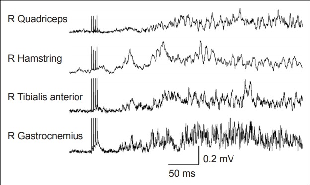
Enhanced exteroceptive reflex in a patient with stiff-person syndrome. The stimulation was delivered to the right tibial nerve, and the EMG signals from the right leg muscles were recorded. Two phases of response were obtained: a brief short latency first phase (about 50 ms), followed by a second phase with a longer latency and duration of more than 100 ms. Adapted from Espay and Chen[70]. R: right.
Celiac ataxia
Celiac disease can manifest as myoclonus ataxia syndrome associated with gluten-related antibodies. Electrophysiological studies usually show giant SEPs and cortical discharges preceding jerk onset [71]. EEG signals frequently reveal occipital sharp waves, but some cases show centro-parietal polyspikes [72] or para-sagittal oscillatory activities. The cortical myoclonus may be related to primary cerebellar pathology, which leads to hyperexcitability of the motor cortex [40]. This is consistent with TMS paired pulse studies showing reduced short interval intracortical inhibition and enhanced intracortical facilitation in patients with celiac ataxia [73].
CONCLUSIONS
Electrophysiological studies are helpful in many situations to precisely and objectively delineate movement disorder phenomena to provide a “laboratory supported” diagnosis of movement disorders. Sometimes it can be used to evaluate the effects of treatment and can be used in follow-up assessments. However, for many tests, the sensitivity and specificity have not been determined. Limitations of the test should be recognized, and the findings should be interpreted together with clinical observations.
Footnotes
Conflict of Interest
The authors have no financial conflicts of interest.
Author Contributions
Conceptualization: All authors. Funding acquisition: Robert Chen. Supervision: Robert Chen. Writing—original draft: Kai-Hsiang Stanley Chen. Writing—review & editing: All authors.
Supplementary Materials
The online-only Data Supplement is available with this article at https://doi.org/10.14802/jmd.19064.
Needle electromyography (EMG) recording of diaphragmatic myoclonus. The first two lines show normal rhythmic inspiratory EMG activity (4–5 Hz). The onset of diaphragmatic myoclonus interrupted the normal inspiratory rhythm, which is marked by the black arrow. The open arrow points to the cardiac pacemaker artifacts. Diaphragmatic myoclonus was not related to the pacemaker activities. Adapted from Chen et al.[1]
Example of the Bereitschaftspotential in functional jerks. Recordings of electroencephalography (EEG) signals from the C3 electrode (upper trace) and EMG signals from the right biceps muscle (bottom trace) from a patient with functional right arm jerks. Forty trials were averaged. The EEG recordings show a Bereitschaftspotential, which is a slow, negative potential, with an onset about 1,500 ms before EMG onset. Time 0 = EMG onset. Adapted from Phielipp and Chen[5].
Distraction in a patient with functional tremor. Accelerometer and EMG recordings from a patient with functional tremor of the right foot. The right foot tremor ceased when the patient started left foot tapping at 2 Hz. The black arrow indicates the start of left foot tapping, and the red arrows indicate the suppression of the tremors. This individual with right foot functional tremor also presents with variable tremor frequencies and EMG burst amplitudes. The top trace corresponds to the accelerometer signals recorded from the left foot and the middle trace corresponds to the accelerometer signals from the right foot. The bottom trace is the rectified EMG signal from the right tibialis anterior muscle.
Tonic coactivation sign in a patient with functional tremor. A: EMG signals recorded from the right wrist extensor and flexor muscles in a patient with essential tremor. There was no coactivation of the agonist and antagonist muscles before tremor onset (red arrows). B: EMG signals recorded from the left wrist extensor and flexor muscles in a patient with functional tremor. The black double-headed arrow indicates simultaneous EMG activation in both the agonist and antagonist muscles before tremor onset (single-headed black arrow).
Example of cortical myoclonus. The EMG burst duration was usually shorter than 70 ms. Adapted from Phielipp and Chen[5]. ECR: extensor carpi radialis muscle. R: right, L: left, FDI: first dorsal interosseous muscle.
Giant somatosensory evoked potentials in a patient with cortical myoclonus. Somatosensory evoked potentials recorded from a patient with cortical myoclonus. Recordings from both Cp3-Fz from right median nerve stimulation and Cp4-Fz from left median nerve stimulation both showed giant P25-N30 waves with amplitudes larger than 10 µV.
Propriospinal myoclonus (PSM) with a secondary cause. EMG recording from a case of PSM induced by ciprofloxacin. The duration of the EMG burst varies from 80 to 400 ms. The dashed line indicates a rostral and caudal order of recruitment starting in the rectus abdominis muscle. The orbicularis oculi muscle was not involved in the jerks. Adapted from Post et al.[54]
REFERENCES
- 1.Chen R, Remtulla H, Bolton CF. Electrophysiological study of diaphragmatic myoclonus. J Neurol Neurosurg Psychiatry. 1995;58:480–483. doi: 10.1136/jnnp.58.4.480. [DOI] [PMC free article] [PubMed] [Google Scholar]
- 2.Mima T, Hallett M, Shibasaki H. Chapter 7. Coherence, cortico-muscular. In: Hallett M, editor. Handbook of Clinical Neurophysiology. Vol 1. Elsevier; 2003. pp. 87–94. [Google Scholar]
- 3.Mauguière F. Chapter 5. Somatosensory evoked responses. In: Hallett M, editor. Handbook of Clinical Neurophysiology. Vol 1. Elsevier; 2003. pp. 45–75. [Google Scholar]
- 4.Pratt H, Politoske D, Starr A. Mechanically and electrically evoked somatosensory potentials in humans: effects of stimulus presentation rate. Electroencephalogr Clin Neurophysiol. 1980;49:240–249. doi: 10.1016/0013-4694(80)90218-7. [DOI] [PubMed] [Google Scholar]
- 5.Phielipp NM, Chen R. Chapter 10. Neurophysiologic assessment of movement disorders in humans, in movement disorders. In: LeDoux MS, editor. Movement disorders. 2nd ed. Boston: Academic Press; 2015. pp. 171–186. [Google Scholar]
- 6.van der Salm SM, Tijssen MA, Koelman JH, van Rootselaar AF. The bereitschaftspotential in jerky movement disorders. J Neurol Neurosurg Psychiatry. 2012;83:1162–1167. doi: 10.1136/jnnp-2012-303081. [DOI] [PubMed] [Google Scholar]
- 7.Cassim F, Szurhaj W, Sediri H, Devos D, Bourriez J, Poirot I, et al. Brief and sustained movements: differences in event-related (de)synchronization (ERD/ERS) patterns. Clin Neurophysiol. 2000;111:2032–2039. doi: 10.1016/s1388-2457(00)00455-7. [DOI] [PubMed] [Google Scholar]
- 8.Leocani L, Toro C, Manganotti P, Zhuang P, Hallett M. Event-related coherence and event-related desynchronization/synchronization in the 10 Hz and 20 Hz EEG during self-paced movements. Electroencephalography and Clinical Neurophysiology/Evoked Potentials Section. 1997;104:199–206. doi: 10.1016/s0168-5597(96)96051-7. [DOI] [PubMed] [Google Scholar]
- 9.Pfurtscheller G, Lopes da Silva FH. Event-related EEG/MEG synchronization and desynchronization: basic principles. Clin Neurophysiol. 1999;110:1842–1857. doi: 10.1016/s1388-2457(99)00141-8. [DOI] [PubMed] [Google Scholar]
- 10.Beudel M, Zutt R, Meppelink AM, Little S, Elting JW, Stelten BML, et al. Improving neurophysiological biomarkers for functional myoclonic movements. Parkinsonism Relat Disord. 2018;51:3–8. doi: 10.1016/j.parkreldis.2018.03.029. [DOI] [PMC free article] [PubMed] [Google Scholar]
- 11.Deuschl G, Eisen A. Long-latency reflexes following electrical nerve stimulation. The Inter national Federation of Clinical Neurophysiology. Electroencephalogr Clin Neurophysiol Suppl. 1999;52:263–268. [PubMed] [Google Scholar]
- 12.Floeter MK. Chapter 16. Spinal reflexes. In: Hallett M, editor. Handbook of Clinical Neurophysiology. Vol 1. Elsevier; 2003. pp. 231–246. [Google Scholar]
- 13.Chen R, Ashby P. Reflex responses in upper limb muscles to cutaneous stimuli. Can J Neurol Sci. 1993;20:271–278. doi: 10.1017/s0317167100048174. [DOI] [PubMed] [Google Scholar]
- 14.Jenner JR, Stephens JA. Cutaneous reflex responses and their central nervous pathways studied in man. J Physiol. 1982;333:405–419. doi: 10.1113/jphysiol.1982.sp014461. [DOI] [PMC free article] [PubMed] [Google Scholar]
- 15.Chen KH, Lin PC, Chen YJ, Yang BS, Lin CH. Development of method for quantifying essential tremor using a small optical device. J Neurosci Methods. 2016;266:78–83. doi: 10.1016/j.jneumeth.2016.03.014. [DOI] [PubMed] [Google Scholar]
- 16.Bhatia KP, Bain P, Bajaj N, Elble RJ, Hallett M, Louis ED, et al. Consensus Statement on the classification of tremors. from the task force on tremor of the International Parkinson and Movement Disorder Society. Mov Disord. 2018;33:75–87. doi: 10.1002/mds.27121. [DOI] [PMC free article] [PubMed] [Google Scholar]
- 17.Deuschl G, Bain P, Brin M. Consensus statement of the Movement Disorder Society on Tremor. Ad Hoc Scientific Committee. Mov Disord. 1998;13 Suppl 3:2–23. doi: 10.1002/mds.870131303. [DOI] [PubMed] [Google Scholar]
- 18.Hess CW, Pullman SL. Tremor: clinical phenomenology and assessment techniques. Tremor Other Hyperkinet Mov (N Y) 2012:2:tre-02-65-365–1. doi: 10.7916/D8WM1C41. [DOI] [PMC free article] [PubMed] [Google Scholar]
- 19.Pal PK. Electrophysiologic evaluation of psychogenic movement disorders. J Mov Disord. 2011;4:21–32. doi: 10.14802/jmd.11004. [DOI] [PMC free article] [PubMed] [Google Scholar]
- 20.Nisticò R, Pirritano D, Salsone M, Novellino F, Del Giudice F, Morelli M, et al. Synchronous pattern distinguishes resting tremor associated with essential tremor from rest tremor of Parkinson’s disease. Parkinsonism Relat Disord. 2011;17:30–33. doi: 10.1016/j.parkreldis.2010.10.006. [DOI] [PubMed] [Google Scholar]
- 21.Nisticò R, Fratto A, Vescio B, Arabia G, Sciacca G, Morelli M, et al. Tremor pattern differentiates drug-induced resting tremor from Parkinson disease. Parkinsonism Relat Disord. 2016;25:100–103. doi: 10.1016/j.parkreldis.2016.02.002. [DOI] [PubMed] [Google Scholar]
- 22.Papengut F, Raethjen J, Binder A, Deuschl G. Rest tremor suppression may separate essential from parkinsonian rest tremor. Parkinsonism Relat Disord. 2013;19:693–697. doi: 10.1016/j.parkreldis.2013.03.013. [DOI] [PubMed] [Google Scholar]
- 23.Deuschl G, Raethjen J, Lindemann M, Krack P. The pathophysiology of tremor. Muscle Nerve. 2001;24:716–735. doi: 10.1002/mus.1063. [DOI] [PubMed] [Google Scholar]
- 24.Hassan A, van Gerpen JA. Orthostatic tremor and orthostatic myoclonus: weight-bearing hyperkinetic disorders: a systematic review, new insights, and unresolved questions. Tremor Other Hyperkinet Mov (N Y) 2016;6:417. doi: 10.7916/D84X584K. [DOI] [PMC free article] [PubMed] [Google Scholar]
- 25.McAuley JH, Britton TC, Rothwell JC, Findley LJ, Marsden CD. The timing of primary orthostatic tremor bursts has a task-specific plasticity. Brain. 2000;123:254–266. doi: 10.1093/brain/123.2.254. [DOI] [PubMed] [Google Scholar]
- 26.Rigby HB, Rigby MH, Caviness JN. Orthostatic tremor: a spectrum of fast and slow frequencies or distinct entities? Tremor Other Hyperkinet Mov (N Y) 2015;5:324. doi: 10.7916/D8S75FHK. [DOI] [PMC free article] [PubMed] [Google Scholar]
- 27.Schwingenschuh P, Katschnig P, Seiler S, Saifee TA, Aguirregomozcorta M, Cordivari C, et al. Moving toward “laboratory-supported” criteria for psychogenic tremor. Mov Disord. 2011;26:2509–2515. doi: 10.1002/mds.23922. [DOI] [PMC free article] [PubMed] [Google Scholar]
- 28.Zeuner KE, Shoge RO, Goldstein SR, Dambrosia JM, Hallett M. Accelerometry to distinguish psychogenic from essential or parkinsonian tremor. Neurology. 2003;61:548–550. doi: 10.1212/01.wnl.0000076183.34915.cd. [DOI] [PubMed] [Google Scholar]
- 29.McAuley J, Rothwell J. Identification of psychogenic, dystonic, and other organic tremors by a coherence entrainment test. Mov Disord. 2004;19:253–267. doi: 10.1002/mds.10707. [DOI] [PubMed] [Google Scholar]
- 30.Kumru H, Valls-Solé J, Valldeoriola F, Marti MJ, Sanegre MT, Tolosa E. Transient arrest of psychogenic tremor induced by contralateral ballistic movements. Neurosci Lett. 2004;370:135–139. doi: 10.1016/j.neulet.2004.08.009. [DOI] [PubMed] [Google Scholar]
- 31.Hallett M. Psychogenic parkinsonism. J Neurol Sci. 2011;310:163–165. doi: 10.1016/j.jns.2011.03.019. [DOI] [PMC free article] [PubMed] [Google Scholar]
- 32.Deuschl G, Köster B, Lücking CH, Scheidt C. Diagnostic and pathophysiological aspects of psychogenic tremors. Mov Disord. 1998;13:294–302. doi: 10.1002/mds.870130216. [DOI] [PubMed] [Google Scholar]
- 33.van der Stouwe AM, Elting JW, van der Hoeven JH, van Laar T, Leenders KL, Maurits NM, et al. How typical are ‘typical’ tremor characteristics? Sensitivity and specificity of five tremor phenomena. Parkinsonism Relat Disord. 2016;30:23–28. doi: 10.1016/j.parkreldis.2016.06.008. [DOI] [PubMed] [Google Scholar]
- 34.Kamble NL, Pal PK. Electrophysiological evaluation of psychogenic movement disorders. Parkinsonism Relat Disord. 2016;22:S153–S158. doi: 10.1016/j.parkreldis.2015.09.016. [DOI] [PubMed] [Google Scholar]
- 35.Schwingenschuh P, Saifee TA, Katschnig-Winter P, Macerollo A, Koegl-Wallner M, Culea V, et al. Validation of “laboratory-supported” criteria for functional (psychogenic) tremor. Mov Disord. 2016;31:555–562. doi: 10.1002/mds.26525. [DOI] [PubMed] [Google Scholar]
- 36.Brown P, Thompson PD. Electrophysiological aids to the diagnosis of psychogenic jerks, spasms, and tremor. Mov Disord. 2001;16:595–599. doi: 10.1002/mds.1145. [DOI] [PubMed] [Google Scholar]
- 37.Ugawa Y, Shimpo T, Mannen T. Physiological analysis of asterixis: silent period locked averaging. J Neurol Neurosurg Psychiatry. 1989;52:89–93. doi: 10.1136/jnnp.52.1.89. [DOI] [PMC free article] [PubMed] [Google Scholar]
- 38.Kojovic M, Cordivari C, Bhatia K. Myoclonic disorders: a practical approach for diagnosis and treatment. Ther Adv Neurol Disord. 2011;4:47–62. doi: 10.1177/1756285610395653. [DOI] [PMC free article] [PubMed] [Google Scholar]
- 39.Shibasaki H, Hallett M. Electrophysiological studies of myoclonus. Muscle Nerve. 2005;31:157–174. doi: 10.1002/mus.20234. [DOI] [PubMed] [Google Scholar]
- 40.Tijssen MA, Thom M, Ellison DW, Wilkins P, Barnes D, Thompson PD, et al. Cortical myoclonus and cerebellar pathology. Neurology. 2000;54:1350–1356. doi: 10.1212/wnl.54.6.1350. [DOI] [PubMed] [Google Scholar]
- 41.Chen R, Ashby P, Lang AE. Stimulus-sensitive myoclonus in akineticrigid syndromes. Brain. 1992;115(Pt 6):1875–1888. doi: 10.1093/brain/115.6.1875. [DOI] [PubMed] [Google Scholar]
- 42.Beudel M, Elting JWJ, Uyttenboogaart M, van den Broek MWC, Tijssen MAJ. Reticular myoclonus: it really comes from the brainstem! Mov Disord Clin Pract. 2014;1:258–260. doi: 10.1002/mdc3.12054. [DOI] [PMC free article] [PubMed] [Google Scholar]
- 43.Deuschl G, Lücking CH. Physiology and clinical applications of hand muscle reflexes. Electroencephalogr Clin Neurophysiol Suppl. 1990;41:84–101. doi: 10.1016/b978-0-444-81352-7.50012-1. [DOI] [PubMed] [Google Scholar]
- 44.Li JY, Cunic DI, Paradiso G, Gunraj C, Pal PK, Lang AE, et al. Electrophysiological features of myoclonus-dystonia. Mov Disord. 2008;23:2055–2061. doi: 10.1002/mds.22273. [DOI] [PubMed] [Google Scholar]
- 45.Brown P, Rothwell JC, Thompson PD, Britton TC, Day BL, Marsden CD. New observations on the normal auditory startle reflex in man. Brain. 1991;114(Pt 4):1891–1902. doi: 10.1093/brain/114.4.1891. [DOI] [PubMed] [Google Scholar]
- 46.Brown P, Rothwell JC, Thompson PD, Britton TC, Day BL, Marsden CD. The hyperekplexias and their relationship to the normal startle reflex. Brain. 1991;114(Pt 4):1903–1928. doi: 10.1093/brain/114.4.1903. [DOI] [PubMed] [Google Scholar]
- 47.van de Warrenburg BP, Cordivari C, Brown P, Bhatia KP. Persisting hyperekplexia after idiopathic, self-limiting brainstem encephalopathy. Mov Disord. 2007;22:1017–1020. doi: 10.1002/mds.21411. [DOI] [PubMed] [Google Scholar]
- 48.Roze E, Bounolleau P, Ducreux D, Cochen V, Leu-Semenescu S, Beaugendre Y, et al. Propriospinal myoclonus revisited: clinical, neurophysiologic, and neuroradiologic findings. Neurology. 2009;72:1301–1309. doi: 10.1212/WNL.0b013e3181a0fd50. [DOI] [PubMed] [Google Scholar]
- 49.Brown P, Thompson PD, Rothwell JC, Day BL, Marsden CD. Axial myoclonus of propriospinal origin. Brain. 1991;114(Pt 1A):197–214. [PubMed] [Google Scholar]
- 50.Espay AJ, Ashby P, Hanajima R, Jog MS, Lang AE. Unique form of propriospinal myoclonus as a possible complication of an enteropathogenic toxin. Mov Disord. 2003;18:942–948. doi: 10.1002/mds.10453. [DOI] [PubMed] [Google Scholar]
- 51.Brown P, Rothwell JC, Thompson PD, Marsden CD. Propriospinal myoclonus: evidence for spinal “pattern” generators in humans. Mov Disord. 1994;9:571–576. doi: 10.1002/mds.870090511. [DOI] [PubMed] [Google Scholar]
- 52.Williams DR, Cowey M, Tuck K, Day B. Psychogenic propriospinal myoclonus. Mov Disord. 2008;23:1312–1313. doi: 10.1002/mds.22072. [DOI] [PubMed] [Google Scholar]
- 53.van der Salm SM, Erro R, Cordivari C, Edwards MJ, Koelman JH, van den Ende T, et al. Propriospinal myoclonus: clinical reappraisal and review of literature. Neurology. 2014;83:1862–1870. doi: 10.1212/WNL.0000000000000982. [DOI] [PMC free article] [PubMed] [Google Scholar]
- 54.Post B, Koelman JH, Tijssen MA. Propriospinal myoclonus after treatment with ciprofloxacin. Mov Disord. 2004;19:595–597. doi: 10.1002/mds.10717. [DOI] [PubMed] [Google Scholar]
- 55.Kang SY, Sohn YH. Electromyography patterns of propriospinal myoclonus can be mimicked voluntarily. Mov Disord. 2006;21:1241–1244. doi: 10.1002/mds.20927. [DOI] [PubMed] [Google Scholar]
- 56.Erro R, Bhatia KP, Edwards MJ, Farmer SF, Cordivari C. Clinical diagnosis of propriospinal myoclonus is unreliable: an electrophysiologic study. Mov Disord. 2013;28:1868–1873. doi: 10.1002/mds.25627. [DOI] [PubMed] [Google Scholar]
- 57.van der Salm SM, Koelman JH, Henneke S, van Rootselaar AF, Tijssen MA. Axial jerks: a clinical spectrum ranging from propriospinal to psychogenic myoclonus. J Neurol. 2010;257:1349–1355. doi: 10.1007/s00415-010-5531-6. [DOI] [PMC free article] [PubMed] [Google Scholar]
- 58.Sanger TD, Chen D, Fehlings DL, Hallett M, Lang AE, Mink JW, et al. Definition and classification of hyperkinetic movements in childhood. Mov Disord. 2010;25:1538–1549. doi: 10.1002/mds.23088. [DOI] [PMC free article] [PubMed] [Google Scholar]
- 59.Tijssen MA, Marsden JF, Brown P. Frequency analysis of EMG activity in patients with idiopathic torticollis. Brain. 2000;123(Pt 4):677–686. doi: 10.1093/brain/123.4.677. [DOI] [PubMed] [Google Scholar]
- 60.Nijmeijer SW, de Bruijn E, Forbes PA, Kamphuis DJ, Happee R, Koelman JH, et al. EMG coherence and spectral analysis in cervical dystonia: discriminative tools to identify dystonic muscles? J Neurol Sci. 2014;347:167–173. doi: 10.1016/j.jns.2014.09.041. [DOI] [PubMed] [Google Scholar]
- 61.De Bruijn E, Nijmeijer SWR, Forbes PA, Koelman JHTM, Van Der Helm FCT, Tijssen MAJ, et al. Dystonic neck muscles show a shift in relative autospectral power during isometric contractions. Clinical Neurophysiology. 2017;128:1937–1945. doi: 10.1016/j.clinph.2017.06.258. [DOI] [PubMed] [Google Scholar]
- 62.Yianni J, Wang SY, Liu X, Bain PG, Nandi D, Gregory R, et al. A dominant bursting electromyograph pattern in dystonic conditions predicts an early response to pallidal stimulation. J Clin Neurosci. 2006;13:738–746. doi: 10.1016/j.jocn.2005.07.022. [DOI] [PubMed] [Google Scholar]
- 63.Quartarone A. Transcranial magnetic stimulation in dystonia. Handb Clin Neurol. 2013;116:543–553. doi: 10.1016/B978-0-444-53497-2.00043-7. [DOI] [PubMed] [Google Scholar]
- 64.Espay AJ, Morgante F, Purzner J, Gunraj CA, Lang AE, Chen R. Cortical and spinal abnormalities in psychogenic dystonia. Ann Neurol. 2006;59:825–834. doi: 10.1002/ana.20837. [DOI] [PubMed] [Google Scholar]
- 65.Quartarone A, Rizzo V, Terranova C, Morgante F, Schneider S, Ibrahim N, et al. Abnormal sensorimotor plasticity in organic but not in psychogenic dystonia. Brain. 2009;132(Pt 10):2871–2877. doi: 10.1093/brain/awp213. [DOI] [PMC free article] [PubMed] [Google Scholar]
- 66.Aramideh M, Ongerboer de Visser BW. Brainstem reflexes: electrodiagnostic techniques, physiology, normative data, and clinical applications. Muscle Nerve. 2002;26:14–30. doi: 10.1002/mus.10120. [DOI] [PubMed] [Google Scholar]
- 67.Matsumoto JY, Caviness JN, McEvoy KM. The acoustic startle reflex in stiff-man syndrome. Neurology. 1994;44:1952–1955. doi: 10.1212/wnl.44.10.1952. [DOI] [PubMed] [Google Scholar]
- 68.Balint B, Meinck HM, Bhatia KP. Axial dystonia mimicking stiff person syndrome. Mov Disord Clin Pract. 2015;3:176–179. doi: 10.1002/mdc3.12249. [DOI] [PMC free article] [PubMed] [Google Scholar]
- 69.Khasani S, Becker K, Meinck HM. Hyperekplexia and stiff-man syndrome: abnormal brainstem reflexes suggest a physiological relationship. J Neurol Neurosurg Psychiatry. 2004;75:1265–1269. doi: 10.1136/jnnp.2003.018135. [DOI] [PMC free article] [PubMed] [Google Scholar]
- 70.Espay AJ, Chen R. Rigidity and spasms from autoimmune encephalomyelopathies: stiff-person syndrome. Muscle Nerve. 2006;34:677–690. doi: 10.1002/mus.20653. [DOI] [PubMed] [Google Scholar]
- 71.Pennisi M, Bramanti A, Cantone M, Pennisi G, Bella R, Lanza G. Neurophysiology of the “Celiac Brain”: disentangling gut-brain connections. Front Neurosci. 2017;11:498. doi: 10.3389/fnins.2017.00498. [DOI] [PMC free article] [PubMed] [Google Scholar]
- 72.Bhatia KP, Brown P, Gregory R, Lennox GG, Manji H, Thompson PD, et al. Progressive myoclonic ataxia associated with coeliac disease: the myoclonus is of cortical origin, but the pathology is in the cerebellum. Brain. 1995;118(Pt 5):1087–1093. doi: 10.1093/brain/118.5.1087. [DOI] [PubMed] [Google Scholar]
- 73.Pennisi G, Lanza G, Giuffrida S, Vinciguerra L, Puglisi V, Cantone M, et al. Excitability of the motor cortex in de novo patients with celiac disease. PloS One. 2014;9:e102790. doi: 10.1371/journal.pone.0102790. [DOI] [PMC free article] [PubMed] [Google Scholar]
Associated Data
This section collects any data citations, data availability statements, or supplementary materials included in this article.
Supplementary Materials
Needle electromyography (EMG) recording of diaphragmatic myoclonus. The first two lines show normal rhythmic inspiratory EMG activity (4–5 Hz). The onset of diaphragmatic myoclonus interrupted the normal inspiratory rhythm, which is marked by the black arrow. The open arrow points to the cardiac pacemaker artifacts. Diaphragmatic myoclonus was not related to the pacemaker activities. Adapted from Chen et al.[1]
Example of the Bereitschaftspotential in functional jerks. Recordings of electroencephalography (EEG) signals from the C3 electrode (upper trace) and EMG signals from the right biceps muscle (bottom trace) from a patient with functional right arm jerks. Forty trials were averaged. The EEG recordings show a Bereitschaftspotential, which is a slow, negative potential, with an onset about 1,500 ms before EMG onset. Time 0 = EMG onset. Adapted from Phielipp and Chen[5].
Distraction in a patient with functional tremor. Accelerometer and EMG recordings from a patient with functional tremor of the right foot. The right foot tremor ceased when the patient started left foot tapping at 2 Hz. The black arrow indicates the start of left foot tapping, and the red arrows indicate the suppression of the tremors. This individual with right foot functional tremor also presents with variable tremor frequencies and EMG burst amplitudes. The top trace corresponds to the accelerometer signals recorded from the left foot and the middle trace corresponds to the accelerometer signals from the right foot. The bottom trace is the rectified EMG signal from the right tibialis anterior muscle.
Tonic coactivation sign in a patient with functional tremor. A: EMG signals recorded from the right wrist extensor and flexor muscles in a patient with essential tremor. There was no coactivation of the agonist and antagonist muscles before tremor onset (red arrows). B: EMG signals recorded from the left wrist extensor and flexor muscles in a patient with functional tremor. The black double-headed arrow indicates simultaneous EMG activation in both the agonist and antagonist muscles before tremor onset (single-headed black arrow).
Example of cortical myoclonus. The EMG burst duration was usually shorter than 70 ms. Adapted from Phielipp and Chen[5]. ECR: extensor carpi radialis muscle. R: right, L: left, FDI: first dorsal interosseous muscle.
Giant somatosensory evoked potentials in a patient with cortical myoclonus. Somatosensory evoked potentials recorded from a patient with cortical myoclonus. Recordings from both Cp3-Fz from right median nerve stimulation and Cp4-Fz from left median nerve stimulation both showed giant P25-N30 waves with amplitudes larger than 10 µV.
Propriospinal myoclonus (PSM) with a secondary cause. EMG recording from a case of PSM induced by ciprofloxacin. The duration of the EMG burst varies from 80 to 400 ms. The dashed line indicates a rostral and caudal order of recruitment starting in the rectus abdominis muscle. The orbicularis oculi muscle was not involved in the jerks. Adapted from Post et al.[54]


