Abstract
Rectus femoris muscle belly tears have not been reported in the literature to our knowledge. This is a case of an isolated rectus femoris intrasubstance tear in a healthy college football kicker possibly caused by the eccentric and concentric load cycles associated with kicking activities. Dynamic ultrasound was crucial in establishing a diagnosis and investigating the mechanism behind this rare injury.
Keywords: Eccentric strengthening, magnetic resonance imaging, quadriceps imaging, quadriceps stretching, quadriceps tear, rectus femoris tear, ultrasound
Rectus femoris (RF) tendinopathies and associated tendon ruptures are well studied.1,2 The current standard for diagnostic imaging of these pathologies is magnetic resonance imaging.3 However, there are few studies and reports that investigate isolated RF muscle belly tears. To our knowledge, this is the first report of an isolated RF muscle belly tear in a healthy, active individual that was diagnosed using ultrasound.
CASE DESCRIPTION
A 19-year-old right hand–dominant male National Collegiate Athletic Association football kicker (1.854 m [6 ft 1 in.], 79.4 kg [175 lb], body mass index 23.09 kg/m2) presented with a 9-day history of worsening acute right mid-thigh pain and weakness. He characterized the pain as dull, 5/10 upon presentation but 7 to 8/10 on average. The pain was exacerbated by activity of punting and kicking field goals but not during kickoff. Overall, he stated that the pain was worsening and only minimally alleviated with rest, ibuprofen, and naproxen.
On physical exam at the initial visit, the patient had a normal gait pattern. His sensation was intact in all lower-extremity distributions. The motor exam was significant for 4/5 muscle strength in the right knee extensors and 4/5 strength in the gluteus medius muscles bilaterally. Ely’s test for RF spasticity/tightness was positive bilaterally. Diagnostic ultrasound was carried out with the probe positioned to evaluate the right proximal RF in long axis. Imaging revealed a hypoechoic region 14 cm distal to the anterior superior iliac spine over the right RF, consistent with an RF midsubstance incomplete partial thickness tear (Figure 1).
Figure 1.
Focused diagnostic ultrasound revealing a right rectus femoris midsubstance tear in the (a) long axis view and (b) short axis view. (c) Short axis view of scar tissue in the rectus femoris muscle belly.
Conservative treatment was favored because it was deemed likely that the tear would heal without intervention. The patient was advised to cease all kicking during field goals. He was allowed to participate during kickoff due to the absence of pain during that activity and the use of a more linear kick that would minimally aggravate the tear compared to field goal kicks. In addition, he was advised to maintain a consistent stretching and warm-up routine along with hip flexor biomechanics and stretching exercises. One such quadriceps stretch modality involved the patient lying on one side and pulling the ankle toward the ipsilateral buttock. The patient also informed his university athletic trainers of the injury and worked with them to pursue other quadriceps stretching exercises in order to adequately engage the muscle fibers prior to activity. The patient was advised to use nonsteroidal anti-inflammatory drugs (i.e., ibuprofen or naproxen) with or without icing if the pain returned.
At the 6-week follow-up visit, the patient returned with minimal to resolved pain at rest but persistent pain in the right mid-thigh region with kicking activities. He stated that 2 weeks after the last visit, he tried warming up with field goal kicks and felt pain immediately. This pain was exacerbated with leg raises. After these events, he continued to punt for his team but stopped kicking field goals. The patient inquired about additional treatment options, because conservative management had failed. Football season ended 1 week before this visit and he wanted to be ready for spring training. He elected to have a leukocyte-rich, platelet-rich plasma injection. The patient was sent home with instructions not to ice the area or use any nonsteroidal anti-inflammatory drugs to prevent antagonizing the healing, pro-inflammatory effects of the injection.4 He was also placed on a rehabilitation program that employed the use of an eccentric strengthening protocol.
Two weeks after the leukocyte-rich, platelet-rich plasma injection, the patient stated that his thigh pain had almost completely resolved. He had not engaged in any leg workouts in the interim. Two weeks after this, the patient stated his thigh pain had completely resolved, and he was subsequently cleared to return to his sport without restrictions.
DISCUSSION
The direct and indirect heads of the RF originate from the anterior inferior iliac spine and acetabular ridge, respectively, and insert into the tibial tuberosity. The muscle elongates distally and inserts into the tibial tuberosity via the patellar tendon.5 Biomechanically, as the RF crosses the hip and knee joints, it is used to slow down the knee flexion and hip extension by undergoing eccentric contraction. The muscle also undergoes concentric contraction in the swing phase as the hip flexes and the knee is extended.6 Based on the clinical evidence, we surmise that the RF tear occurred during the terminal windup phase of kicking during maximal eccentric load.
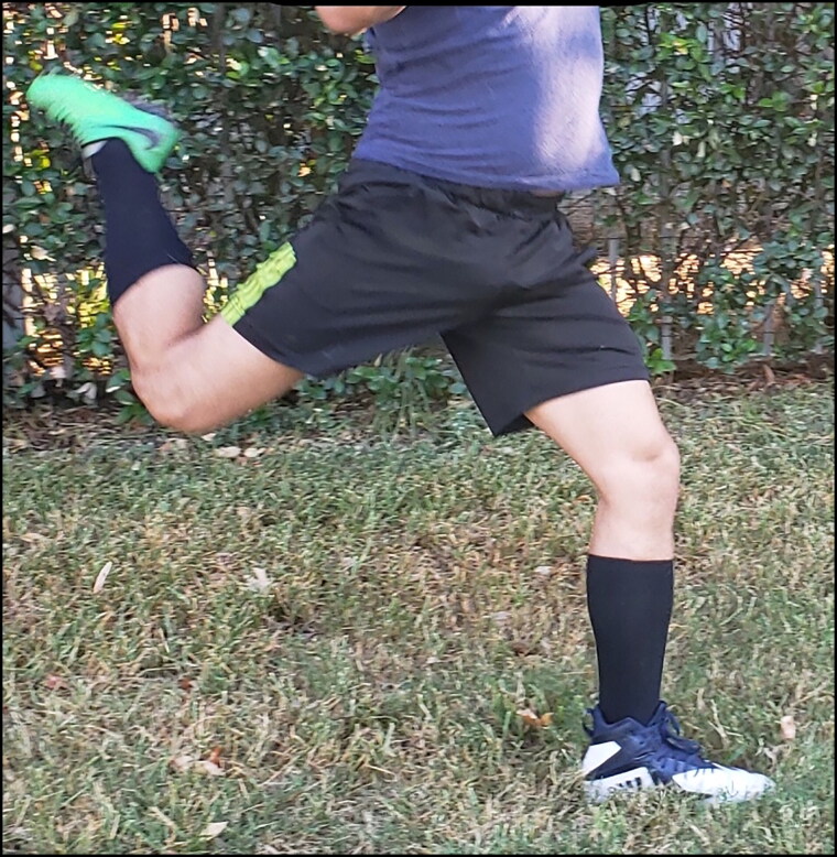
The patient experienced more pain while placekicking but was able to punt without pain. This is most likely due to the location of the tear and the decreased stress on the RF while punting. While placekicking, the patient has a larger swing phase, which includes extension at the hip and flexion at the knee (Figure 2). During the swing-back phase, the RF undergoes eccentric contraction in order to slow the leg down. Immediately after eccentric contraction, the RF performs concentric contraction in order to flex the hip and extend the knee.6 This puts a large amount of stress on the RF muscle due to strong eccentric contraction at the knee and hip joints and the subsequent transition into concentric contraction. During punting, there is less hip extension and no run-up phase. This leads to decreased eccentric contraction in the RF muscle, because it is not being stretched at both joints to the same degree. We surmise that this is the reason that the patient was able to punt without pain, unlike placekicking.
Figure 2.
Demonstration of the rectus femoris eccentric contraction phase. In this photograph, the individual is bringing his hip into extension during the windup phase of kicking as he would for kickoff or a field goal.
Risk of RF injury in kickers can be decreased by implementing a preventive exercise program involving stretching, strengthening, and core stability to decrease stress on the muscle and increase its ability to absorb force during activity. Specifically, the hip flexors should be strengthened using concentric (direct loading) exercises and the knee extensors should be strengthened using eccentric (resistance) exercises. Strengthening of core muscles has also been found to relieve the stress placed on the RF.7
Ultrasound was used as an important imaging study in this case because of its ability to diagnose musculoskeletal injuries both quickly and cost-effectively. In this case, ultrasound was also used in a dynamic manner to assess how the RF tear transformed in response to different phases of muscle engagement and relaxation. This was critical both in understanding the mechanism of injury and in providing targeted recommendations for which activities to discontinue. For these reasons, we recommend using standard and dynamic ultrasound as first-line imaging modalities in diagnosing suspected muscle intrasubstance tears.
References
- 1.Figved W, Grindem H, Aaberg M, et al. Muscle strength measurements and functional outcome of an untreated complete distal rectus femoris muscle tear. BMJ Case Rep. 2014;2014. doi: 10.1136/bcr-2013-203191. [DOI] [PMC free article] [PubMed] [Google Scholar]
- 2.Pesquer L, Poussange N, Sonnery-Cottet B, et al. Imaging of rectus femoris proximal tendinopathies. Skeletal Radiol. 2016;45:889–897. doi: 10.1007/s00256-016-2345-3. [DOI] [PubMed] [Google Scholar]
- 3.Ouellette H, Thomas BJ, Nelson E, et al. MR imaging of rectus femoris origin injuries. Skeletal Radiol. 2006;35:665–672. doi: 10.1007/s00256-006-0162-9. [DOI] [PubMed] [Google Scholar]
- 4.Galliera E, Corsi MM, Banfi G. Platelet rich plasma therapy: inflammatory molecules involved in tissue healing. J Biol Regul Homeost Agents. 2012;26(Suppl 1):35S–42S. [PubMed] [Google Scholar]
- 5.Parvizi J, Kim GK. Anatomy of the knee. In: High Yield Orthopaedics. Philadelphia, PA: Elsevier; 2010:20–22. doi: 10.1016/B978-1-4160-0236-9.00019-5. [DOI] [Google Scholar]
- 6.Barfield WR. The biomechanics of kicking in soccer. Clin Sports Med. 1998;17:711–728, vi. [DOI] [PubMed] [Google Scholar]
- 7.Mendiguchia J, Alentorn-Geli E, Idoate F, et al. Rectus femoris muscle injuries in football: a clinically relevant review of mechanisms of injury, risk factors and preventive strategies. Br J Sports Med. 2013;47:359–366. doi: 10.1136/bjsports-2012-091250. [DOI] [PubMed] [Google Scholar]


