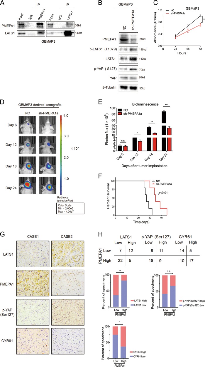
Fig. 7.
PMEPA1a knockdown inhibits glioma growth in GBM#P3 cells in vitro and in vivo. a Co-immunoprecipitations to demonstrate association of PMEPA1a with LATS1 in GBM#P3 cells. b Western blot analysis to evaluate components of the Hippo kinase pathway in lysates prepared from GBM#P3-NC and -sh-PMEPA1a cells. β-tubulin was used as loading control. c Absorbance values (450 nm) obtained from the CCK8 assay performed on GBM#P3-NC and -sh-PMEPA1a cells. Data are represented as the mean ± SEM. d, e In vivo bioluminescent images and quantification of GBM#P3-NC and -sh-PMEPA1a derived xenografts at the indicated time points. f Kaplan–Meier survival analysis performed with survival data from mice implanted with GBM#P3-NC and -sh-PMEPA1a cells. Log-rank test, P < 0.01. g Representative images of IHC staining of PMEPA1 and LATS1 in primary human glioma tissue samples. Scale bars, 50 μm. h Correlation analysis for PMEPA1 with LATS1 in primary human glioma samples based on IHC scoring. IHC scores are indicated in parentheses. χ2-test, P = 0.0020. Student’s t-test: n.s. = not significant, *P < 0.05, **P < 0.01, ***P < 0.001

