To the Editor,
Urethral catheterization, an invasive procedure with risks of infection, used to be regarded as the gold standard for measuring residual urine volume. It has now been superseded by portable ultrasound scanners performed by nurses at the patient’s bedside. The benefits include fewer invasive catheterizations and increased patient comfort. However, caution has to be exercised when using automated bladder scanners to identify urine volumes in complex cases such as patients with ascites or in the presence of other pelvic pathology. On the other hand, use of Point of care ultrasonography (POCUS) is rapidly gaining popularity in the field of nephrology [1, 2]. POCUS is essentially bedside ultrasound performed by the clinician to answer focused questions (e.g. “does this patient have hydronephrosis?”). Herein, we report an illustrative case of pelvic ascites where nephrologist-performed POCUS has facilitated the correct diagnosis.
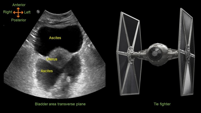
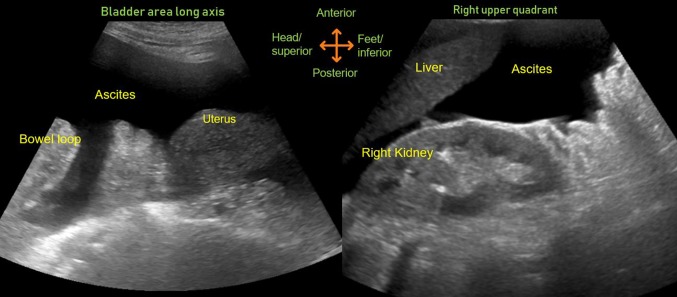
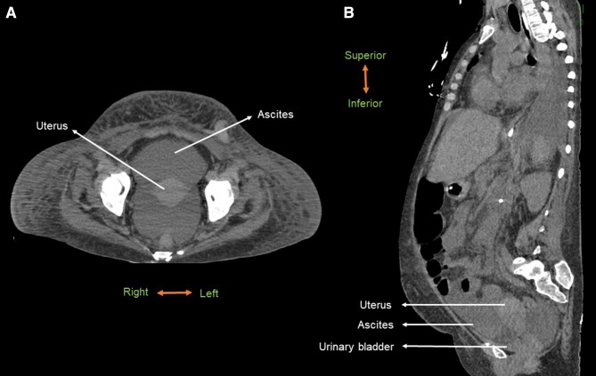
A 45-year-old woman with a history of liver cirrhosis was admitted for failure to thrive. Her hospital course was complicated by decompensated cirrhosis, septic shock and acute respiratory failure requiring mechanical ventilation. She presented with a serum creatinine of 2.52 mg/dL (baseline ~ 0.9 mg/dL), which worsened to 3.6 mg/dL during hospitalization with development of oliguria requiring renal replacement therapy. A routine bladder scan to monitor renal recovery revealed a urine volume of approximately 800 mL. However, there was no urine return on insertion of a urethral catheter and urology consult was requested to assist with the catheter placement. In the meanwhile, we performed a POCUS examination, which demonstrated large amount of anechoic (black) fluid in the pelvis that was ‘continuous’ with the peritoneal cavity in the longitudinal plane indicating that it is pelvic ascites and not the urinary bladder. In the transverse plane, uterus was seen floating in the pelvic ascites and together with ovarian ligaments, giving the appearance of a “TIE fighter” (‘Star Wars’ fictional Starfighter) (Figs. 1 , 2) [3, 4]. Moreover, a computed tomography scan obtained later confirmed decompressed urinary bladder deep in the pelvis (Fig. 3), which could not be visualized on the transabdominal ultrasound.
Fig. 1.
Transverse plane ultrasound image of the bladder area demonstrating uterus floating in pelvic ascites resembling a TIE fighter
Fig. 2.
Longitudinal ultrasound images of the bladder area and right upper quadrant demonstrating ascites. The fluid is continuous around the uterus confirming that the anterior anechoic structure seen in the transverse plane is ascites and not the urinary bladder
Fig. 3.
Transverse (a) and sagittal (b) views of CT scan of the abdomen demonstrating relationship of the ascitic fluid, uterus and the urinary bladder
The blind nature of automated bladder scanner measurement does not allow differentiation of bladder from other fluid collections [5]. Therefore, clinicians, particularly nephrologists should perform POCUS and need to be aware of the pelvic anatomy in patients prone to ascites, which likely avoids unnecessary consultations and catherizations. In addition, scanning in both longitudinal and transverse planes is important to differentiate urinary bladder from pelvic ascites as both urine and ascites appear anechoic on a sonogram.
Funding
None.
Compliance with ethical standards
Conflict of interest
The authors declare that they have no conflict of interest.
Statement of human and animal rights
This article does not contain any studies with human or animal subjects performed by any of the authors.
Informed consent
Obtained from the patient for their anonymized data to be published.
Permissions
The image of ‘TIE figher’ was obtained from imgbin.com
Footnotes
Publisher's Note
Springer Nature remains neutral with regard to jurisdictional claims in published maps and institutional affiliations.
References
- 1.Koratala A, Segal MS, Kazory A. integrating point-of-care ultrasonography into nephrology fellowship training: a model curriculum. Am J Kidney Dis. 2019;74(1):1–5. doi: 10.1053/j.ajkd.2019.02.002. [DOI] [PubMed] [Google Scholar]
- 2.Koratala A. Focus on POCUS: it is time for the kidney doctors to upgrade their physical examination. Clin Exp Nephrol. 2019;23(7):982–984. doi: 10.1007/s10157-019-01707-8. [DOI] [PubMed] [Google Scholar]
- 3.Botz B. TIE fighter sign of pelvic free fluid. https://radiopaedia.org/cases/tie-fighter-sign-of-pelvic-free-fluid. Last accessed: 7/27/2019.
- 4.Singhapricha T, Ander D. Tie fighter sign. http://www.em.emory.edu/ultrasound/ImageWeek/Trauma/TIE%20fighter.html. Last accessed: 7/27/2019.
- 5.Prentice DM, Sona C, Wessman BT, et al. Discrepancies in measuring bladder volumes with bedside ultrasound and bladder scanning in the intensive care unit: a pilot study. J Intensive Care Soc. 2018;19(2):122–126. doi: 10.1177/1751143717740805. [DOI] [PMC free article] [PubMed] [Google Scholar]


