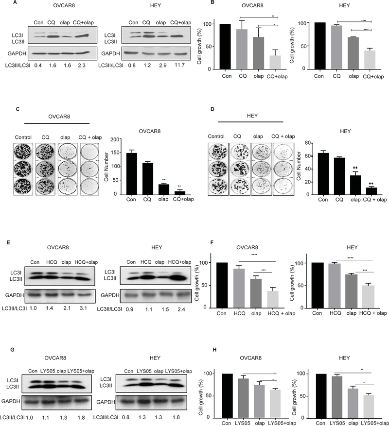
Figure 2. Inhibition of olaparib-induced autophagy decreases cell viability.
Cells were pretreated for 4 hours with 12 μM of chloroquine (CQ), with or without 5 μM of olaparib (olap) or both for 5 days. A. Whole cell lysates were analyzed for conversion of LC3I to LC3II by western blot. B. 4,000 cells/well were plated in 96-well plate, then sequentially treated with olaparib (5 μM) or chloroquine (12 μM) and incubated for 5 days before fixation and staining by sulforhodamine. C and D. OVCAR8 and HEY cells were seeded, in a 6 well plate and cultured with olaparib (5 μM), CQ (10 μM), or olaparib + CQ for 14 days and stained with Coomassie blue. Cells were pretreated for 4 hours with hydroxychloroquine (HCQ) (10 μM) or LYS05 (2 μM) with or without 5 μM of olaparib (olap) or both for 5 days. E and G. Conversion of LC3I to LC3II was analyzed by western blot. F and H. 4,000 cells/well were plated in 96-well plate, then sequentially treated with olaparib (5 μM) or chloroquine (12 μM) and incubated for 5 days before fixation and staining by sulforhodamine. Data represents results from at least 3 experiments.

