Version Changes
Revised. Amendments from Version 1
The article has been updated to include follow up of 80 weeks. Following progression to stage IV disease after one year of cetuximab, combination therapy with the addition of PD-1 blocker nivolumab was administered for another six months, achieving disease stability. This choice reflects recent advancements in the treatment scenario of advanced cSCC, with anti-PD-1 monoclonal antibody cemiplimab now established as the standard of care in immune-competent patients.
Abstract
We present the case of a 60-year-old man with unresectable cutaneous squamous cell carcinoma (cSCC) of the sternal area, which was not amenable to radiation therapy (stage III, T3N0M0). The treatment history of this patient is remarkable as the disease had progressed through all lines of conventional therapy established in the literature. The patient was treated with epidermal growth factor receptor (EGFR) inhibitor cetuximab for 35 cycles and restaged after 12 months of therapy with a whole body CT scan, documenting stage IV disease (T3N2bM1). The use of cetuximab as a single agent was effective for a limited time and we decided to initiate combination therapy with cetuximab and nivolumab. Restaging after six months of this combination regimen documented stable disease.
Keywords: cutaneous squamous cell carcinoma, cetuximab, EGFR, non-melanoma skin cancer
Introduction
This case describes the effective use of cetuximab and nivolumab in an extensive thoracic cutaneous squamous cell carcinoma resistant to all previous lines of chemotherapy.
Non-melanoma skin cancer (NMSC) is the most common malignant neoplasm affecting Caucasian individuals, the main types of which are basal cell carcinoma (BCC) and squamous cell carcinoma (SCC). SCC has a lower incidence than BCC and the gold standard of treatment is surgical excision. Between 1–5% of SCCs exhibit biologically aggressive behavior and are resistant to surgery 1.
The management of metastatic or locally advanced cutaneous SCC (cSCC), traditionally relying on conventional radiotherapy (alone or in combination with surgery) and systemic chemotherapy, benefited from the promising addition of targeted inhibitors of the epidermal growth factor receptor (EGFR) pathway and dramatically changed following the introduction of immunotherapy with checkpoint inhibitors 2. Anti-EGFR monoclonal antibody cetuximab, at the standard weekly dosage of 250mg/m2, provides an off-label treatment option with potential clinical value in advanced cSCC 3.
In 2018, a study by Migden et al. dramatically changed the previous scenario establishing the new standard of care with PD-1 blockade in immunocompetent patients, in the absence of contraindications to immunotherapy 4. Anti-PD1 monoclonal antibody cemiplimab was consequently approved for use in Europe in July 2019.
Case presentation
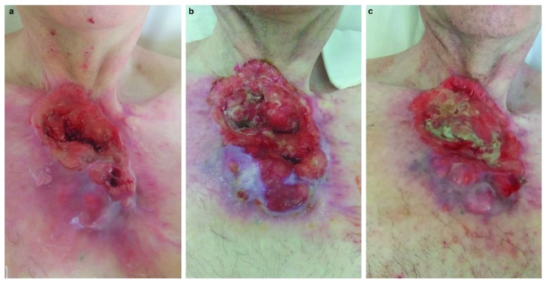
A 60-year-old Caucasian man, currently unemployed, presented to our dermatology department complaining of the recurrence of a thoracic cSCC. Physical examination revealed an extensive ulcerative skin lesion of the sternal area covered by necrotic and fibrinous tissue. The patient reported intermittent pain and bleeding ( Figure 1).
Figure 1.
Clinical presentation before cetuximab ( a) and after six ( b) and 12 weeks of therapy ( c).
The onset of a nodular skin lesion in the same site dated back to 2000, but an initial diagnosis of BCC was made only in 2013, when a single biopsy was performed (see Table 1 for timeline). A computerized tomography (CT) scan followed, demonstrating a high local burden of disease, with destructive osteo-muscular infiltration, preventing a surgical or radiation approach, and the patient was treated with vismodegib (150 mg daily). After 12 months of apparent clinical remission, a local relapse was observed, and the histologic examination of an excisional biopsy diagnosed SCC. Surgical removal of the tumor was not radical, and the patient was referred for adjuvant chemotherapy, failing four consecutive cytotoxic regimens, until the personal decision of the patient to withdraw from treatment.
Table 1. Timeline of interventions and outcomes.
| Timeline | Medical history and past interventions | |
|---|---|---|
| No family history of skin cancer 1999: total gastrectomy for gastric adenocarcinoma | ||
| Diagnostic testing and interventions | ||
| Past
Interventions 2013 – 2017 |
2000: Patient reports onset of nodular skin lesion
2013: Incisional biopsy: BCC CT scan (23-Jan-2013): high local burden of disease (50mm AP diameter), with destructive osteo-muscular infiltration - vismodegib 150mg daily from Feb to Nov-2013 2014: relapse of nodular skin lesion Excision biopsy (Feb-2014): SCC Wide surgical excision (22-May-2014): not radical Adjuvant chemotherapy: - cisplatinum 100mg/m2 day 1 with fluorouracil 1000mg/m2 for four days of 21-day-cycles, Aug – Sep-2014 - radio-chemotherapy with gemcitabine Dec-2014 – Jan-2015 - cisplatinum 100mg/m2 day 1 with docetaxel 75mg/m2 day 1 of 21-day-cycles Aug-2016 – Nov-2016 - gemcitabine monotherapy 3000mg/m2 on day 1 and 15 of 28-day-cycles Dec-2016 – Jul-2017 |
|
| 31-Jan-2018 | Baseline assessment stage III T3N0M0,
ECOG 0 |
Immunohistochemistry: low/no PD-L1 expression CT scan (31-Jan-2018):
DT×DAP×DL 120×62×110mm |
| 19-Apr-2018 | Cetuximab monotherapy 35 cycles | -
cetuximab initial single dose of 400mg/m2 followed by
- cetuximab 250mg/m2 weekly for seven cycles followed by - cetuximab 250mg/m2 every two weeks CT scan (28-May-2018): DAP 72mm CT scan (10-Sep-2018): DT×DAP×DL 135×70×110mm CT scan (12-Dec-2018): DT×DAP×DL 153×70×120mm |
| 20-Feb-2019 | Restaging stage IV T3N2bM1 ECOG 1 | CT scan (20-Feb-2019) unchanged dimension, development of
lymphadenopathies, the greater of which in the right paratracheal region DT 16mm and in the right supra- and sub-clavicular region DT 15mm, right axillary lymph node 6×6mm, development of secondary osteolytic lesions of D8 and D9 vertebral bodies |
| 22-May-2019 | Cetuximab/nivolumab combination
therapy 13 cycles of each agent |
-
cetuximab single dose of 250mg/m2 Q2W and
nivolumab single fixed
dose of 240mg Q2W administered at alternating weeks CT scan (13-May-2019) unchanged dimensions (DAP 60mm), slight reduction of previous lymphadenopathies DM 13mm and DM 10mm respectively, increased right axillary lymph node 10×8mm, stable secondary lesions of D8 and D9 vertebral bodies with marked reduction of pre- and paravertebral tissue involvement compared to previous exam CT scan (06-Aug-2019) slight dimensional increase with DL×DT×DAP 59×43×67mm |
| 18-Nov-2019 | Restaging stage IV T3N2bM1 ECOG 1 | CT scan (18-Nov-2019) DL×DT×DAP 78×60×85mm, unchanged
lymphadenopathies DM 13mm and DM 10mm respectively, stable secondary lesions of D8 and D9 vertebral bodies |
[i] BCC, basal cell carcinoma; CT, computerized tomography; DAP, anterior-posterior diameter; DL, longitudinal diameter; DM, maximum diameter; DT, transverse diameter; PDL-1, programmed cell death ligand-1; SCC, squamous cell carcinoma.
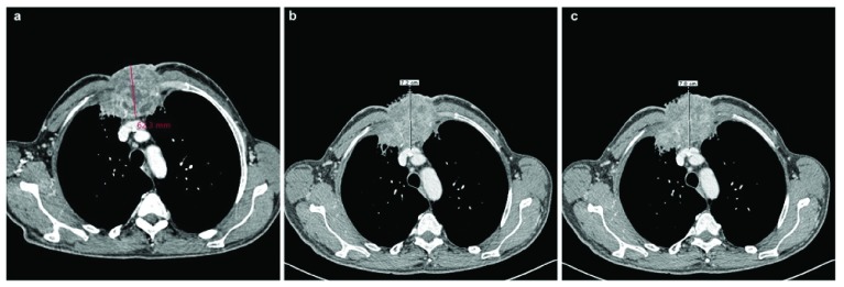
A stage III-disease (T3N0M0, Figure 2a) 5 advised the use of anti-PD-1 therapy. Even if PDL-1 testing is not required, immunohistochemistry was performed on the previous biopsy sample documenting no/low expression of PDL-1. Being cemiplimab not yet available, we resorted to cetuximab, the use of which is off-label for cSCC. We administered cetuximab at an initial single dose of 400mg/m2, followed by 250mg/m2 every week, for seven cycles, and every two weeks, for 35 total cycles. Follow-up assessment with periodic CT at three ( Figure 2b and 2c) and six months documented stable locally advanced disease. Therapy was well tolerated, with the only complaint of an acneiform eruption, which began after one week of treatment and was managed with clindamycin 1% gel twice a day and oral minocycline 100mg twice a day for four weeks.
Figure 2.
CT scan performed at baseline ( a), after six ( b) and 12 weeks of therapy ( c), highlighting the anterior-posterior diameter of the tumor.
The patient was restaged after 44 weeks with a whole-body CT scan that demonstrated progression to stage IV disease (T3N2bM1) with metastatic involvement. Combination therapy with the addition of PD-1 blocker was planned and we employed locally available anti-PD1 monoclonal antibody nivolumab according to the following scheme: cetuximab single dose of 250mg/m2 Q2W and nivolumab single fixed dose of 240mg Q2W administered at alternating weeks. Sequential CT assessments after 12 and 26 weeks showed stable disease at best, with slight increase of the primitive lesion and unchanged nodal and metastatic localizations.
Discussion
In our report, response to cetuximab as a single agent and in combination with nivolumab was assessed for as long as 80 weeks.
We were challenged to select an effective treatment in this advanced case and resorted to EGFR inhibitor therapy. Cetuximab is approved for the treatment of locally or regional advanced SCC of the head and neck region (in combination with radiation) or for recurrent or metastatic disease (alone or in association with platinum). Its use in cSCC of other regions is currently off-label but our choice of drug was extensively supported by evidence in published literature 3. A phase II study of unresectable cSCC treated with cetuximab for at least six weeks registered 25% objective response and 42% disease stabilization 6. A diffuse papulopustular acneiform eruption is the most common cutaneous reaction pattern to EGFR inhibitors, reported in over two-thirds of treated subjects but severe in only 5–10% of cases. Cutaneous toxicity is suggested to be a proxy for response to cetuximab 7.
Immune checkpoint inhibition revolutionized the management of advanced cSCC and anti-PD-1 monoclonal antibody cemiplimab is currently the preferred first line therapy, following registration for this specific indication in the US in July 2018 and in Europe in July 2019. Response to cemiplimab was reported in 50% of patients in the expansion cohorts of the phase 1 study and in 47% of patients in the metastatic-disease cohort of the phase 2 study, with response exceeding 6 months in 57% of cases 4, 8.
Nivolumab monotherapy is indicated for the treatment of recurrent or metastatic SCC of the head and neck in adults progressing on or after platinum-based therapy. In a patient without access to cemiplimab clinical trials, nivolumab was our agent of choice due to the biological similarity to SCC of the head-neck district 9.
Recent experience from the literature attributes long-term remission and good tolerability to PD-1 checkpoint inhibition with nivolumab in cSCC. A series of three patients with advanced cSCC treated with nivolumab reported partial response in two subjects and stable disease in the third 10.
Chen et al. recently reported a case of clinical regression of invasive cSCC after six months of dual treatment with cetuximab weekly and nivolumab biweekly and hypothesized the mechanisms underlying a synergistic action of these two agents 11.
Conclusions
Serial biopsies are mandatory for advanced BCC candidates prior to vismodegib treatment to exclude foci of multiple differentiation 12.
Prior to the introduction of cemiplimab, no drugs were approved specifically for cSCC.
The efficacy of cetuximab is limited as a single agent, with modest durations for stable disease.
Low PDL-1 expression does not preclude the efficacy of checkpoint inhibitors; in fact, cemiplimab is approved without requirement for testing.
PD-1 blockade is the new standard of care in advanced cSCC in immunocompetent patients.
Data availability
All data underlying the results are available as part of the article and no additional source data are required.
Consent
Written informed consent for publication of their clinical details and clinical images was obtained from the patient.
Funding Statement
Associazione Romana Ricerca Dermatologica covered the publication fees of this article as support to the authors.
The funders had no role in study design, data collection and analysis, decision to publish, or preparation of the manuscript.
[version 2; peer review: 2 approved]
References
- 1. Que SKT, Zwald FO, Schmults CD: Cutaneous squamous cell carcinoma: Management of advanced and high-stage tumors. J Am Acad Dermatol.United States;2018;78(2):249–61. 10.1016/j.jaad.2017.08.058 [DOI] [PubMed] [Google Scholar]
- 2. Stratigos A, Garbe C, Lebbe C, et al. : Diagnosis and treatment of invasive squamous cell carcinoma of the skin: European consensus-based interdisciplinary guideline. Eur J Cancer.England;2015;51(14):1989–2007. 10.1016/j.ejca.2015.06.110 [DOI] [PubMed] [Google Scholar]
- 3. Jalili A, Pinc A, Pieczkowski F, et al. : Combination of an EGFR blocker and a COX-2 inhibitor for the treatment of advanced cutaneous squamous cell carcinoma. J Dtsch Dermatol Ges.Wiley/Blackwell (10.1111);2008;6(12):1066–9. 10.1111/j.1610-0387.2008.06861.x [DOI] [PubMed] [Google Scholar]
- 4. Migden MR, Rischin D, Schmults CD, et al. : PD-1 Blockade with Cemiplimab in Advanced Cutaneous Squamous-Cell Carcinoma. N Engl J Med. 2018;379(4):341–51. 10.1056/NEJMoa1805131 [DOI] [PubMed] [Google Scholar]
- 5. Amid M, editor: Part II Head and Neck. In: AJCC Cancer Staging Manual. 8th ed. New York, NY, USA: Springer;2018;53. [Google Scholar]
- 6. Maubec E, Petrow P, Scheer-Senyarich I, et al. : Phase II study of cetuximab as first-line single-drug therapy in patients with unresectable squamous cell carcinoma of the skin. J Clin Oncol.United States;2011;29(25):3419–26. 10.1200/JCO.2010.34.1735 [DOI] [PubMed] [Google Scholar]
- 7. Jatoi A, Green EM, Rowland KM, Jr, et al. : Clinical predictors of severe cetuximab-induced rash: observations from 933 patients enrolled in north central cancer treatment group study N0147. Oncology. 2009;77(2):120–3. 10.1159/000229751 [DOI] [PMC free article] [PubMed] [Google Scholar]
- 8. Papadopoulos KP, Owonikoko TK, Johnson ML, et al. : REGN2810: A fully human anti-PD-1 monoclonal antibody, for patients with unresectable locally advanced or metastatic cutaneous squamous cell carcinoma (CSCC)—Initial safety and efficacy from expansion cohorts (ECs) of phase I study. J Clin Oncol. American Society of Clinical Oncology. 2017;35(15_suppl):9503 10.1200/JCO.2017.35.15_suppl.9503 [DOI] [Google Scholar]
- 9. Malm IJ, Bruno TC, Fu J, et al. : Expression profile and in vitro blockade of programmed death-1 in human papillomavirus-negative head and neck squamous cell carcinoma. Head Neck.United States;2015;37(8):1088–95. 10.1002/hed.23706 [DOI] [PMC free article] [PubMed] [Google Scholar]
- 10. Blum V, Müller B, Hofer S, et al. : Nivolumab for recurrent cutaneous squamous cell carcinoma: three cases. Eur J Dermatol. 2018;28(1):78–81. [DOI] [PubMed] [Google Scholar]
- 11. Chen A, Ali N, Boasberg P, et al. : Clinical Remission of Cutaneous Squamous Cell Carcinoma of the Auricle with Cetuximab and Nivolumab. J Clin Med. 2018;7(1): pii: E10. 10.3390/jcm7010010 [DOI] [PMC free article] [PubMed] [Google Scholar]
- 12. Zhu GA, Sundram U, Chang AL: Two different scenarios of squamous cell carcinoma within advanced Basal cell carcinomas: cases illustrating the importance of serial biopsy during vismodegib usage. JAMA Dermatol.United States;2014;150(9):970–3. 10.1001/jamadermatol.2014.583 [DOI] [PubMed] [Google Scholar]


