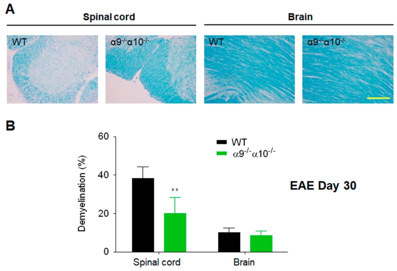
Figure 4.
Reduced demyelination in EAE α9/α10 DKO mice. (A) Luxol blue staining reveals the intensity of myelin integrity in brain or spinal cord sections from wild-type (WT) or nAChR α9/α10 subunit DKO mice (α9−/−α10−/−) at day 30 after immunization. Scale bar: 50 µm. (B) Quantified data show reduced demyelination in spinal cords of α9/α10 DKO mice. N = 3 mice per group, at least four sections examined per mouse, n ≥ 12. Mean ± S.E.M.; unpaired t-test; ** p < 0.01.

