Abstract
Background
Yam tuber is a storage organ, derived from the modified stem. Tuber expansion is a complex process, and depends on the expressions of genes that can be influenced by environmental and endogenous factors. However, little is known about the regulatory mechanism of tuber expansion. In order to identify the genes and miRNAs involved in tuber expansion, we examined the mRNAs and small RNAs in Dioscorea opposita (Chinese yam) cv. Guihuai 16 tuber during its initiation and expansion stages.
Results
A total of 14,238 differentially expressed genes in yam tuber at its expansion stage were identified by using RNA sequencing technology. Among them, 5723 genes were up-regulated, and 8515 genes were down-regulated. Functional analysis revealed the coordination of tuber plant involved in processes of cell events, metabolism, biosynthesis, and signal transduction pathways at transcriptional level, suggesting that these differentially expressed genes are somehow involved in response to tuber expansion, including CDPK, CaM, CDL, SAUR, DELLA, SuSy, and expansin. In addition, 541 transcription factor genes showed differential expression during the expansion stage at transcriptional level. MADS, bHLH, and GRAS were involved in cell differentiation, division, and expansion, which may relate to tuber expansion. Noteworthy, data analysis revealed that 22 known tuber miRNAs belong to 10 miRNA families, and 50 novel miRNAs were identified. The integrated analysis of miRNA-mRNA showed that 4 known miRNAs and 11 genes formed 14 miRNA-target mRNA pairs were co-expressed in expansion stage. miRNA160, miRNA396, miRNA535 and miRNA5021 may be involved in complex network to regulate cell division and differentiation in yam during its expansion stage.
Conclusion
The mRNA and miRNA datasets presented here identified a subset of candidate genes and miRNAs that are putatively associated with tuber expansion in yam, a hypothetical model of genetic regulatory network associated with tuber expansion in yam was put forward, which may provide a foundation for molecular regulatory mechanism researching on tuber expansion in Dioscorea species.
Keywords: Yam tuber, Dioscorea opposita, Expansion, mRNA, Small RNA
Background
Yams (Dioscorea spposita) are monocotyledonous plants belonging to the family Dioscoreaceae, and tuber is its harvested organ. Tuber originates from the expansion of underground stem, is suitable for nutrients storage, with many large parenchyma cells. Tuber morphogenesis and starch, along with accumulated proteins are two main processes of tuber growth [1]. The tuber morphogenesis of yam can be divided into three stages: initiation stage, expansion stage, and maturation stage. The expansion stage can be divided into three periods: early expansion stage, middle expansion stage, late expansion stage [2, 3]. Tuber morphogenesis is a complex physiological process regulated by heredity, environment, and hormones [4]. Great efforts have been made to explore the physiological factors affecting the morphogenesis of yam tubers. The short-day treatment tended to promote tuber growth at the primary tuber growth stage of the plant, and bulbil development at the rapid tuber growth, but the responses varied among species and cultivar [5, 6]. Endogenous hormones including gibberellins (GA), acetic acid (IAA) and abscisic acid (ABA) performed a key role at the beginning of tuber expansion stage, and trans-zeatin (tZ), jasmonic acid (JA) were also involved in tuber expansion [2, 7, 8]. Exogenous hormones have been used to study the mechanism of tuber expansion, GAs could promote tuber expansion and yield through in vitro and in vivo treatment [9, 10]. Exogenous GA application combined with ABA has promoted microtuber growth and expansion [11]. Exogenous JA was found to be essential for yam tuberization, and induced an increase in the number of tubers in vitro and in vivo [12, 13]. However, fundamental knowledges of endogenous metabolic networks are poor in tuber expansion.
The induction and growth of microtubers in vitro were controlled by nutrients, and sucrose concentration was the most crucial factor affecting tuberization and frequency of proliferation in yam [7, 14]. Yam tuber morphology was significantly correlated with nutrient accumulation and enzymatic activity. Sucrose, soluble sugars, and proteins increased significantly during tuber expansion stage, then subsequently decreased at maturity stage. Starch content increased throughout tuber morphogenesis, and sucrose synthase, sucrose phosphate synthase, and AGPase were significantly correlated with nutrient accumulation [15]. Although many DNA molecular markers have been used to uncover the genetic diversity and relationship among yam germplasms [16–18], little is known about specific genes involved in tuber morphogenesis. The sucrose synthase 4 and sucrose-phosphate synthase 1 were associated with the earliest stages of starch biosynthesis and storage; a SCARECROW-LIKE gene was involved in the formation of adventitious roots [19]. PE2.1 and PE53 are the members of pectinesterase (PE) superfamily, which may be involved in the regulation of starch and sucrose metabolism and signaling pathways. Therefore, they may play an essential role in microtuber formation [20]. Tuber morphogenesis is a complex biological process involving many specific genes and proteins, especially at yam tuber expansion stage. Transcriptome techniques can efficiently find and detect these genes and proteins. Potato is a tuber crop. Several transcriptome analyses revealed that numerous genes are regulated in early stages of stolon-to-tuber transitions, or tuberization by nutrients, photoperiodic conditions, exogenous hormones, and stress in potato tuber [21–24]. Former transcriptomic study revealed that some putative genes were involved in dioscin biosynthesis [25], along with this chalcone isomerase (CHS), flavanone 3-hydroxylase (F3H), flavonoid 3′-monooxygenase (F3’H), dihydroflavonol 4-reductase (DFR), leucoanthocyanidin dioxygenase (LDOX), and flavonol 3-O-glucosyltransferase (UF3GT) were significantly expressed in flavonoid biosynthesis [26]. However, there are no reports of transcriptome study on tuber expansion.
microRNAs (miRNAs) are small, endogenous, non-coding RNAs that have essential functions in many biological processes, such as the regulations of growth and development, stress response, and metabolism. Many studies have shown that miRNAs play essential roles in root and tuber formation or development [27–29]. miR165/166 regulated root growth by determining the fate of root cells in Arabidopsis combined with phytohormone crosstalk, by negatively regulating its target genes auxin response factor ARF10, ARF16 and ARF17 [30]. miRNA172 and miR156 were involved in tuberization process, either as a component or a regulator of long-distance gibberellin signaling pathways [31, 32]. Potato specific miRNA193, miRNA152, and conserved miR172–1, miRNA172–5 showed significant expression during developmental stages of tuberization [28]. However many studies have found that miRNAs are involved in tuber and root development, the miRNA-mediated regulatory network during tuber expansion is still unclear.
Although whole-genome sequencing of the heterozygous diploid Guinea yam (D. rotundata) had been performed for sex determination [33], a detailed comparative mRNA and miRNA analysis during yam tuber expansion stage need to be detected. In this study, to identify and analyze the global gene and miRNA expression dataset in tuber expansion, six libraries prepared from D. opposita (Chinese yam) cv. Guihuai 16 tuber of initiation stage (GH16_I) and expansion stage (GH16_E) were sequenced by using a BGISEQ-500 platform. Furthermore, the association analysis between mRNA and miRNA expression was done, and the elucidation of the regulatory relationship of miRNA and their corresponding mRNA targets was studied for understanding the expansion of tuber.
Results
Overview of RNA-Seq dynamics and small RNA sequencing
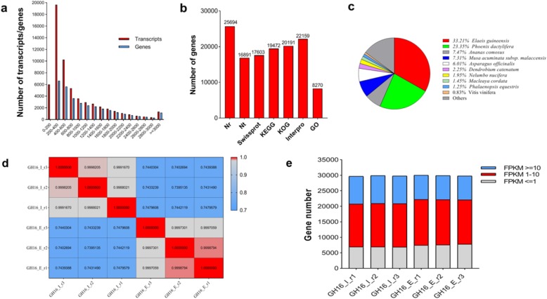
To identify the regulation of mRNA and miRNAs co-regulatory network during tuber expansion, the RNA-Seq and small RNA were examined during tuber initiation stage (GH16_I) and expansion stage (GH16_E) (Fig. 1). Meanwhile, transcriptome library was constructed from a pool of mixed RNA consisting of initiation and expansion stages in order to construct RNA-Seq and small RNA (named Total_1). Approximately 74.71 Mb original data in total were gained from BGISEQ-500 platform at BGI-Shenzhen (Table 1). After filtering low-quality reads and adaptor sequences, 6.67 Gb clean reads were obtained and processed by de novo analysis using Trinity software. The assembly produced a total of 54,781 transcripts. Then, Tgicl software was used on transcripts to remove abundance, and 32,207 genes were gained. The N50 statistic was 1508, which meant that more than 50% of the genes were longer than 1508 bp. The length distribution of all the assembled yam genes shown in Fig. 2a, which indicated that 7.91% of the complete transcripts and 13.00% of the total genes were longer than 2000 bp.
Fig.1.
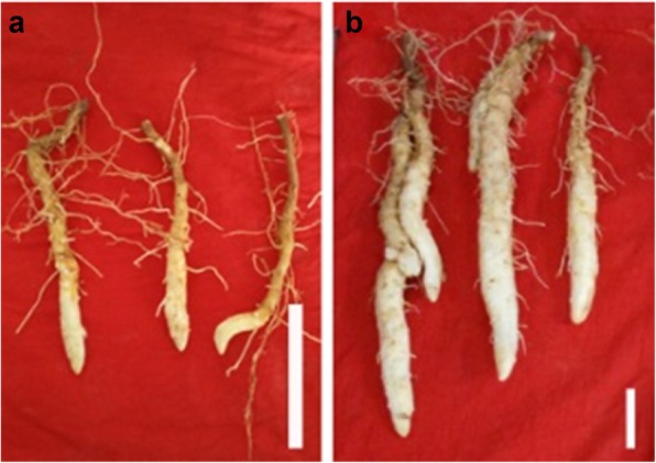
A picture of Guihuai 16 (D. opposita) tuber at different developmental stages. Samples were collected from field-grown cultivar Guihuai 16 (D. opposita) during its initiation and expansion stages. a: Initiation stage, b: Expansion stage, white bar is 5 cm
Table 1.
Statistic analysis of clean reads for mRNA in tuber initiation and expansion stages in yam
| Sample | Total_1 | GH16_I_r1 | GH16_I_r2 | GH16_I_r3 | GH16_E_r1 | GH16_E_r2 | GH16_E_r3 | Sum |
|---|---|---|---|---|---|---|---|---|
| Total Raw Reads (Mb) | 74.71 | 66.43 | 66.42 | 66.52 | 66.43 | 66.43 | 66.35 | – |
| Total Clean Bases (Gb) | 6.67 | 6.57 | 6.57 | 6.58 | 6.56 | 6.57 | 6.56 | – |
| Clean Reads Q20(%) | 96.4 | 98.63 | 98.56 | 98.54 | 98.52 | 98.5 | 98.57 | – |
| Clean Reads Q30(%) | 88.22 | 93.12 | 92.86 | 92.78 | 92.67 | 92.63 | 92.95 | – |
| Clean Reads Ratio(%) | 89.34 | 98.91 | 98.92 | 98.96 | 98.7 | 98.87 | 98.9 | – |
| Total Mapped Reads(%) | – | 83.07 | 83.34 | 83.17 | 82.05 | 81.77 | 82.03 | – |
| Total Expressed Genes | – | 29,658 | 29,905 | 29,839 | 30,012 | 29,893 | 29,811 | 32,026 |
| Total trinity Transcripts | 54,781 | |||||||
| Total Tgicl Genes | 32,207 | |||||||
| GeneGC(%) | 44.63 | |||||||
| GeneN50 | 1508 |
Fig. 2.
The annotation of Guihuai16 (D. opposita) tuber assembled transcriptome and gene expression profiling. a Length distribution of assembled cultivar Guihuai 16 (D. opposite) transcripts and genes, the abscissa represents the length. b Number of genes aligned to different databases. c Distribution of species aligned by assembled cultivar Guihuai 16 (D. opposita) tuber genes. d Correlation analysis between samples replicates. e Distribution of gene number expression concentration in different FPKM intervals of each mRNA libraries, gray, red and blue represents three situations of FPKM (FPKM<=1, FPKM 1–10, FPKM> = 10), respectively
A total of 32,207 genes were functionally annotated with 7 functional database.
(NR, NT, GO, KOG, KEGG, SwissProt, and InterPro), 25,694 (79.78%), 16,891 (52.45%), 17,603 (54.66%), 19,472 (60.46%), 20,191 (62.69%), 22,159 (68.80%), 8270 (25.68%) reads were annotated functionally respectively (Fig. 2b). 13,566 genes were commonly annotated in NR, KOG, KEGG, SwissProt and InterPro databases. Based on the functional annotation results of NR database, the proportions of different species in the notes of genes were calculated, 8533 (33.21%), 5999 (23.35%), 1920 (7.47%) and 1879 (7.31%) genes were aligned to Elaeis guineensis, Phoenix dactylifera, Ananas comosus, Musa acuminata subsp and Malaccensis (Fig. 2c). Similar species distributions were also observed for yam tuber in previous research [25], 8229 (16.2%) genes in D. zingiberensis had the most hits from Elaeis guineensis, followed by Phoenix dactylifera (6857, 13.5%), Musa acuminate (2692, 5.3%).
After filtering low-quality reads and adaptor sequences, 6.57 Gb, 6.57 Gb, 6.58 Gb, 6.56 Gb, 6.57Gb, and 6.56Gb clean reads were obtained in six RNA-Seq analysis libraries (initiation stage named GH16_I, expansion stage named GH16_E) respectively, and a total of 32,026 expressed genes were detected (Table 1). The average mapping rate of transcriptome library (named Total_1) was 82.57%. A heat map cluster showed good correlations among replicates which indicated high repeatability of the data (Fig. 2d). In order to show the number of genes in different FPKM intervals of each mRNA libraries more intuitively, three situations of FPKM (FPKM<=1, FPKM 1–10, FPKM> = 10) were counted the number of genes (Fig. 2e), indicating that most genes were expressed in the FPKM 1–10 ranges in the libraries. Genes with expression levels > 5 FPKM were retained for statistical analysis.
Furthermore, the corresponding six small RNA libraries at the three time-points were also constructed for deep sequencing. Initially, a total of 170,957,171 reads were generated (Table 2). After filtering low-quality reads and adaptor sequences, 157,958,048 clean reads longer than 18 nt for six libraries with an average of 26.32 M clean reads were obtained, and length distribution of clean reads showed that the classes of sRNA were 21-24 nt (Additional file 1: Figure S1). Subsequently, 6,388,211 (25.11%), 5,872,589 (22.36%), 6,086,348 (22.98%) reads in tuber initiation stage and 4,593,044 (17.48%), 5,032,588 (18.58%), 4,642,869 (17.58%) reads in tuber expansion stage were mapped to sRNA database (rRNA, tRNA, snRNA and snoRNA), respectively.
Table 2.
Statistic analysis of clean reads for small RNA sequencing in tuber initiation and expansion stages in yam
| Sample name | Tota reads | Clean reads | Mapped reads | Known miRNA | Novel miRNA | Total miRNA |
|---|---|---|---|---|---|---|
| GH16_I_r1 | 27,687,839 | 25,438,600 | 6,388,211 | 18 | 50 | 68 |
| GH16_I_r2 | 28,663,585 | 26,268,556 | 5,872,589 | 19 | 47 | 66 |
| GH16_I_r3 | 28,610,515 | 26,482,287 | 6,086,348 | 21 | 49 | 70 |
| GH16_E_r1 | 28,256,295 | 26,276,062 | 4,593,044 | 20 | 49 | 69 |
| GH16_E_r2 | 29,343,139 | 27,085,820 | 5,032,588 | 20 | 49 | 69 |
| GH16_E_r3 | 28,395,798 | 26,406,723 | 4,642,869 | 22 | 50 | 72 |
| Sum | 170,957,171 | 157,958,048 |
Differentially expressed genes annotation by GO term and KEGG pathway
To identify differentially regulated genes in tuber expansion stage, DESeq software was used to compare the changes of gene expression between initiation and expansion stages. Among them, 5723 genes were up-regulated, 8515 genes were down-regulated, respectively, and it were differentially expressed in expansion stage (GH16_E), compared to initiation stage (GH16_I) (Additional file 2: Table S1).
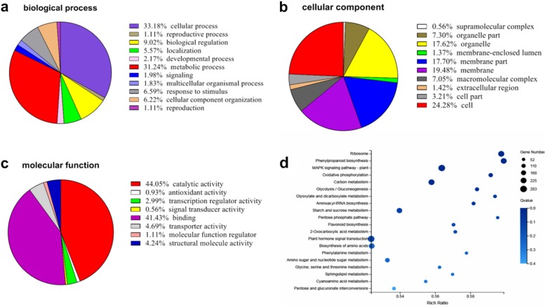
For better comprehension of DEGs functions, 44 GO categories were identified. For biological processes, DEGs associated with cellular process (33%), metabolic process (31%), and biological regulation (9%) were enriched during expansion stage (Fig. 3a). For cellular component, 10 GO categories were enriched in DEGs, including cell (24%), membrane (19%), membrane part (18%), and organelle (18%) (Fig. 3b). The molecular functions of the DEGs were mainly associated with catalytic activity (44%), binding (41%), transporter activity (5%), structural molecular activity (4%) (Fig. 3c). Among the significant GO term analysis, 15 genes were enriched in cell wall polysaccharide metabolic process (GO:0010383), 15 genes were involved in hemicellulose metabolic process (GO:0010410), and 13 genes were related to xyloglucan metabolic process (GO:0010411) related to cell wall formation during expansion stage (Table 3). Besides, the results also revealed several significant expression genes involved in tissue development, root morphogenesis, root system development, and root development (Table 4).
Fig. 3.
Gene Ontology and KEGG pathway annotation of Guihuai16 (D. opposita) tuber assembled genes. a, b and c represent the biological process, cellular component, and molecular function, respectively. d Top 20 significant KEGG pathways
Table 3.
GO enrichment analysis of DEGs
| Type | ID | Term | Gene Number | Rich Ratio | P value |
|---|---|---|---|---|---|
| Molecular function | GO:0005198 | Structural molecule activity | 183 | 0.62 | 1.83867E-07 |
| GO:0003735 | Structural constituent of ribosome | 143 | 0.61 | 1.71036E-05 | |
| GO:0003700 | DNA binding transcription factor activity | 116 | 0.59 | 0.001099451 | |
| Cellular component | GO:0005576 | Extracellular region | 91 | 0.70 | 2.22612E-07 |
| GO:0043228 | Non-membrane-bounded organelle | 285 | 0.58 | 1.68443E-06 | |
| GO:0043232 | Intracellular non-membrane-bounded organelle | 285 | 0.58 | 1.68443E-06 | |
| GO:0005840 | Ribosome | 157 | 0.61 | 1.90586E-05 | |
| GO:0005618 | Cell wall | 49 | 0.72 | 4.36888E-05 | |
| GO:0030312 | External encapsulating structure | 49 | 0.72 | 4.36888E-05 | |
| GO:0048046 | Apoplast | 32 | 0.78 | 0.000075892 | |
| GO:0022625 | Cytosolic large ribosomal subunit | 22 | 0.81 | 0.000355453 | |
| Biological process | GO:0044262 | Cellular carbohydrate metabolic process | 77 | 0.69 | 1.68372E-05 |
| GO:0005975 | Carbohydrate metabolic process | 171 | 0.60 | 0.00025216 | |
| GO:0044264 | Cellular polysaccharide metabolic process | 53 | 0.70 | 0.000285538 | |
| GO:0045786 | Negative regulation of cell cycle | 11 | 1.00 | 0.000452873 | |
| GO:0006073 | Cellular glucan metabolic process | 50 | 0.69 | 0.000495823 | |
| GO:0010383 | Cell wall polysaccharide metabolic process | 15 | 0.88 | 0.001069447 | |
| GO:0010410 | Hemicellulose metabolic process | 15 | 0.88 | 0.001069447 | |
| GO:0010411 | Xyloglucan metabolic process | 13 | 0.93 | 0.000841262 |
Table 4.
Functional classification and pathway assignment of differentially expressed DEG by GO in expansion stage
| Tissue development [GO:0009888] | Gene ID | log2(GH16_E/GH16_I) |
|---|---|---|
| Actin-related protein 3 | CL1179.Contig2_Total_1 | −2.00 |
| Anaphase-promoting complex subunit 10 | CL1997.Contig4_Total_1 | 1.82 |
| Alpha-tubulin | CL28.Contig3_Total_1 | −5.08 |
| Tubulin alpha chain | CL3054.Contig1_Total_1 | −1.81 |
| Glutamine synthetase nodule isozyme | Unigene128_Total_1 | −4.98 |
| Phosphoenolpyruvate carboxylase 2 | Unigene13453_Total_1 | −1.23 |
| Phosphoenolpyruvate carboxylase 2 | Unigene13455_Total_1 | 1.32 |
| Anaphase-promoting complex subunit 10 | Unigene18039_Total_1 | − 1.15 |
| Anaphase-promoting complex subunit 6 | Unigene2600_Total_1 | −1.06 |
| Actin-related protein 2 | Unigene4771_Total_1 | −1.10 |
| ATPase ASNA1 homolog | Unigene5972_Total_1 | −1.32 |
| Homeobox protein knotted-1-like 3 | Unigene6778_Total_1 | 3.48 |
| Root morphogenesi, root system development, root development [GO:0009888,GO:0022622,GO:0048364] | ||
| Cytoplasmic tRNA 2-thiolation protein 2 | CL1237.Contig1_Total_1 | −1.02 |
| Cytoplasmic tRNA 2-thiolation protein 2 | CL1237.Contig2_Total_1 | −2.28 |
| Mediator of RNA polymerase II transcription subunit 32 | CL2787.Contig2_Total_1 | 2.02 |
| Succinate dehydrogenase assembly factor 2 | CL3034.Contig1_Total_1 | −1.04 |
| Guanine nucleotide-binding protein | Unigene18752_Total_1 | −3.18 |
| Enhanced ethylene response protein 5 | Unigene2193_Total_1 | −1.75 |
| ATPase ASNA1 | Unigene5972_Total_1 | −1.32 |
KEGG is a signal pathway database with vibrant signal pathway map, 20 pathways were identified during yam tuber expansion stage. Interestingly, KEGG pathway analysis showed that plant hormone signal transduction (ko04075), biosynthesis of amino acids (ko01230) were enriched with DEGs during expansion stage (Fig. 3d). Other pathways such as MAPK signaling pathway (ko04016), starch and sucrose metabolism (ko00500), and carbon metabolism (ko01200) were also identified as involving 283, 204, and 236 DEGs, respectively. The metabolic pathways may be closely related to the development of tuber expansion and bioactive compound synthesis.
Comprehensive analysis of differentially expressed genes in expansion stage
Compared with initiation stage, there were a large number of DEGs in tuber expansion stage using NR, GO, and KEGG annotation. Signal transduction, cell wall, cell division, starch, and sucrose metabolism were selected for profiling during the expansion of yam tuber.
Hormone signal
A total of 242 DEGs were identified to be highly similar to many plant hormone signal pathways, including 131 down-regulated and 111 up-regulated DEGs in expansion stage (Additional file 2: Table S1). Interestingly, most plant hormone-related genes in GA, IAA, and ABA signal pathways were discovered during expansion stage.
In auxin transduction pathway, the transcriptional level of auxin influx carrier /auxin-responsive protein IAA (AUX/IAA) and small auxin up RNA (SAUR) were significantly down-regulated during the expansion stage, while auxin-responsive GH3 gene family (GH3) was up-regulated. In contrast, two auxin response factor ARFs (CL2135.Contig1_Total_1, and Unigene5660_Total_1) were shown high expression level during expansion stage, while other two ARFs (CL2887.Contig2_Total_1, Unigene5486_Total_1) were low expression level during expansion stage (Additional file 3: Table S2).
In gibberellin transduction pathway, the expression of gibberellin receptor GID2 was low expression during expansion stage. In contrast, DELLA proteins were highly expressed during the expansion stage. Meanwhile, protein phosphatase 2C(PP2C) was highly expressed during expansion stage.
MAPK and calcium signaling
Regulation of genes related to MAPK and calcium signaling during the expansion stage were also investigated. Six mitogen-activated protein kinases (MAPK) genes were up-regulated during expansion stage, while MPK6 and MPK8 were down-regulated. In summary, 48 DETs were homologous with calcium signal-related genes (Additional file 2: Table S1), including calcium-dependent protein kinases (CDPKs), calcium-binding proteins (CBPs), and calreticulin (CBL). It is worth noticing that CBLs were down-regulated during expansion stage (Additional file 3: Table S2).
Cell wall and cell cycle
A total of 98 transcripts homologous to the genes associated with cell wall and cell cycle were observed as differentially regulated during expansion stage (Additional file 2: Table S1), including xyloglucan endotransglucosylase/hydrolase (XTH), expansin, extension, cyclin-dependent kinases (CKS), cell division protease (ftsHs), cell division cycle 5-like protein (CDC5), cell division control protein (CDC), cyclin-dependent kinases (CDKs), and cyclin-dependent kinase inhibitor (CDKIs). All of the expansin, extension, cell wall synthesis, and CKS genes were down-regulated during expansion stage. Meanwhile, most of the cell cytoskeleton and XTH were down-regulated during expansion stage in yam (Additional file 3: Table S2).
Starch and sucrose metabolism
The major constituents of starch and sucrose metabolism during expansion stage are sucrose synthase genes (SuSy), sucrose phosphate synthase genes (SPS), starch synthase (SS), and invertase genes (INV) (Additional file 2: Table S1). Among them, SuSy were down-regulated during expansion stage. Interestingly, dioscorins, the major storage proteins in yam tubers, were significantly up-regulated during the expansion stage (Additional file 3: Table S2). These results indicated that many functional genes were involved in expansion stage of yam tuber.
Transcription factor
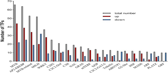
A total of 541 TF-encoding genes belonging to 48 TF families were differentially expressed during expansion stage, MYB, MYB-related, and AP2-EREP were enriched (Fig. 4). 286 TF encoding genes were up-regulated and 255 TF encoding genes were down-regulated, respectively (Additional file 4: Table S3). The most abundant TF gene families with the highest number of expressions during expansion stage were depicted by heat map (Fig. 5). Moreover, these genes were involved in circadian rhythm pathway, starch and sucrose metabolism pathway, and GA pathway by KEGG analysis respectively.
Fig. 4.
The numbers of up-regulated and down-regulated transcription factors during expansion stage
Fig. 5.
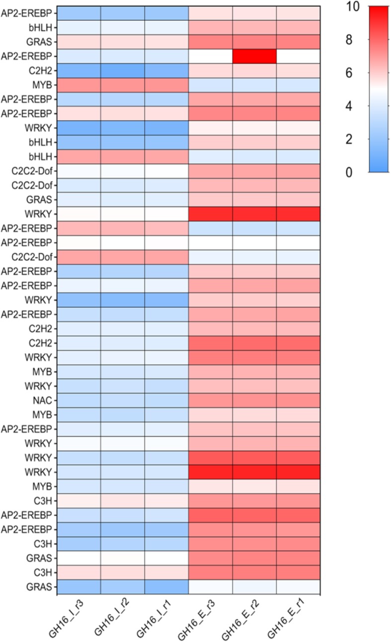
Heat map representing the levels of highly expressed transcription factors. The color scale on the right represents log-transformed FPKM value. Every row shows a different TF gene. Red, white, and blue indicate slow, medium and high levels of mRNA expression, respectively
Detection of known and novel miRNAs expressed in tuber initiation and expansion stages
The investigation of both known miRNA and novel putative miRNAs was performed by miRDeep2 program. This program combined the position and frequency of small RNAs with the secondary structure of miRNA precursor to provide novel miRNA that can accurately be in the tubers. To compare miRNA expression in six libraries, the number of clean reads was used as background for normalization, and transcripts per million reads (TPM) was used to present the expression levels of miRNAs. Data analysis showed that there were 22 known miRNAs (21 and 20 in tuber initiation and expansion stage, respectively) and 50 novel miRNAs in yam tuber (Additional file 5: Table S4) and 68, 66, 70 total miRNAs were detected in tuber initiation stage (GH16_I), and 69, 69, 72 total miRNAs were detected in tuber expansion stage (GH16_E), respectively (Table 2). Distribution of normalized miRNAs expression showed that approximately 75–81% of the total detected miRNA expression exceeded 10 TPM in six libraries (Additional file 5: Table S4).
Further analysis revealed that 22 known miRNAs belonging to 10 miRNA families, miRNA168, miRNA396, and novel miRNA160 were the most extensively represented families. All miRNAs were analyzed to detect differential miRNA expressions (DEMs). The results showed that miRNAs expression was dynamically regulated during expansion stages. A total of 44 differentially expressed miRNAs were identified including 11 known and 33 novel miRNAs; it showed that 40 miRNAs were down-regulated, and 4 miRNAs were up-regulated during expansion stage (Table 5). Interestingly, miR160 family (miRNA160, miRNA160a-5p, miRNA160b-1, miRNA160h-1) and miR535a_1 were down-regulated during expansion stage, miRNA396b and miRNA168 were up-regulated compared to other miRNA families (Table 5).
Table 5.
Differentially expressed miRNAs involved in expansion stage
| miRNA id | Read count (GH16_I) | Read count (GH16_E) | Expression (GH16_I) | Expression (GH16_E) | log2Ratio (GH16_E/GH16_I) |
P value |
|---|---|---|---|---|---|---|
| novel_mir43 | 54,974 | 33,041 | 3682.20 | 1731.72 | − 1.97 | 0.00000 |
| novel_mir35 | 52,437 | 32,873 | 3515.56 | 1728.51 | −1.91 | 0.00000 |
| miR160a-5p | 18,032 | 121 | 1171.81 | 6.42 | −8.45 | 0.00000 |
| miR168a-5p | 55,653 | 59,949 | 3708.90 | 3153.82 | −1.13 | 0.00000 |
| miR168 | 20,208 | 106,873 | 1334.10 | 5618.38 | 1.17 | 0.00000 |
| novel_mir1 | 16,275 | 7760 | 1085.59 | 407.58 | −2.30 | 0.00000 |
| miR396b | 1097 | 21,915 | 72.84 | 1176.08 | 3.09 | 0.00000 |
| miR535a_1 | 24,978 | 25,221 | 1654.31 | 1315.07 | −1.22 | 0.00000 |
| miR396a-5p | 12,295 | 7317 | 816.34 | 374.44 | −1.98 | 0.00000 |
| novel_mir33 | 6408 | 4269 | 425.49 | 224.55 | −1.82 | 0.00000 |
| novel_mir15 | 9666 | 10,433 | 642.81 | 549.12 | −1.12 | 0.00000 |
| novel_mir11 | 4852 | 3769 | 323.07 | 199.13 | −1.60 | 0.00000 |
| novel_mir25 | 5833 | 6318 | 386.43 | 333.34 | −1.12 | 0.00000 |
| novel_mir32 | 3196 | 2381 | 212.72 | 125.72 | −1.66 | 0.00000 |
| novel_mir5 | 2039 | 1094 | 135.31 | 58.03 | −2.13 | 0.00000 |
| novel_mir50 | 2777 | 2281 | 183.85 | 120.27 | −1.52 | 0.00000 |
| novel_mir10 | 3197 | 3009 | 213.02 | 158.49 | −1.32 | 0.00000 |
| novel_mir44 | 3381 | 3299 | 224.65 | 173.72 | −1.27 | 0.00000 |
| novel_mir36 | 1859 | 1246 | 124.13 | 65.29 | −1.81 | 0.00000 |
| novel_mir31 | 1632 | 1049 | 108.56 | 55.06 | −1.87 | 0.00000 |
| novel_mir18 | 1701 | 1305 | 114.05 | 68.55 | −1.62 | 0.00000 |
| novel_mir19 | 1491 | 1071 | 99.15 | 56.35 | −1.71 | 0.00000 |
| novel_mir12 | 1666 | 1308 | 111.28 | 69.05 | −1.58 | 0.00000 |
| novel_mir45 | 1478 | 1093 | 99.12 | 57.63 | −1.67 | 0.00000 |
| miR160 | 926 | 451 | 62.61 | 24.59 | −2.27 | 0.00000 |
| novel_mir48 | 1107 | 690 | 74.52 | 36.21 | −1.92 | 0.00000 |
| novel_mir26 | 1488 | 1201 | 99.79 | 63.01 | −1.54 | 0.00000 |
| miR160b_1 | 463 | 30 | 30.27 | 1.62 | −5.18 | 0.00000 |
| novel_mir46 | 1268 | 926 | 83.82 | 48.75 | −1.69 | 0.00000 |
| novel_mir42 | 694 | 275 | 46.22 | 14.50 | −2.57 | 0.00000 |
| novel_mir49 | 676 | 265 | 45.07 | 13.82 | −2.59 | 0.00000 |
| novel_mir24 | 1045 | 779 | 69.79 | 40.94 | −1.66 | 0.00000 |
| novel_mir23 | 377 | 109 | 25.16 | 5.68 | −3.03 | 0.00000 |
| miR168a-3p | 1330 | 1489 | 87.83 | 78.66 | −1.07 | 0.00000 |
| novel_mir20 | 374 | 2206 | 24.76 | 116.77 | 1.33 | 0.00000 |
| novel_mir30 | 964 | 999 | 63.39 | 52.50 | −1.18 | 0.00000 |
| novel_mir37 | 173 | 45 | 11.52 | 2.37 | −3.18 | 0.00000 |
| novel_mir47 | 90 | 10 | 6.03 | 0.50 | −4.40 | 0.00000 |
| novel_mir4 | 394 | 447 | 25.80 | 23.70 | −1.05 | 0.00000 |
| novel_mir34 | 10 | 200 | 0.68 | 10.70 | 3.09 | 0.00000 |
| novel_mir8 | 197 | 207 | 13.12 | 10.72 | −1.16 | 0.00000 |
| miR168_1 | 30 | 5 | 1.98 | 0.26 | −3.82 | 0.00000 |
| novel_mir9 | 123 | 142 | 8.24 | 7.48 | −1.03 | 0.00000 |
| miR160h_1 | 7 | 0 | 0.46 | 0.00 | −5.04 | 0.00041 |
Identification of target genes of differentially expressed miRNA in tuber expansion compared with initiation stage
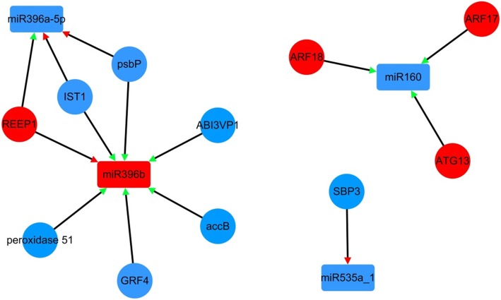
The miRNA regulates target mRNA through translational repression or mRNA degradation. To identify the correlation between the expression of DEMs and DEGs, a total of 11 DEMs were putatively targeted to 34 DEGs (Additional file 6: Table S5). Notably, 4 known miRNAs and 11genes formed 14 miRNA-target mRNA pairs with co-expressed expression in expansion stage (Fig. 6). Furthermore, based on GO and KEGG analysis of the targets, it was showed that differentially expressed miRNAs were involved in multiple pathways. Some of target genes of miRNA were annotated as transcription factors, such as two auxin response factor ARFs (ARF17 and ARF18, targeted by miRNA160), growth-regulating factor 4 (GRF, targeted by miRNA396b), B3 domain-containing protein (ABI3VP1, targeted by miRNA396b), and squamosa promoter-binding-like protein 3 (SBP3, targeted by miR535a_1).
Fig. 6.
Regulatory network from the integrated analysis of miRNA-mRNA data. Co-expressed miRNA-mRNA interactions visualized as a network using Cytoscape. Triangle represents the expression type of miRNA (red triangle represents positive correlation, green triangle represents negative correlation in each miRNA-mRNA), red represents up-regulation and blue represents down-regulation in network, the circle represents mRNA, the rectangle represents miRNA
On the other hand, some target genes were revealed to be involved in transport and catabolism processes, such as autophagy-related protein 13b (ATG13, targeted by miRNA160), IST1-like protein (IST1, targeted by miR396a-5p and miR396b). Finally, some genes were annotated as other functional proteins, such as receptor expression-enhancing protein1 (REEP1, targeted by miR396a-5p and miR396b), photosystem II oxygen-evolving enhancer protein 2 (psbP, targeted by miR396a-5p and miR396b), and acetyl-CoA carboxylase biotin carboxyl carrier protein (accB, targeted by miR396b).
Validation of the DEGs and DEMs data using RT-qPCR
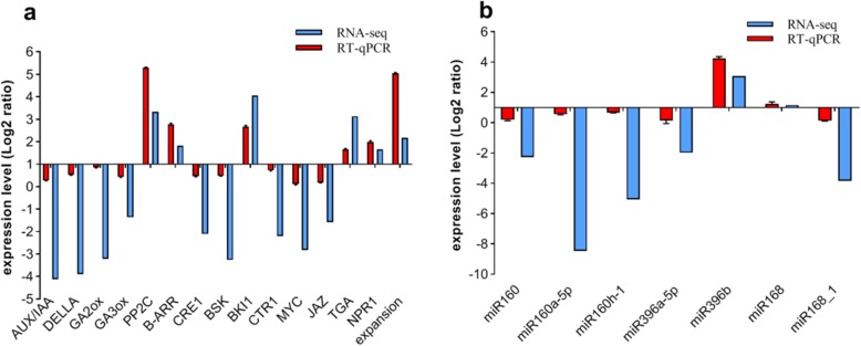
To identify the accuracy and reliability of RNA-Seq and miRNA data, RT-qPCR was used to measure the expressions of some DEGs and DEMs, including 15 mRNAs, and 7 miRNAs, specific primers were designed (Additional files 7 and 8: Tables S6 and S7). All mRNA and miRNA expressions have confirmed the accuracy of RNA-Seq and small RNA data. Overall, these results showed that 15 mRNAs showed similar expression patterns compared to DEGs analysis (Fig. 7a), and 7 miRNAs also showed very similar patterns compared to DEMs analysis (Fig. 7b). These results indicated that the RNA-Seq and small RNA data were reliable.
Fig. 7.
Verification of (a) DEGs and (b) miRNAs by qRT-PCR
Discussion
In tuber plants, tuber development is based on successive gene expression. Recently, the gene expression analysis of tuber development has been studied by transcriptome analysis, such as Solanum tuberosum, Miscanthus lutarioriparius, Raphanus sativus, Nelumbo nucifera and Aconitum heterophyllum [28, 34–37]. Yam is one of the most commercial stem tuber crops. Yam tuber expansion stage is an attractive theoretical model for studying the development of underground organs because it is the primary storage organ as nutrients reservoir. Therefore, the understanding of candidate genes involved in tuber expansion stage is supreme importance. In this study, 14,238 genes were identified that have shown significant differential expression during expansion stage.
Multiple signaling pathways regulation
KEGG enrichment analysis revealed that a large number of genes were involved in pathways related to MAPK signaling pathway, plant hormone signal transduction, starch and sucrose metabolism, and carbon metabolism (Fig. 3d). These genes are involved in cell wall biosynthesis, cell proliferation, cell expansion, nutrient accumulation, primary metabolism, and hormone signal transduction [34, 38, 39]. Cellular processes are triggered by specific stimuli and hormones that are involved in a series of signaling pathways. Calcium transduction pathway is a primary essential signaling transduction category required for normal growth and development of plants. As a second messenger, it plays an important role in many fundamental cellular processes such as cytoplasmic streaming, thigmotropism, gravitropism, cell division, cell differentiation, photomorphogenesis, plant defense, and various stress responses [40–42]. A potato Ca2+-dependent protein kinase, StCDPK1, which was reported to be expressed during tuberization, possibly be involved in potato tuberization through transcriptional activation of certain genes [40, 43]. In addition, StCDPK1 was affected by JA, ABA, GA treatment [43–45], suggested that CDPK possibly be a mediator of hormone-to-tuber stimulation.
Moreover, majority of studies have demonstrated that mRNA abundance of CBL, CBP, and GaM were affected by cell division during swelling of potato tubers [40, 43, 46]. In this study, expression of calcium signaling-related genes, including CDPK and CBPs, increased during tuber expansion stage. Interesting, CBLs were down-regulated (Additional file 3: Table S2). The role of CBL in root development is characterized by a robust gene expression during cell proliferation in the root, and its expression in root is regulated by auxin and cytokinin [47, 48]. These results demonstrated that calcium signal is regulated during tuber expansion stage. Besides, MAPK signal pathways are known to play a central role in cell proliferation, differentiation and hormones, but it is not known to be involved in tuber or root formation [49]. In this study, MAPK signal-related genes were up-regulated during expansion stage, and it means that MAPK signal pathways take part in yam tuber expansion. Cell number and cell size are two key determinants of plant and organ size, while involved in cell division, cell expansion and cell cycle [50, 51]. In this study, XTH, expansin, extension, CKS, ftsHs, CDC5, CDC, CDKs, and CDKIs involved in cell division, cell expansion and cell cycles were differentially regulated during expansion stage. These differentially regulated genes involved in cell wall and cell cycle metabolism were identified in Arabidopsis, Rehmannia glutinosa, and radish [35, 36, 52].
Transcription factors regulation
Transcription factors have been identified to play an essential role in the regulation of plant growth, development, and secondary metabolism. Most transcription factors have been identified to play critical roles in organogenesis, including MADS, bHLH, and GRAS. In sweet potato, MADS were preferentially expressed in root at initiation of tuberization, and in the vascular cambium region where the active cell proliferation regulated by jasmonic acid, cytokinins and stress response [53, 54]. In Arabidopsis, bHLH family, such as PIF3, PIF4, and PIF5 are key regulators for cell elongation during seedling development, antagonistically regulated by light and gibberellins [55]. GRAS transcription factor family specifically regulated the initiation of axillary meristem in Arabidopsis, such as SCARECROW (SCR) is expressed in cortex/endodermal initial cells in the root system and played a crucial role in regulating the radial organization of the root [56]. DELLA, such as RGA and GAI, controls cell expansion and cell division in hypocotyl, shoot, root, and floral induction [57]. In this study, 12 MADS, 52 bHLH, and 17 GRAS were found to be expressed differently during expansion stage at transcript level, which plays key regulatory functions in cell differentiation, division and expansion (Fig. 4). Zinc finger CCCH domain-containing protein 14 (C3H) has shown to be involved in growth and development, hormone response, and response to biotic and biotic stresses [58]. All data suggest that these transcription factors may be potentially involved in the expansion stage.
Hormonal signaling regulation
A set of transcriptome evidence suggests that hormone signal pathway-related genes involved in regulating of storage organ formation have been identified in sweet potato, potato, radish, lotus, and carrot, while may be potentially involved in cell division, differentiation, and expansion [1, 35, 37, 59, 60]. In the KEGG pathway annotation, hormone signal pathway was the most enriching one that was involved in eight signal pathways (Fig. 3d). In radish, most Aux/IAA, ARFs, and SAUR genes were abundantly expressed in the root cortex splitting and expansion stage, implying that these transcripts may be involved in cambium cell expansion [35]. Among auxin signal-related genes, Aux/IAA, ARFs were highly expressed in expansion stage in this study, and auxin has been identified to regulate cell division and expansion by altering genes expression [1, 38], suggesting that Aux/IAA, ARFs may offer excellent candidates during expansion stage.
Interestingly, in this study, DELLA genes were up-regulated, while GIDI, like GID2, were down-regulated during expansion stage (Additional file 3: Table S2). DELLA proteins are nuclear transcriptional regulators that repress GA signaling and may restrict plant growth presumably by causing transcriptional reprogramming. Binding of GA to GID1 enhances the interaction between GID1 and DELLA, resulting in rapid degradation of DELLAs via the ubiquitin-proteasome pathway. The GA-GID1-DELLA signal control hypocotyl root elongation by reducing GA levels and DELLA interacts directly with PIF4, PIF3, and SCL to mediate crosstalk between GA and light signals [55, 61]. DELLA is significantly expressed in GA-mediated rhizomes, suggesting that DELLAs can regulate rhizomatic expansion [37]. Besides, GA, IAA, and ethylene affect cell growth in roots via DELLA proteins [57]. Overall, these results indicate that hormone signaling-related genes have a complex regulatory network involved in tuber expansion.
Starch and sucrose metabolism regulation
Starch and sucrose are considered to be one of the significant carbohydrates source, in which expansion tubers are highly coordinated with starch and sucrose metabolism genes in potato, radish, and lotus identified by transcriptome analysis [22, 35, 37]. In this study, alpha-amylase, beta-amylase, and isoamylase were up-regulated (Additional file 2: Table S1), which is similar to increase in beta-amylase activity in swollen taproot in radish [35], where starch content decreased during the tuber expansion stage [15], and some sucrose metabolism genes were detected during expansion stage, including SuSy, SPS, INV, and invertase inhibitor (Additional file 2: Table S1). Evidence shows that sucrose can be converted to starch in storage root by SuSy and AGPase, which means that they play a vital role in the early stage of radish expansion [35]. The SuSy gene was down-regulated during expansion stage. The results were similar to the taproot expansion stage in radish [35], implying that they may play a significant role in tuber expansion stage of yam. Yam tuber morphology is significantly correlated with sucrose and starch [15]. Therefore, these starch and sucrose metabolism genes are necessary for tuber expansion.
miRNAs regulation by targeting the potential genes
miRNA mediated gene regulation has been extensively studied in root and tuber development through transcriptional and post-transcriptional levels, which provides a better understanding of molecular regulatory network during tuber expansion stage. In this study, most miRNAs were down-regulated during expansion stage (Table 5). In maize leaves, miR160 was significantly up-regulated in meristem relative to the elongation and mature zones [62]. However, miR160 was down-regulated during yam tuber expansion stage (Table 5). In general, miR160 expression is different in different plants. In this study, some miRNA-mRNA pairs were observed during expansion stage (Fig. 6), miR160 was involved in root cell division and differentiation by regulating auxin response factors (ARFs) affecting root development in Arabidopsis thaliana [30]. miRNA160 was down-regulated during expansion stage, auxin response factor ARF18 and ARF17 were up-regulated during expansion stage (Fig. 6), whereas, overexpression of miR160 in transgenic rice not only down-regulated the expressions of ARF10, ARF16, and ARF17, but also inhibited root-cap cells differentiation, lost control of cell division and led to ectopic expansion of the apical stem cell populations [63]. Previous studies have shown that the miRNA160-targeted ARF has been identified to be involved in cell expansion and cell differentiation in radish root and potato tuber [28]. In addition, miRNA396 plays an essential role in root growth and inhibits leaf cell division by UV-B radiation [64, 65]. miR535 is expressed in fruit development [66]. miRNA160, miRNA396, and miR535 may be involved in complex a network of regulating cell division and differentiation during expansion stage.
Regulatory networks associated with tuber expansion
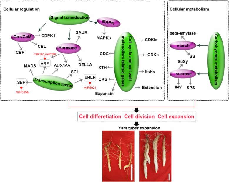
Tuber and root development and environmental responses involve gene regulatory networks [21, 24, 35, 37]. miRNA and target genes have been extensively studied in tuber development [21, 28], which significantly advanced our understanding of molecular regulatory networks underlying during yam tuber expansion. In this study, based on our RNA-Seq and small RNA analysis, previous transcriptomics analysis and results of other tuber crops, a putative model of regulatory network associated with yam tuber expansion was proposed (Fig. 8). During vegetative growth, environmental factors are the first signal to stimulate tuber growth, and cells development through some signal transduction pathways (hormone, calcium and, MAPK signaling) and metabolism processes (cell wall, starch and sucrose metabolism). Some specific genes promote cell differentiation division and expansion at tuber expansion. In detail, it was found that CBL, CBP, and GaM may affect cell division and proliferation during potato tubers expansion [40, 43, 46]. Moreover, MAPK signal pathway is known to play a central role in cell proliferation, differentiation and hormone signaling [50, 51]. XTH, expansin, extension, CKS, ftsHs, CDC5, CDC, CDKs, and CDKIs involved in cell division, cell expansion and cell cycles were differentially regulated during expansion stage. A finding similar to these differences in cell wall and cell cycle regulatory genes are involved in cell wall metabolism in Arabidopsis, Rehmannia glutinosa, radish [35, 36, 52]. DELLA controls cell expansion and cell division in hypocotyl, shoot, root, and floral induction [57]. DELLAs were significantly expressed in GA-mediated rhizome development process, suggesting that these DELLAs can regulate rhizomatic expansion [37]. In sweet potato, MADS were preferentially expressed in root at the initiation of the tuber, and in the vascular cambium region where the active cell proliferation was regulated by jasmonic acid, cytokinins, and stress response [53, 54]. Most Aux/IAA, ARFs and SAUR genes were enriched and expressed in the root cortex during splitting and expanding stage in radish, suggesting that these transcripts may be involved in cell expansion in the cambium [35]. SuSy, SPS, INV, SS, and beta-amylase, were highly correlated with tuber expansion, such as potato, radish and lotus [22, 35, 37]. In this study, some miRNA and target genes were observed during yam tuber expansion stage, bHLH (target by miR5021), SBP (target by miR535a), and ARFs (targeted by miRNA160 and miR396), were identified to be involved in cell expansion and cell differentiation in radish root and potato tuber [28, 35]. Taken together, the result suggested that these DEGs may play a vital role in the regulatory network of tuber expansion.
Fig.8.
A proposed model of genetic and molecular interactions in the regulatory network during tuber expansion in yam. Arrows represents genes were involved in signal transduction pathways or carbohydrate metabolism, red circle represents target genes of miRNA
Conclusion
A total of 14,238 genes expressed differentially during yam tuber expansion stage were identified first time using RNA-Seq technology. These results reveal the coordination of tuber involved in the processes of cellular events, metabolism, biosynthesis, and signal transduction pathways at transcriptional levels. It is worth noticing that the integrated analysis of miRNA-mRNA was identified to be differentially expressed in tuber expansion. The mRNA and miRNA datasets presented here identify a subset of candidate genes and miRNAs putatively associated with tuber expansion in yam, and therefore propose a hypothetical model of the genetic regulatory network associated with tuber expansion in yam, which can provide the basis for molecular regulatory mechanism to study the expansion development in Dioscorea opposita species.
Methods
Plant material
Dioscorea opposita cultivar Guihuai 16 was planted in April at the farm of Guangxi University in Nanning, Guangxi Province, China (22°53′06.7〞N 108°21′36.6〞E). Its healthy tubers germination, planting patterns and tuber growth stages were consistent as described by Gong [2] in 2016. All tuber growth stages were proved to have stable agronomic characteristics on three years of field observation. Tuber samples were collected at tuber initiation stage after field planting approximately forty days (initiation stage); additional samples were collected at sixty days (expansion stage). Anatomical characteristics of Guihuai 16 tuber at different developmental stages, including initiation and expansion stages were studied by Luo [67], and its histological characteristics consist of epidermis, parenchyma, and scattered vascular bundle. Five plants were selected randomly from every repetition each time. Then, the distal ends (5 mm long) of five fresh tubers from a repetition were washed with distilled water, cut down into pieces, and mixed as a biological repetition. All samples were immediately frozen in liquid nitrogen, and stored at − 80 °C until further use for the construction of various libraries. Two tuber developmental stages, including initiation and expansion stage, were used for the construction of various libraries. Three types of libraries were constructed, including one transcriptome de novo analysis libraries for mixed sample of initiation and expansion stage (named Total_1), six RNA-Seq analysis libraries (initiation stage named GH16_I, expansion stage named GH16_E), and small RNA analysis (initiation stage named C, and expansion stage named T in original data, in order to be consistent with the RNA-Seq data analyze, we renamed GH16_I, GH16_E for initiation and expansion stage, respectively). Each sample had three biological replicates with two technical replicates for each biological replicate.
Construction and sequencing of transcriptome de novo, RNA-Seq and small RNA libraries
Information about the sample pool can be found on the subsections of plant materials above. Total RNA was isolated from distal ends of tuber samples using MiniBEST reagent (TaKaRa, Dalian, China), and RNA integrity was assessed by an Agilent 2100 BioAnalyzer. Construction of the transcriptome de novo and RNA-Seq libraries were performed at the Beijing Genome Institute (BGI; Shenzhen, China). Briefly, mRNA was purified from total RNA using poly-T oligo-attached magnetic beads, fragmented, and reverse transcribed into cDNA, and the full-length cDNA was used T4 DNA ligase (Invitrogen, USA), and products were enriched by PCR to generate the cDNA library. Finally, the cDNA library was examined using an Agilent 2100 Bioanalyzer before sequencing on BGISEQ-500 system platform at BGI-Shenzhen. Paired-end reads were generated with a length of 90 bp for each cDNA libraries.
For the small RNA libraries, RNA bands of around 18-30 nt (small RNAs) in length were isolated. Then small RNAs were ligated to a 5′-adaptor and a 3′-adaptor, and small RNAs were transcribed into cDNA. 14 rounds of PCR amplification were performed to enrich the cDNA fragments. After target fragments (100~120 bp) were purified, it was used for cluster generation and sequenced using BGISEQ-500 system platform at BGI-Shenzhen. BGISEQ-500 is a next-generation sequencing (NGS) technology platform developed by the Beijing Genomics Institute (BGI) in 2016 [68]. It is based on combinatorial Probe-Anchor Synthesis and improved DNA Nanoballs technology to generate short reads at a large scale. This platform has already been used for genome sequencing, RNA-Seq, and small RNA analysis.
Raw reads from mRNAs were subjected to remove the reads with adaptors or containing more than 5% unknown (‘N’) bases. Then, the low-quality reads (defined as reads having more than 20% of bases with quality≤15) were trimmed. The filtered clean reads were saved in FASTQ file, which has been submitted to Sequence Read Archive (SRA) of NCBI. Next, the Trinity software [69] was used to perform de novo assembly of the high-quality cleaned reads, and Tgicl software [70] was assembled to remove the redundant Trinity generated contigs. Final assembled datasets were performed to annotate genes function and expression.
Small RNA reads were also screened from raw sequencing reads by removing adaptors, poly-A sequences, and low-quality bases. Sequences shorter than 18 nt or longer than 32 nt were removed after trimming. Final filtered clean reads were saved in the FASTQ file, which was used for sequential analysis.
Processing and mapping of mRNA-Seq and small RNA libraries sequencing data
After stringent quality filering, a mixed sample of initiation and expansion stage library and six RNA-Seq libraries clean reads were used to map the two reference genome sequences (D. rotundata and D. alata, bioproject accession numbers were PRJDB3383 and PRJEB10904, respectively). Among which, 40.91%~ 43.41% of reads were mapped to the D. rotundata genome, and 57.58%~ 61.16% to the D. alata genome (Additional file 9: Table S8). The two reference genomes were not plausible used for genes function and expression analysis. Hence functional databases NT, NR, GO, KOG, KEGG, SwissProt, and InterPro were used to annotate genes function, while Blastn, Blastx, Diamond, Blast2GO, and InterProScan5 were used to align genes. Six RNA-Seq reads were mapped to the transcriptome de novo analysis data (named Total_1) using bowtie2 results with default parameters after pre-processing mRNA-Seq data [71]. Gene expression levels were presented as FPKM (fragments per kilobase of exon per million fragments mapped) values [72], and the six small RNA clean reads were submitted to the sRNA databases by using Bowtie, Cmsearch, and Rfam software [73]. The clean reads aligned to the sRNA databases with more than 90% matched length and a maximum alignment score of 90 were used from further analysis.
Identification of known and novel miRNAs, and predicting the targets of miRNAs
To identify the isoforms of known miRNA, cleaned sRNA reads were performed using the miRProf tool, and conserved miRNAs were identified using known plant miRNAs registered in miRBase [74]. To identify novel miRNAs and their precursors, unique reads were submitted to the RIPmiR [75]. Potential miRNA targets were predicted using the psRoot and Target Finder [76].
Identification and functional annotation of differentially expressed genes and miRNAs
The expression levels of mRNA genes were measured as FPKM, and genes with expression levels > 5 FPKM were retained for statistical analysis. miRNA read counts were normalized to reads per million (TRM). Differentially expressed genes (DEGs) and miRNAs (DEMs) were identified by DEGseq [77]. Therefore, DEGs and DEMs were identified by stringent thresholds (|the FDR < 0.01, |log2 (fold change)| ≥ 1 and P-value < 0.005). All DEGs were subjected to gene ontology and KEGG pathway analysis. In addition, enrichment GO and KEGG pathway analyses were implemented using p ≤ 0.05 and q-value < 0.9 as the threshold. Enrichment analysis of DEGs was analyzed using GO and KEGG databases to obtain a detailed description of the DEGs. Each DEG in tuber was predicted by aligning the gene sequences against the Plant Transcriptional Factor Database. The DEGs were classified according to their TF families. A miRNA-target gene regulatory network was constructed using Cytoscape_v3. 2. 1 program.
Validation of the DEGs and DEMs data using RT-qPCR
To identify the accuracy and reliability of mRNA and miRNA data, RT-qPCR was used to measure the expressions of DEGs and DEMs. Total RNA used for RNA-Seq and small RNA analysis previously was reversely transcribed into cDNA with PrimeScript™ RT reagent kit and Mir-X™ miRNA first-strand synthesis kit (Takara, Dalian, China), separately, according to the manufacture methods. RT-qPCR was performed using primers (Additional files 7 and 8: Tables S6 and S7) on real-time PCR detection system (BIO-RAD). Quantification expression for DEGs were detected by using the TB Green™ Premix Ex Taq™ II (Tli RNaseH Plus) (Takara, Dalian, China), 10 μl reaction solution containing 1x Green TB Premix Ex Taq II, 10 μM primer.
One-third dilution of the cDNA sample was used, and the reaction conditions were: 30s at 95 °C followed by 40 cycles of 30s at 95 °C and 30s at 60 °C. Quantification expression for DEMs were detected by the Mir-X miRNA qRT-PCR SYBR kit using 25 μl reaction solution, containing 2 × SYBR advantage premix, 50 × ROX dye, 10 μM miRNA-specific Primer, 2 μl cDNA samples were used and the reaction conditions were: 10s at 95 °C followed by 40 cycles of 5 s at 95 °C and 20s at 60 °C. All mRNAs and miRNA expressions had three biological replicates with three technical replicates for each of biological replicate, ACTIN was used as reference gene, the relative expression levels were collected by the comparative Ct protocol.
Supplementary information
Additional file 1: Figure S1. Length distribution of small RNA sequences in small RNA libraries.
Additional file 2: Table S1. Differentially expressed genes (DEGs) in expansion stage compared with initiation stage.
Additional file 3: Table S2. The differentially expressed genes involved in signal transduction pathways in expansion stage (FPKM> 5).
Additional file 4: Table S3. The differentially expressed transcription factors involved in expansion stage.
Additional file 5: Table S4. Analysis of clean reads for small RNA sequencing in initiation and expansion stages.
Additional file 6: Table S5. Identification of miRNAs target genes in expansion stage.
Additional file 7: Table S6. The primers of 15 DEGs validated by RT-qPCR analysis.
Additional file 8: Table S7. The primers of 7 DEMs validated by RT-qPCR analysis.
Additional file 9: Table S8. Summary of alignment statistics of RNA-Seq libraries mapped to reference genome.
Abbreviations
- B-ARR
myb family transcription factor
- BKI1
BRI1 kinase inhibitor 1
- BSK
BR-signaling kinase
- CRE1
Farnesyl diphosphate synthase
- CTR1
Serine/threonine-protein kinase CTR1
- DEG
Differentially expressed gene
- DEM
Differentially expressed microRNA
- GA2ox
Gibberellin 2-beta-dioxygenase
- GA3ox
Gibberellin 3-beta-dioxygenase
- GO
Gene ontology
- JAZ
Jasmonate ZIM domain-containing protein
- KEGG
Kyoto encyclopedia of genes and genomes
- miRNA
MicroRNA
- MYC
Transcription factor MYC
- NPR1
Regulatory protein NPR1
- qRT-PCR
Real-time quantitative RT-PCR
- RNA-Seq
RNA sequencing
- SnRK2
Serine/threonine-protein kinase
- PP2C
Protein phosphatase 2C
- TF
Transcription factor
- TGA
transcription factor TGA
Authors’ contributions
DX and LFH have designed experiments. YYZ, SZL, and Saba have analyzed the sequencing data for transcriptome assembly. JZ and AQW have developed qPCR experiments. YYZ and LFH have written the manuscript.
Funding
This work was supported by the National Natural Science Foundation of China (Grant No. 81860670), the Guangxi Innovation Team Project of Tubers of Modern Agricultural Industrial Technology System of China (nycytxgxcxtd-11-01), and the Guangxi Natural Science Foundation of China (Grant No. 2018GXNSFDA281029).
Availability of data and materials
The materials of this study were provided by the College of Agriculture at Guangxi University. Correspondence and requests for materials should be addressed to Longfei He (lfhe@gxu.edu.cn). The raw sequencing data have submitted to the NCBI SRA database (PRJNA533985). The assembled genes data also were submitted to GenBank TSA database (GHUN00000000).
Ethics approval and consent to participate
Not applicable.
Consent for publication
Not applicable.
Competing interests
We declare that the research was conducted in the absence of any commercial or financial relationships that could be a potential conflict of interest.
Footnotes
Publisher’s Note
Springer Nature remains neutral with regard to jurisdictional claims in published maps and institutional affiliations.
Contributor Information
Aiqin Wang, Email: waiqing1966@126.com.
Longfei He, Email: lfhe@gxu.edu.cn.
Supplementary information
Supplementary information accompanies this paper at 10.1186/s12864-020-6492-5.
References
- 1.Aksenova NP, Konstantinova TN, Golyanovskaya SA, Sergeeva LI, Romanov GA. Hormonal regulation of tuber formation in potato plants. Russ J Plant Physl. 2012;59(4):451–466. doi: 10.1134/S1021443712040024. [DOI] [Google Scholar]
- 2.Gong MX, Luo HL, Wang AQ, Zhou YY, Huang WJ, Zhu PL, He LF. Phytohormone profiling during tuber development of Chinese yam by ultra-high performance liquid chromatography–triple quadrupole tandem mass spectrometry. J Plant Growth Regul. 2016;36(2):362–373. doi: 10.1007/s00344-016-9644-8. [DOI] [Google Scholar]
- 3.Matsumoto R, Kikuno H, Shiwachi H, Toyohara H, Takebayashi Y, Jikumaru Y, Kamiya Y. Growth of vine cuttings and fuctuations of concentrations of endogenous plant hormones in water yam (Dioscorea alata L.) Trop Agric Dev. 2013;57(1):23–30. [Google Scholar]
- 4.Yoshida Y, Takahashi H, Kanda H, Kanahamaz K. Effect of seed tuber weights on the development of tubers and flowering spikes in Japanese yams (Dioscorea japonica) grown under different photoperiods and with plant growth regulators. J Jpn Soc Hortic Sci. 2007;76(3):230–236. doi: 10.2503/jjshs.76.230. [DOI] [Google Scholar]
- 5.Chen SW, Shiwachi H, Sanada A, Toyohara H. Theobroxide and day-length effects on the growth of yam (Dioscorea spp.) J.ISSAAS. 2010;16(1):22–30. [Google Scholar]
- 6.Shiwachi H, Ayankanmi T, Asiedu R. Effect of day length on the development of tubers in yams (Dioscorea spp.) Trop Sci. 2002;42:162–170. [Google Scholar]
- 7.Ovono PO, Kevers C, Dommes J. Axillary proliferation and tuberisation of Dioscorea cayenensis-D-rotundata complex. Plant Cell Tiss Org. 2007;91(2):107–114. doi: 10.1007/s11240-007-9238-z. [DOI] [Google Scholar]
- 8.Chen FQ, Fu Y, Wang DL, Gao X, Wang L. The effect of plant growth regulators and sucrose on the micropropagation and microtuberization of Dioscorea nipponica Makino. J Plant Growth Regul. 2007;26(1):38–45. doi: 10.1007/s00344-005-0147-2. [DOI] [Google Scholar]
- 9.Gong MX, Luo HL, Yuan HJ, Wei SQ, Yang XD, He LF. Effects of exogenous gibberellin and paclobutrazol on tuber expansion and bulbil formation of Chinese yam. Acta Horticulturae Sinica. 2015;42(6):1175–1184. [Google Scholar]
- 10.Behera KK, Pani D, Sahoo S, Maharana T, Sethi BK. Effect of GA3 and urea treatments on improvement of microtuber production and productivity of different types of planting material in greater yam (Dioscorea alata L.) Not Bot Horti Agrobot. 2009;37(2):81–84. [Google Scholar]
- 11.Hamadina EI, Craufurd PQ, Battey NH, Asiedu R. In vitro micro-tuber initiation and dormancy in yam. Ann Appl Biol. 2010;157(2):203–212. doi: 10.1111/j.1744-7348.2010.00418.x. [DOI] [Google Scholar]
- 12.Bazabakana R, Wattiez R, Baucher M, Diallo B, Jaziri M. Effect of jasmonic acid on developmental morphology during in vitro tuberization of Dioscorea alata (L.) Plant Growth Regul. 2003;40(3):229–237. doi: 10.1023/A:1025087111148. [DOI] [Google Scholar]
- 13.Kim SK, Kim JT, Jang SW, Lee SC, Lee BH, Lee IJ. Exogenous effect of gibberellins and jasmonate on tuber enlargement of Dioscorea opposita. Agron Res. 2005;3(1):39–44. [Google Scholar]
- 14.Yan H, Yang L, Li Y. Axillary shoot proliferation and tuberization of Dioscorea fordii Prain et Burk. Plant Cell Tiss Org. 2011;104(2):193–198. doi: 10.1007/s11240-010-9818-1. [DOI] [Google Scholar]
- 15.Liang RF, Li CZ, Zhang J, He LF, Wei BH, Gan XQ, He HY. Changes of matter accumulation and relative enzymatic activity during yam tuber development. Acta Agron Sin. 2011;37(5):903–910. doi: 10.3724/SP.J.1006.2011.00903. [DOI] [Google Scholar]
- 16.Loko YL, Bhattacharjee R, Agre AP, Dossou-Aminon I, Orobiyi A, Djedatin GL, Dansi A. Genetic diversity and relationship of Guinea yam (Dioscorea cayenensis lam.–D.rotundata Poir.Complex) germplasm in Benin (West Africa) using microsatellite markers. Genet Resour Crop Ev. 2017;64(6):1205–1219. doi: 10.1007/s10722-016-0430-z. [DOI] [Google Scholar]
- 17.Girma G, Gedil M, Spillane C. Morphological, SSR and ploidy analysis of water yam (Dioscorea alata L.) accessions for utilization of aerial tubers as planting materials. Genet Resour Crop Ev. 2017;64(2):291–305. doi: 10.1007/s10722-015-0351-2. [DOI] [Google Scholar]
- 18.Tamiru M, Yamanaka S, Mitsuoka C, Babil P, Takagi H, Lopez-Montes A, Sartie A, Asiedu R, Terauchi R. Development of genomic simple sequence repeat markers for yam. Crop Sci. 2015;55(5):2191–2200. doi: 10.2135/cropsci2014.10.0725. [DOI] [Google Scholar]
- 19.Akakpo R, Scarcelli N, Chair H, Dansi A, Djedatin G, Thuillet AC, Rhone B, Francois O, Alix K, Vigouroux Y. Molecular basis of African yam domestication: analyses of selection point to root development, starch biosynthesis, and photosynthesis related genes. BMC Genomics. 2017;18(1):782. doi: 10.1186/s12864-017-4143-2. [DOI] [PMC free article] [PubMed] [Google Scholar]
- 20.Zhao Xiting, Zhang Xiaoli, Guo Xiaobo, Li Shujie, Han Linlin, Song Zhihui, Wang Yunying, Li Junhua, Li Mingjun. Identification and Validation of Reference Genes for qRT-PCR Studies of Gene Expression inDioscorea opposita. BioMed Research International. 2016;2016:1–13. doi: 10.1155/2016/3089584. [DOI] [PMC free article] [PubMed] [Google Scholar]
- 21.Kondhare KR, Malankar NN, Devani RS, Banerjee AK. Genome-wide transcriptome analysis reveals small RNA profiles involved in early stages of stolon-to-tuber transitions in potato under photoperiodic conditions. BMC Plant Biol. 2018;18(1):284. doi: 10.1186/s12870-018-1501-4. [DOI] [PMC free article] [PubMed] [Google Scholar]
- 22.Van Harsselaar JK, Lorenz J, Senning M, Sonnewald U, Sonnewald S. Genome-wide analysis of starch metabolism genes in potato (Solanum tuberosum L.) BMC Genomics. 2017;18(1):37. doi: 10.1186/s12864-016-3381-z. [DOI] [PMC free article] [PubMed] [Google Scholar]
- 23.Gong L, Zhang HX, Gan XY, Zhang L, Chen YC, Nie FJ, Shi L, Li M, Guo ZQ, Zhang GH, Song YX. Transcriptome profiling of the potato (Solanum tuberosum L.) plant under drought stress and water-stimulus conditions. Plos One. 2015;10(5):e0128041. doi: 10.1371/journal.pone.0128041. [DOI] [PMC free article] [PubMed] [Google Scholar]
- 24.Shan J, Song W, Zhou J, Wang X, Xie C, Gao X, Xie T, Liu J. Transcriptome analysis reveals novel genes potentially involved in photoperiodic tuberization in potato. Genomics. 2013;102(4):388–396. doi: 10.1016/j.ygeno.2013.07.001. [DOI] [PubMed] [Google Scholar]
- 25.Li J, Liang Q, Li CF, Liu MD, Zhang YS. Comparative transcriptome analysis identifies putative genes involved in dioscin biosynthesis in Dioscorea zingiberensis. Molecules. 2018;23(2):454. doi: 10.3390/molecules23020454. [DOI] [PMC free article] [PubMed] [Google Scholar]
- 26.Wu ZG, Jiang W, Mantri N, Bao XQ, Chen SL, Tao ZM. Transciptome analysis reveals flavonoid biosynthesis regulation and simple sequence repeats in yam (Dioscorea alata L.) tubers. BMC Genomics. 2015;16:346. doi: 10.1186/s12864-015-1547-8. [DOI] [PMC free article] [PubMed] [Google Scholar]
- 27.Liu C, Liu XX, Xu WL, Fu WM, Wang FD, Gao JW, Li QY, Zhang ZG, Li JJ, Wang SF. Identification of miRNAs and their targets in regulating tuberous root development in radish using small RNA and degradome analyses. 3 Biotech. 2018;8(7):311. doi: 10.1007/s13205-018-1330-z. [DOI] [PMC free article] [PubMed] [Google Scholar]
- 28.Lakhotia N, Joshi G, Bhardwaj AR, Katiyar-Agarwal S, Agarwal M, Jagannath A, Goel S, Kumar A. Identification and characterization of miRNAome in root, stem, leaf and tuber developmental stages of potato (Solanum tuberosum L.) by high-throughput sequencing. BMC Plant Biol. 2014;14:6. doi: 10.1186/1471-2229-14-6. [DOI] [PMC free article] [PubMed] [Google Scholar]
- 29.Sun YY, Qiu Y, Zhang XH, Chen XH, Shen D, Wang HP, Li XX. Genome-wide identification of microRNAs associated with taproot development in radish (Raphanus sativus L.) Gene. 2015;569(1):118–126. doi: 10.1016/j.gene.2015.05.044. [DOI] [PubMed] [Google Scholar]
- 30.Mallory AC, Bartel DP, Bartel B. MicroRNA-directed regulation of Arabidopsis AUXIN RESPONSE FACTOR17 is essential for proper development and modulates expression of early auxin response genes. Plant Cell. 2005;17(5):1360–1375. doi: 10.1105/tpc.105.031716. [DOI] [PMC free article] [PubMed] [Google Scholar]
- 31.Sarkar D. Photoperiodic inhibition of potato tuberization: an update. Plant Growth Regul. 2010;62(2):117–125. doi: 10.1007/s10725-010-9502-9. [DOI] [Google Scholar]
- 32.Martin A, Adam H, Diaz-Mendoza M, Zurczak M, Gonzalez-Schain ND, Suarez-Lopez P. Graft-transmissible induction of potato tuberization by the microRNA miR172. Development. 2009;136(17):2873–2881. doi: 10.1242/dev.031658. [DOI] [PubMed] [Google Scholar]
- 33.Tamiru M, Natsume S, Takagi H, White B, Yaegashi H, Shimizu M, Yoshida K, Uemura A, Oikawa K, Abe A. Genome sequencing of the staple food crop white Guinea yam enables the development of a molecular marker for sex determination. BMC Biol. 2017;15(1):86. doi: 10.1186/s12915-017-0419-x. [DOI] [PMC free article] [PubMed] [Google Scholar]
- 34.Hu RB, Yu CJ, Wang XY, Jia CL, Pei SQ, He K, He G, Kong YZ, Zhou GK. De novo transcriptome analysis of Miscanthus iutarioriparius identifies candidate genes in rhizome development. Front Plant Sci. 2017;8:492. doi: 10.3389/fpls.2017.00492. [DOI] [PMC free article] [PubMed] [Google Scholar]
- 35.Yu RG, Wang J, Xu L, Wang Y, Wang RH, Zhu XW, Sun XC, Luo XB, Xie Y, Everlyne M, et al. Transcriptome profiling of taproot reveals complex regulatory networks during taproot thickening in radish (Raphanus sativus L.) Front Plant Sci. 2016;7:1210. doi: 10.3389/fpls.2016.01210. [DOI] [PMC free article] [PubMed] [Google Scholar]
- 36.Malhotra N, Sood H, Chauhan RS. Transcriptome-wide mining suggests conglomerate of genes associated with tuberous root growth and development in Aconitum heterophyllum Wall. 3 Biotech. 2016;6(2):152. doi: 10.1007/s13205-016-0466-y. [DOI] [PMC free article] [PubMed] [Google Scholar]
- 37.Yang M, Zhu LP, Pan C, Xu LM, Liu YL, Ke WD, Yang PF. Transcriptomic analysis of the regulation of rhizome formation in temperate and tropical lotus (Nelumbo nucifera) Sci Rep. 2015;5:13059. doi: 10.1038/srep13059. [DOI] [PMC free article] [PubMed] [Google Scholar]
- 38.Ursache R, Nieminen K, Helariutta Y. Genetic and hormonal regulation of cambial development. Physiol Plant. 2013;147(1):36–45. doi: 10.1111/j.1399-3054.2012.01627.x. [DOI] [PubMed] [Google Scholar]
- 39.Bhalerao RP, Fischer U. Environmental and hormonal control of cambial stem cell dynamics. J Exp Bot. 2017;68(1):79–87. doi: 10.1093/jxb/erw466. [DOI] [PubMed] [Google Scholar]
- 40.Nookaraju A, Pandey SK, Upadhyaya CP, Heung JJ, Kim HS, Chun SC, Kim DH, Park SW. Role of Ca2+-mediated signaling in potato tuberization: an overview. Bot Stud. 2012;53(2):177–189. [Google Scholar]
- 41.Chen T, Wu X, Chen Y, Li X, Huang M, Zheng M, Baluska F, Samaj J, Lin J. Combined proteomic and cytological analysis of Ca2+-calmodulin regulation in Picea meyeri pollen tube growth. Plant Physiol. 2009;149(2):1111–1126. doi: 10.1104/pp.108.127514. [DOI] [PMC free article] [PubMed] [Google Scholar]
- 42.Feng JL, Yang ZJ, Chen SP, El-Kassaby YA, Chen H. Signaling pathway in development of Camellia oleifera nurse seedling grafting union. Trees-Struct Funct. 2017;31(5):1543–1558. doi: 10.1007/s00468-017-1568-9. [DOI] [Google Scholar]
- 43.Gargantini PR, Giammaria V, Grandellis C, Feingold SE, Maldonado S, Ulloa RM. Genomic and functional characterization of StCDPK1. Plant Mol Biol. 2009;70(1–2):153–172. doi: 10.1007/s11103-009-9462-5. [DOI] [PubMed] [Google Scholar]
- 44.Yu XC, Li MJ, Gao GF, Feng HZ, Geng XQ, Peng CC, Zhu SY, Wang XJ, Shen YY, Zhang DP. Abscisic acid stimulates a calcium-dependent protein kinase in grape berry. Plant Physiol. 2006;140(2):558–579. doi: 10.1104/pp.105.074971. [DOI] [PMC free article] [PubMed] [Google Scholar]
- 45.Ulloa RM, Raices M, MacIntosh GC, Maldonado S, Tellez-Inon MT. Jasmonic acid affects plant morphology and calcium-dependent protein kinase expression and activity in Solanum tuberosum. Physiol Plant. 2002;115(3):417–427. doi: 10.1034/j.1399-3054.2002.1150312.x. [DOI] [PubMed] [Google Scholar]
- 46.Kim MS, Kim HS, Kim YS, Baek KH, Oh HW, Hahn KW, Bae RN, Lee IJ, Joung H, Jeon JH. Superoxide anion regulates plant growth and tuber development of potato. Plant Cell Rep. 2007;26(10):1717–1725. doi: 10.1007/s00299-007-0380-1. [DOI] [PubMed] [Google Scholar]
- 47.Kumar Meena M, Kumar Vishwakarma N, Tripathi V, Chattopadhyay D. CBL-interacting protein kinase 25 contributes to root meristem development. J Exp Bot. 2019;70(1):133–147. doi: 10.1093/jxb/ery334. [DOI] [PMC free article] [PubMed] [Google Scholar]
- 48.Cui XY, Du YT, Fu JD, Yu TF, Wang CT, Chen M, Chen J, Ma YZ, Xu ZS. Wheat CBL-interacting protein kinase 23 positively regulates drought stress and ABA responses. BMC Plant Biol. 2018;18:93. doi: 10.1186/s12870-018-1306-5. [DOI] [PMC free article] [PubMed] [Google Scholar]
- 49.Hirt H. Connecting oxidative stress, auxin, and cell cycle regulation through a plant mitogen-activated protein kinase pathway. P Natl Acad Sci USA. 2000;97(6):2405–2407. doi: 10.1073/pnas.97.6.2405. [DOI] [PMC free article] [PubMed] [Google Scholar]
- 50.Polyn S, Willems A, De Veylder L. Cell cycle entry, maintenance, and exit during plant development. Curr Opin Plant Biol. 2015;23:1–7. doi: 10.1016/j.pbi.2014.09.012. [DOI] [PubMed] [Google Scholar]
- 51.Guo M, Simmons CR. Cell number counts-the fw2.2 and CNR genes and implications for controlling plant fruit and organ size. Plant Sci. 2011;181(1):1–7. doi: 10.1016/j.plantsci.2011.03.010. [DOI] [PubMed] [Google Scholar]
- 52.Paque S, Mouille G, Grandont L, Alabadi D, Gaertner C, Goyallon A, Muller P, Primard-Brisset C, Sormani R, Blazquez MA, Perrot-Rechenmann C. AUXIN BINDING PROTEIN1 links cell wall remodeling, auxin signaling, and cell expansion in Arabidopsis. Plant Cell. 2014;26(1):280–295. doi: 10.1105/tpc.113.120048. [DOI] [PMC free article] [PubMed] [Google Scholar]
- 53.Dong TT, Song WH, Tan CT, Zhou ZL, Yu JW, Han RP, Zhu MK, Li ZY. Molecular characterization of nine sweet potato (Ipomoea batatas lam.) MADS-box transcription factors during storage root development and following abiotic stress. Plant Breed. 2018;137(5):790–804. doi: 10.1111/pbr.12613. [DOI] [Google Scholar]
- 54.Song WH, Tan CT, Zhu MK, Xu T, Dong TK, Li ZY. Cloning and expression analysis of stress treatments of a MADS-box transcription factor in sweet potato. Bulletin of Botanical Research. 2017;37(4):587–595,627. [Google Scholar]
- 55.de Lucas M, Daviere JM, Rodriguez Falcon M, Pontin M, Iglesias Pedraz JM, Lorrain S, Fankhauser C, Blazquez MA, Titarenko E, Prat S. A molecular framework for light and gibberellin control of cell elongation. Nature. 2008;451(7177):480–484. doi: 10.1038/nature06520. [DOI] [PubMed] [Google Scholar]
- 56.Heo J, Chang KS, Kim IA, Lee M, Lee SA, Song S, Lee MM, Lim J. Funneling of gibberellin signaling by the GRAS transcription regulator SCARECROW-LIKE 3 in the Arabidopsis root. P Natl Acad Sci USA. 2011;108(5):2166–2171. doi: 10.1073/pnas.1012215108. [DOI] [PMC free article] [PubMed] [Google Scholar]
- 57.Daviere JM, Achard P. A pivotal role of DELLAs in regulating multiple hormone signals. Mol Plant. 2016;9(1):10–20. doi: 10.1016/j.molp.2015.09.011. [DOI] [PubMed] [Google Scholar]
- 58.Li WT, He M, Wang J, Wang YP. Zinc finger protein (ZFP) in plants-a review. Plant Omics. 2013;6(6):474–480. [Google Scholar]
- 59.Que F, Wang GL, Li T, Wang YH, Xu ZS, Xiong AS. Genome-wide identification, expansion, and evolution analysis of homeobox genes and their expression profiles during root development in carrot. Functional & Integrative Genomics. 2018;18(6):685–700. doi: 10.1007/s10142-018-0624-x. [DOI] [PubMed] [Google Scholar]
- 60.Ponniah SK, Thimmapuram J, Bhide K, Kalavacharla V, Manoharan M. Comparative analysis of the root transcriptomes of cultivated sweetpotato (Ipomoea batatas L. Lam) and its wild ancestor (Ipomoea trifida Kunth G. Don) BMC Plant Biology. 2017;17:9. doi: 10.1186/s12870-016-0950-x. [DOI] [PMC free article] [PubMed] [Google Scholar]
- 61.Zhang Z, Ogawa M, Fleet CM, Zentella R, Hu J, Heo JO, Lim J, Kamiya Y, Yamaguchi S, Sun T. SCARECROW-LIKE 3 promotes gibberellin signaling by antagonizing master growth repressor DELLA in Arabidopsis. P Natl Acad Sci USA. 2011;108(5):2160–2165. doi: 10.1073/pnas.1012232108. [DOI] [PMC free article] [PubMed] [Google Scholar]
- 62.Aydinoglu F, Lucas SJ. Identification and expression profiles of putative leaf growth related microRNAs in maize (Zea mays L.) hybrid ADA313. Gene. 2019;690:57–67. doi: 10.1016/j.gene.2018.12.042. [DOI] [PubMed] [Google Scholar]
- 63.Yang SN, Wang J, Chen ZX, Tian XJ, Zhang JW, Long Y, Pei XW, Yuan QH. Clone and function analysis miR160f in common wild rice (Oryza rufipogon Griff.) Biotechnol Bull. 2014;11:114–118. [Google Scholar]
- 64.Bao ML, Bian HW, Zha YL, Li FY, Sun YZ, Bai B, Chen ZH, Wang JH, Zhu MY, Han N. miR396a-mediated masic helix-loop-helix transcription factor bHLH74 repression acts as a regulator for root growth in Arabidopsis seedlings. Plant Cell Physiol. 2014;55(7):1343–1353. doi: 10.1093/pcp/pcu058. [DOI] [PubMed] [Google Scholar]
- 65.Casadevall R, Rodriguez RE, Debernardi JM, Palatnik JF, Casati P. Repression of growth regulating factors by the microRNA396 inhibits cell proliferation by UV-B radiation in Arabidopsis leaves. Plant Cell. 2013;25(9):3570–3583. doi: 10.1105/tpc.113.117473. [DOI] [PMC free article] [PubMed] [Google Scholar]
- 66.Shi M, Hu X, Wei Y, Hou X, Yuan X, Liu J, Liu Y. Genome-wide profiling of small RNAs and degradome revealed conserved regulations of miRNAs on auxin-responsive genes during fruit enlargement in peaches. Int J Mol Sci. 2017;18(12):2599. doi: 10.3390/ijms18122599. [DOI] [PMC free article] [PubMed] [Google Scholar]
- 67.Luo HL, Gong MX, Zhou YY, He LF. Analysis of starch accumulation and differentially expressed proteins during the development of Chinese yam tuber. Journal of South China Agricultural University. 2018;39(6):61–69. [Google Scholar]
- 68.Goodwin S, McPherson JD, McCombie WR. Coming of age: ten years of next-generation sequencing technologies. Nat Rev Genet. 2016;17(6):333–351. doi: 10.1038/nrg.2016.49. [DOI] [PMC free article] [PubMed] [Google Scholar]
- 69.Grabherr MG, Haas BJ, Yassour M, Levin JZ, Thompson DA, Amit I, Adiconis X, Fan L, Raychowdhury R, Zeng QD, et al. Full-length transcriptome assembly from RNA-Seq data without a reference genome. Nat Biotechnol. 2011;29(7):644. doi: 10.1038/nbt.1883. [DOI] [PMC free article] [PubMed] [Google Scholar]
- 70.Pertea G, Huang X, Liang F, Antonescu V, Sultana R, Karamycheva S, Lee Y, White J, Cheung F, Parvizi B, et al. TIGR gene indices clustering tools (TGICL): a software system for fast clustering of large EST datasets. Bioinformatics. 2003;19(5):651. doi: 10.1093/bioinformatics/btg034. [DOI] [PubMed] [Google Scholar]
- 71.Cock PJ, Fields CJ, Goto N, Heuer ML, Rice PM. The sanger FASTQ file format for sequences with quality scores, and the Solexa/Illumina FASTQ variants. Nucleic Acids Res. 2010;38(6):1767–1771. doi: 10.1093/nar/gkp1137. [DOI] [PMC free article] [PubMed] [Google Scholar]
- 72.Langmead B, Salzberg SL. Fast gapped-read alignment with bowtie 2. Nat Methods. 2012;9(4):357–359. doi: 10.1038/nmeth.1923. [DOI] [PMC free article] [PubMed] [Google Scholar]
- 73.Nawrocki EP, Eddy SR. Infernal 1.1: 100-fold faster RNA homology searches. Bioinformatics. 2013;29(22):2933–2935. doi: 10.1093/bioinformatics/btt509. [DOI] [PMC free article] [PubMed] [Google Scholar]
- 74.Nawrocki EP, Burge SW, Bateman A, Daub J, Eberhardt RY, Eddy SR, Floden EW, Gardner PP, Jones TA, Tate J, et al. Rfam 12.0: updates to the RNA families database. Nucleic acids research.2015;43(Database issue):D130–D137. [DOI] [PMC free article] [PubMed]
- 75.Breakfield NW, Corcoran DL, Petricka JJ, Shen J, Sae-Seaw J, Rubio-Somoza I, Weigel D, Ohler U, Benfey PN. High-resolution experimental and computational profiling of tissue-specific known and novel miRNAs in Arabidopsis. Genome Res. 2012;22(1):163–176. doi: 10.1101/gr.123547.111. [DOI] [PMC free article] [PubMed] [Google Scholar]
- 76.Wu HJ, Ma YK, Chen T, Wang M, Wang XJ. PsRobot: a web-based plant small RNA meta-analysis toolbox. Nucleic Acids Res. 2012;40(W1):W22–W28. doi: 10.1093/nar/gks554. [DOI] [PMC free article] [PubMed] [Google Scholar]
- 77.Wang L, Feng Z, Wang X, Zhang X. DEGseq: an R package for identifying differentially expressed genes from RNA-seq data. Bioinformatics. 2010;26(1):136–138. doi: 10.1093/bioinformatics/btp612. [DOI] [PubMed] [Google Scholar]
Associated Data
This section collects any data citations, data availability statements, or supplementary materials included in this article.
Supplementary Materials
Additional file 1: Figure S1. Length distribution of small RNA sequences in small RNA libraries.
Additional file 2: Table S1. Differentially expressed genes (DEGs) in expansion stage compared with initiation stage.
Additional file 3: Table S2. The differentially expressed genes involved in signal transduction pathways in expansion stage (FPKM> 5).
Additional file 4: Table S3. The differentially expressed transcription factors involved in expansion stage.
Additional file 5: Table S4. Analysis of clean reads for small RNA sequencing in initiation and expansion stages.
Additional file 6: Table S5. Identification of miRNAs target genes in expansion stage.
Additional file 7: Table S6. The primers of 15 DEGs validated by RT-qPCR analysis.
Additional file 8: Table S7. The primers of 7 DEMs validated by RT-qPCR analysis.
Additional file 9: Table S8. Summary of alignment statistics of RNA-Seq libraries mapped to reference genome.
Data Availability Statement
The materials of this study were provided by the College of Agriculture at Guangxi University. Correspondence and requests for materials should be addressed to Longfei He (lfhe@gxu.edu.cn). The raw sequencing data have submitted to the NCBI SRA database (PRJNA533985). The assembled genes data also were submitted to GenBank TSA database (GHUN00000000).


