

Abstract
Small intercondylar notch size is associated with increased risk of anterior cruciate ligament (ACL) injuries and increased difficulty of ACL reconstruction. When encountering a small notch during surgery, some surgeons may resort to a notchplasty, which has been shown to have associated morbidity. The ability to predict notch size on preoperative imaging could allow the orthopaedic surgeon to anticipate surgical difficulty such as an oversized graft and graft impingement and possibly avoid a notchplasty. Many methods have been proposed for measuring intercondylar notch size, but they do not correlate with intraoperative measurements or they utilize computed tomography scanning, which is not readily obtained before ACL reconstruction. The purpose of this study was to develop a method of notch measurement on preoperative radiography and magnetic resonance imaging that match intraoperative arthroscopic measurements. The method presented here can be used to identify narrow intercondylar notches, prepare for potential intraoperative challenges, and formulate surgical plans such as for graft choice in individualized ACL reconstruction.
Small intercondylar notch size is associated with increased risk of anterior cruciate ligament (ACL) injuries1, 2 and increased difficulty of ACL reconstruction. When encountering a small notch during surgery, some surgeons may resort to notchplasty, which has been shown to have associated morbidity.3, 4 The ability to predict notch size on preoperative imaging could allow the orthopaedic surgeon to anticipate surgical difficulty such as an oversized graft and graft impingement and possibly avoid a notchplasty. Many methods have been proposed for measuring intercondylar notch size, but they do not correlate with intraoperative measurements or use computed tomography (CT) scanning, which is not readily obtained before ACL reconstruction. The purpose of this study was to develop a method of notch measurement on preoperative radiography and magnetic resonance imaging (MRI) that matches intraoperative arthroscopic measurements. The method presented here can be used to identify narrow intercondylar notches, prepare for potential intraoperative challenges, and formulate surgical plans such as for graft choice in individualized ACL reconstruction.
Technique
Arthroscopic Notch Measurement
Intraoperative notch measurement during arthroscopy has been demonstrated in the literature.5, 6, 7 The notch is typically viewed from the standard lateral portal (Video) during diagnostic arthroscopy with the patient supine and the knee in 90° of flexion. An arthroscopic ruler (shown is the Trukor depth gauge; Smith & Nephew, Andover, MA) is inserted into the knee through the medial portal and turned parallel to the joint line (Fig 1A). The first measurement taken is the width of the base of the intercondylar notch (Fig 1B). Next, the width of the notch at one-third and two-thirds up the height of the notch is measured as well (Fig 1C and D). The ruler is then turned perpendicular to the joint line, and the height of the lateral and medial walls is measured (Fig 1E and F). This method is easily reproducible, cost-effective, and quick because all measurements take less than 1 minute.
Fig 1.

Arthroscopic view of a left intercondylar notch from a standard lateral portal with the patient supine and knee at 90° flexion. Intraoperative arthroscopic notch measurements with the arthroscopic ruler (Trukor depth gauge; Smith & Nephew, Andover, MA) parallel to the joint line: (A) ruler inserted into notch via the medial portal; (B) notch width (distance between central margins of condyles) at base; (C) notch width at one-third height; (D) notch width at two-thirds height (arrow demonstrates notch apex); with ruler turned perpendicular to the joint line: (E) medial wall (arrow) height; (F) lateral wall (arrow) height (from the base of the condyle to the lateral margin of roof of the notch).
MRI Notch Measurements
The patient's preoperative MRI is used, and the single axial T2 fluid-sensitive sequence image with the best visualized contour of the entire notch entrance is chosen (Fig 2A), because this closely mirrors the view of the notch during arthroscopy (Video). The ruler function built into the imaging software is then selected. The notch base width is measured as the distance from the medial articular cartilage margin of the lateral femoral condyle to the lateral articular margin of the medial femoral condyle (Fig 2B). The heights of the lateral and medial walls are then measured (Fig 2C), and the average height is calculated. This number is divided by 3 to obtain the height increment at which the one-third and two-thirds mid notch width measurements should be taken (Fig 2D). This method is a quick way for the surgeon to identify patients with small intercondylar notches, which may increase surgical difficulty.
Fig 2.

Notch measurements performed on magnetic resonance imaging. (A) The T2 axial image that demonstrates the full contour of the notch entrance is selected, the ruler function is enabled, and the following are measured: (B) notch width (distance between the central margins of the condyles) at base (arrow), and (C) medial (thin arrow) and lateral wall (thick arrow) height (from the base of the condyle to the medial or lateral margin of the roof). The average of the medial and lateral wall heights is calculated, and this number is divided by 3 to obtain the increment at which the notch width at (D) one-third (thin arrow) and two-thirds (thick arrow) of the height are measured.
X-Ray Notch Measurement
The notch can also be measured on the preoperative posteroanterior (PA) weight-bearing flexion X-ray because this is similar to the view of the notch seen during arthroscopy (Video). The ruler tool is selected, and the notch base width is measured as the distance between the central margins of the subchondral bone of the medial and lateral femoral condyles (Fig 3).
Fig 3.
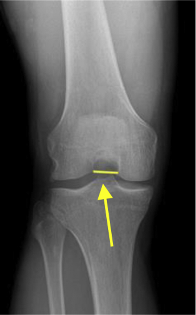
Preoperative weightbearing anteroposterior X-ray film with knee flexed 30° (Rosenberg view). The ruler function is enabled, and the width of the notch at the base is measured as the distance between the medial and lateral femoral condyles closest to the joint line (arrow).
Discussion
This method is a reproducible and cost-effective way of measuring intercondylar notch base width on preoperative imaging that mirrors arthroscopic notch measurements. Because radiography and MRI are routinely performed before ACL reconstruction, this method is useful and practical for orthopaedic surgeons to predict notch size. It also allows the surgeon to adjust the preoperative plan accordingly and account for potential intraoperative challenges.
Many methods of notch size measurements on imaging have been described.2, 8, 9, 10 However, most methods have been used to study the relationship of notch size to ACL injury, and few have tried to reproduce arthroscopic measurements. Zeng et al.11 performed a meta-analysis of 16 studies assessing the correlation between intercondylar notch size and the risk of ACL injury. They found that patients with narrow notch widths were predisposed to ACL injury. However, the methodology in the included studies varied greatly. Some studies measure absolute notch width,2, 9, 12, 13 whereas others measure notch width relative to the size of the femoral condyles.14, 15, 16 Furthermore, different studies have measured these parameters on radiography, MRI, and CT scanning with little standardization. Whitney et al.12 performed a thorough and standardized analysis of notch width and its relation to ACL injury by measuring notch width on coronal oblique images in the plane of the ACL, as well as by measuring the volume of the ACL. They found that decreased ACL volume and decreased notch width correlated with increased risk of ACL injury. However, this measurement method does not correspond to the view of the notch as seen during arthroscopy.
Notch size measurement on arthroscopy has been described as a method of identifying patients with small intercondylar notches.7, 11, 17, 18, 19 Vrooijink et al.6 attempted to correlate notch base width measurements on MRI with those taken during arthroscopy, but did not find a positive correlation. However, these were an average of width measurements on sequential axial images of the MRI and therefore do not represent the most distal notch base width that is measured during arthroscopy.6 Another study attempted to correlate 3-dimensional notch volumes from CT scans with arthroscopic measurements and found a moderate correlation. However, CT scans are rarely performed before ACL reconstruction and do not account for the width of the cartilage at the notch entrance.5
The method presented here is the first to demonstrate a way of measuring notch base width on MRI that closely resembles arthroscopic measurements. MRI is performed before nearly all ACL reconstructions, and most imaging software systems have a ruler tool built in, so this method can be reproduced for most patients. Furthermore, no additional imaging is required, avoiding the risk of radiation and any extra cost. The arthroscopic measurements were performed with a simple arthroscopic ruler, which is inexpensive and available from many companies. Finally, this method is feasible for orthopaedic surgeons because the MRI, X-ray, and arthroscopic measurements can all be performed in less than 1 minute.
The main limitation of this method is the learning curve associated with performing the measurements, particularly on MRI. Some computer knowledge is required to know how to access the ruler tool and to make fine adjustments to the measurements. However, these measurements are similar to those performed for other uses, such as measuring anatomic structures, which orthopaedic surgeons do routinely. Therefore the learning curve is likely not prohibitive (Table 1). Additionally, imaging from different facilities was included in the study, which allowed variation in the radiographic and MRI techniques and quality. However, this may be the most realistic setting in which surgeons would use this technique, because patients often obtain imaging from various sources. The flexion weight-bearing X-rays films have also been found to be dependent on rotation and technique, which may limit the accuracy of the radiographic measurements.20
Table 1.
Advantages and Disadvantages of the Notch Measurement Method
| Advantages |
| No additional imaging |
| Inexpensive equipment |
| Quickly performed |
| Disadvantages |
| Learning curve |
| Dependent on MRI quality |
| X-ray technique variation |
MRI, magnetic resonance imaging.
The method of notch size measurement presented here can help surgeons can identify narrow notches and improve their preoperative plan to allow for more individualized ACL reconstruction. It could also allow surgeons to prevent intraoperative challenges, facilitate individualized graft size, and improve graft placement, which in turn may improve surgical efficiency.
Footnotes
The authors report that they have no conflicts of interest in the authorship and publication of this article. Full ICMJE author disclosure forms are available for this article online, as supplementary material.
Supplementary Data
The first segment of the video displays how arthroscopic measurements of the intercondylar notch were made. The patient is positioned supine on the operating table with the left knee at 90° of flexion. The knee is viewed from a standard lateral portal. The arthroscopic ruler (Trukor depth gauge; Smith & Nephew, Andover, MA) is inserted into the joint through the medial portal. Then the width of the intercondylar notch at the base, at one-third of the height, and at two-thirds of the height is measured. The heights of the medial and lateral walls are then measured. The second segment demonstrates the same notch measurements on magnetic resonance imaging. The T2 axial image that demonstrates the full contour of the notch entrance as would be viewed during arthroscopy is selected, the ruler function is enabled, and the following are measured: notch width at the base and heights of the medial and lateral walls. The average of the medial and lateral wall heights is calculated, and this number is divided by 3 to obtain the increment at which the notch widths at one-third and two-thirds of the height are measured. The final segment shows how notch base width is measured on flexion weightbearing radiographs. The ruler tool is enabled, and the width of the notch between the central margins of the base of the femoral condyles is measured.
References
- 1.Zhang C., Xie G., Fang Z., Zhang X., Huangfu X., Zhao J. Assessment of relationship between three dimensional femoral notch volume and anterior cruciate ligament injury in Chinese Han adults: A retrospective MRI study. Int Orthop. 2019;43:1231–1237. doi: 10.1007/s00264-018-4068-7. [DOI] [PubMed] [Google Scholar]
- 2.Keays S.L., Keays R., Newcombe P.A. Femoral intercondylar notch width size: A comparison between siblings with and without anterior cruciate ligament injuries. Knee Surg Sports Traumatol Arthrosc. 2016;24:672–679. doi: 10.1007/s00167-014-3491-6. [DOI] [PubMed] [Google Scholar]
- 3.Ranuccio F., Familiari F., Tedesco G., La Camera F., Gasparini G. Effects of notchplasty on anterior cruciate ligament reconstruction: A systematic review. Joints. 2017;5:173–179. doi: 10.1055/s-0037-1605551. [DOI] [PMC free article] [PubMed] [Google Scholar]
- 4.Seo Y.J., Yoo Y.S., Kim Y.S. The effect of notchplasty on tunnel widening in anterior cruciate ligament reconstruction. Arthroscopy. 2014;30:739–746. doi: 10.1016/j.arthro.2014.02.024. [DOI] [PubMed] [Google Scholar]
- 5.Van Eck C.F., Martins C.A., Kopf S., Lertwanich P., Fu F.H., Tashman S. Correlation between the 2-dimensional notch width and the 3-dimensional notch volume: A cadaveric study. Arthroscopy. 2011;27:207–212. doi: 10.1016/j.arthro.2010.06.027. [DOI] [PubMed] [Google Scholar]
- 6.Vrooijink S.H., Wolters F., Van Eck C., Fu F. Measurements of knee morphometrics using MRI and arthroscopy: A comparative study between ACL-injured and non-injured subjects. Knee Surg Sports Traumatol Arthrosc. 2011;19(suppl 1):S12–S16. doi: 10.1007/s00167-011-1502-4. [DOI] [PubMed] [Google Scholar]
- 7.Wolters F., Vrooijink S.H., Van Eck C.F., Fu F.H. Does notch size predict ACL insertion site size? Knee Surg Sports Traumatol Arthrosc. 2011;19(suppl 1):S17–S21. doi: 10.1007/s00167-011-1503-3. [DOI] [PubMed] [Google Scholar]
- 8.Bouras T., Fennema P., Burke S., Bosman H. Stenotic intercondylar notch type is correlated with anterior cruciate ligament injury in female patients using magnetic resonance imaging. Knee Surg Sports Traumatol Arthrosc. 2018;26:1252–1257. doi: 10.1007/s00167-017-4625-4. [DOI] [PubMed] [Google Scholar]
- 9.Fujii M., Fennema P., Burke S., Bosman H. Intercondylar notch size influences cyclops formation after anterior cruciate ligament reconstruction. Knee Surg Sports Traumatol Arthrosc. 2015;23:1092–1099. doi: 10.1007/s00167-014-2891-y. [DOI] [PubMed] [Google Scholar]
- 10.Chow R.M., Guzman M.S., Dao Q. Intercondylar notch width as a risk factor for medial femoral condyle osteochondritis dissecans in skeletally immature patients. J Pediatr Orthop. 2016;36:640–644. doi: 10.1097/BPO.0000000000000511. [DOI] [PubMed] [Google Scholar]
- 11.Zeng C., Gao S.G., Wei J. The influence of the intercondylar notch dimensions on injury of the anterior cruciate ligament: A meta-analysis. Knee Surg Sports Traumatol Arthrosc. 2013;21:804–815. doi: 10.1007/s00167-012-2166-4. [DOI] [PubMed] [Google Scholar]
- 12.Whitney D.C., Sturnick D.R., Vacek P.M. Relationship between the risk of suffering a first-time noncontact ACL injury and geometry of the femoral notch and ACL: A prospective cohort study with a nested case-control analysis. Am J Sports Med. 2014;42:1796–1805. doi: 10.1177/0363546514534182. [DOI] [PMC free article] [PubMed] [Google Scholar]
- 13.Good L., Odensten O., Gillquist J. Intercondylar notch measurements with special reference to anterior cruciate ligament surgery. Clin Orthopaed Rel Res. 1989;263:185–189. [PubMed] [Google Scholar]
- 14.Domzalski M., Grzelak P., Gabos P. Risk factors for anterior cruciate ligament injury in skeletally immature patients: Analysis of intercondylar notch width using magnetic resonance imaging. Int Orthop. 2010;34:703–707. doi: 10.1007/s00264-010-0987-7. [DOI] [PMC free article] [PubMed] [Google Scholar]
- 15.Souryal T.O., Freeman T.R. Intercondylar notch size and anterior cruciate ligament injuries in athletes. A prospective study. Am J Sports Med. 1993;21:535–539. doi: 10.1177/036354659302100410. [DOI] [PubMed] [Google Scholar]
- 16.Al-Saeed O., Brown M., Athyal R., Sheikh M. Association of femoral intercondylar notch morphology, width index and the risk of anterior cruciate ligament injury. Knee Surg Sports Traumatol Arthrosc. 2013;21:678–782. doi: 10.1007/s00167-012-2038-y. [DOI] [PubMed] [Google Scholar]
- 17.van Eck C.F., Martins C.A., Lorenz S.G., Fu F.H., Smolinski P. Assessment of correlation between knee notch width index and the three-dimensional notch volume. Knee Surg Sports Traumatol Arthrosc. 2010;18:1239–1244. doi: 10.1007/s00167-010-1131-3. [DOI] [PMC free article] [PubMed] [Google Scholar]
- 18.Sturnick D.R., Vacek P.M., DeSarno M.J. Combined anatomic factors predicting risk of anterior cruciate ligament injury for males and females. Am J Sports Med. 2015;43:839–847. doi: 10.1177/0363546514563277. [DOI] [PMC free article] [PubMed] [Google Scholar]
- 19.Hirtler L., Rohrich S., Kainberger F. The femoral intercondylar notch during life: An anatomic redefinition with patterns predisposing to cruciate ligament impingement. AJR Am J Roentgenol. 2016;207:836–845. doi: 10.2214/AJR.16.16015. [DOI] [PubMed] [Google Scholar]
- 20.Anderson A.F., Anderson C.N., Gorman T.M., Cross M.B., Spindler K.P. Radiographic measurements of the intercondylar notch: Are they accurate? Arthroscopy. 2007;23:261–688. doi: 10.1016/j.arthro.2006.11.003. 268 e1-268 e2. [DOI] [PubMed] [Google Scholar]
Associated Data
This section collects any data citations, data availability statements, or supplementary materials included in this article.
Supplementary Materials
The first segment of the video displays how arthroscopic measurements of the intercondylar notch were made. The patient is positioned supine on the operating table with the left knee at 90° of flexion. The knee is viewed from a standard lateral portal. The arthroscopic ruler (Trukor depth gauge; Smith & Nephew, Andover, MA) is inserted into the joint through the medial portal. Then the width of the intercondylar notch at the base, at one-third of the height, and at two-thirds of the height is measured. The heights of the medial and lateral walls are then measured. The second segment demonstrates the same notch measurements on magnetic resonance imaging. The T2 axial image that demonstrates the full contour of the notch entrance as would be viewed during arthroscopy is selected, the ruler function is enabled, and the following are measured: notch width at the base and heights of the medial and lateral walls. The average of the medial and lateral wall heights is calculated, and this number is divided by 3 to obtain the increment at which the notch widths at one-third and two-thirds of the height are measured. The final segment shows how notch base width is measured on flexion weightbearing radiographs. The ruler tool is enabled, and the width of the notch between the central margins of the base of the femoral condyles is measured.