Abstract
Aging triggers cellular and molecular alterations, including genomic instability and organ dysfunction, which increases the risk of disease in mammals. Recently, due to the markedly growing number of aging dogs in the world, as much as 49% in total number of pet dogs, it is necessary to improve and maintain their quality of life by understanding of the biological effects of aging. Therefore, the aim of this study was to determine specific biomarkers in aging dogs as a means of defining a set of hematological/biochemical biomarkers that influence the aging process. Blood samples were collected from younger (1–3 years) and older (7–10 years) dogs of middle/large size. The hematological/biochemistry analysis was performed to evaluate parameters significantly associated with age. Enzyme-linked immunosorbent assay was used to target growth hormone (GH)/insulin growth factor-1 (IGF-1), one of the main regulators of the aging process. Declining levels of total protein and increased levels of glucose in young dogs was observed regardless of their body size. Notably, a significantly high concentration of GH and IGF-1 in the younger dogs compared to the older dogs was found in middle/large-sized dogs. GH and IGF-1 were also found at significantly high levels in large-sized dogs compared to middle-sized dogs, suggesting a similar trend to that of elderly humans. Consequently, glucose, total protein, GH, and IGF-1 were identified as potential biomarkers for regulating the aging process in large/middle-sized dogs. These findings provide an invaluable insight into the mechanism of aging for the field of aging research.
Keywords: Aging, dog, enzyme-linked immunosorbent assay, hematological test, biomarkers
INTRODUCTION
Recently, many studies have focused on understanding the biochemical basis of aging. As the elderly population increase, incurring high medical costs, it is necessary to understand the process of aging to prevent age-related diseases. It has been proven that biological mechanisms are closely related with longevity in many species [1,2]. Dogs are considered to be the animal with the most similar lifestyle to humans, showing regional adaptations related to their environmental conditions [3,4]. Due to the aging of the pet dogs and the derivative increase in aging-related diseases with their economic burden, it is necessary to promote research on aging in respect of the biological effect as a way to improve healthy and productive longevity for aging population. Although these biological analyses have been well-established and accommodated to the field of human medicine [5,6], the corresponding data in veterinary medicine remains scarce. Moreover, recent comprehensive results for the hematology and serum biochemistry of dogs depending on variations in age have not been well documented. Therefore, the evaluation of how aging affects the physiological characteristics of clinically healthy dogs is necessary. Generally, complete blood counts (CBC) and serum biochemistry parameters are comparatively more objective and specific than physical examinations for the evaluation of physiological conditions [7]. Therefore, we performed overall hematologic and biochemistry analysis for dogs of different ages. In addition, the effects of aging and the rejuvenating potential of the endocrine system have recently received increased attention. In particular, growth hormone (GH) and insulin growth factor-1 (IGF-1) have been considered as potential markers for regulating the aging process [8,9,10]. The reciprocal relationship between GH and aging was previously demonstrated when an age-related decline in the plasma GH levels was observed in laboratory rats, demonstrating that declining levels of GH releasing hormone stimulate the aging process [10]. Interestingly, previous epidemiological studies have indicated that a higher concentration of IGF-1 can decrease the risk of cognitive decline and dementia [11,12], which is intimately linked with age-related process. Likewise, many investigations have indicated that the physiological actions of GH are mediated by IGF-1 [9,13], which suggests that the concentration of IGF-1 may provide a useful measure for GH levels and activity. Therefore, the age-related decline in GH and IGF-1 concentration may contribute to the functional changes observed during aging. The aim of this study was to identify potential biomarkers for physiological changes related to aging using the following experiments: 1) whole screening test for hematology and biochemistry factors in large and middle-size dogs of different ages; 2) enzyme-linked immunosorbent assay (ELISA) to determine the levels of GH and IGF-1 in the serum of dogs in different age groups depending on their size; 3) ELISA to determine the levels of GH and IGF-1 in serum of dogs in different size groups depending on their ages.
MATERIALS AND METHODS
Ethics in animal experiments
In this study, 14 healthy middle-sized beagle dogs (young group: 1–3 years old, old group: 7–10 years old), and 14 healthy large-sized retriever dogs (young group: 1–3 years old, old group: 7–10 years old) without clinical signs of any disease and with normal vitality were used. The mean body weights of middle- and large-sized dogs is 7.4 kg and 28.5 kg, respectively. All dogs were continuously monitored throughout the research period and fed with a consistent amount of commercial adult dry food and water on a daily basis. All experiments were performed in accordance with recommendations described in “The Guide for the Care and Use of Laboratory Animals” published by the Institutional Animal Care and Use Committee of Seoul National University (approval No. SNU-180731-2-1). In this respect, the dog care facilities and the procedures performed met or exceeded the standards established by the Committee for Accreditation of Laboratory Animal Care at Seoul National University.
Blood analysis for CBC
For CBC analysis, blood samples were collected from jugular vein of the dogs in each group and transferred them to ethylenediamine tetraacetic acid tube. Analysis of CBC (Siemens Healthcare, Japan) was performed for total 16 parameters: red blood cell, white blood cell, packed cell volume, hemoglobin, and platelet, neutrophil, basophil, leukocyte, monocyte, lymphocyte, and eosinophil counts. Mean cell volume (MCV), mean cell hemoglobin, mean corpuscular hemoglobin concentration, red cell distribution width, and mean platelet volume (MPV) were subsequently determined.
Blood analysis for serum chemistry parameters
For serum chemistry analysis, blood samples were collected from the jugular vein of dogs in each group. Samples were collected taking aseptic precautions and transferred to them to serum separate tube, then allowed to clot to obtain the serum before centrifuging at 3,000 rpm for 10 min to separate serum. Serum samples were stored at –80°C until further use. A serum chemistry panel test (Hitachi High-Technologies Co., Japan) was performed, which included the measurement of sodium (Na), potassium (K), chlorine, alkaline phosphatase, alanine aminotransferase, aspartate aminotransferase, blood urea nitrogen (BUN), creatinine, glucose, total bilirubin, albumin, total protein, gamma glutamyltransferase, inorganic phosphorus, and calcium levels.
ELISA analysis
For ELISA analysis, blood samples were collected under aseptic conditions and allowed to clot to obtain the serum. The samples were centrifuged at 3,000 rpm for 10 min to separate the serum. The serum samples were then stored at –80°C until further use. The concentrations of IGF-1 (MBS706394; MyBioSource, USA) and GH (CSB-E14962c; CUSABIO, USA) in the serum in each group were measured by ELISA. The assay was performed following the manufacturer's instructions. Briefly, 100 μL of standard and sample was added to each well of the ELISA plates and incubated for 2 h at 37°C. After incubation, the liquid of each well was removed without washing. Then, 100 μL of Biotin-antibody was added to each well and incubated for 1 h at 37°C. The liquid was removed with aspiration and washing was performed with a multi-channel pipette three times using wash buffer (200 μL). Next, 100 μL of HRP-avidin reagent was added to each well and incubated for 1 h at 37°C. The aspiration/wash process was then repeated 5 times using wash buffer. Finally, 90 μL of TMB substrate was added to the plate and incubated for 30 min at 37°C with protection from light. After incubation, 50 μL of stop solution was added to each well. After gently tapping the plate, the optical density of each well was determined using a microplate reader (Tecan Sunrise, Hayward, USA). The spectroscopic absorbance of each well was measured at a wavelength of 480 nm.
Statistical analysis
Descriptive and inductive statistical analysis was performed using GraphPad Prism 5.0 (Graphpad, USA). Differences between the groups were evaluated by Student's t-test, followed by an unpaired t-test. Differences of p < 0.05 were considered significant. The results for blood analysis are expressed as the mean ± standard deviation and for ELISA are expressed as the mean ± standard error of the mean.
RESULTS
Analysis of parameters related to complete blood cell count in middle-sized dogs in different age groups
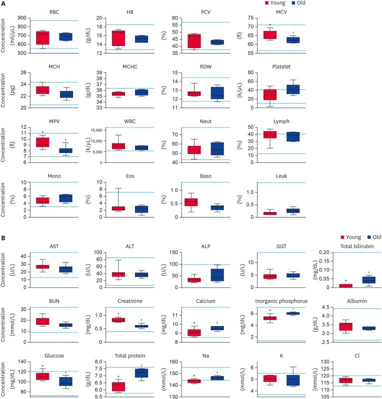
To determine the differences in the physiological characteristics of middle-sized dogs depending on their age, we analyzed the CBC results of each group (Fig. 1A). The CBC results were within normal ranges for all animals except for certain parameters, including platelets and lymph. Moreover, no significant differences were observed between young and old groups in middle-sized dogs except for two parameters, MCV and MPV. The concentration of MCV was significantly increased in younger dogs (65.3 ± 2.5 fl, p < 0.05) compared to older dogs (62.5 ± 1.6 fl, p < 0.05). In addition, the level of MPV was significantly increased in the young group (9.4 ± 0.9 fl, p < 0.05) compared to the old group (8.1 ± 0.7 fl, p < 0.05).
Fig. 1. Evaluation of (A) complete blood cell count and (B) serum biochemistry parameters in middle-sized dogs depending on their age. RBC, HB, PCV, MCV, MCH, MCHC, RDW, MPV, WBC, Neut, Lymph, Mono, Eos, Baso, Leuk, AST, ALT, ALP, GGT, BUN, Na, K, and Cl levels were analyzed. Bars represent normal reference intervals. The blood sample has been analyzed for one time per dog.
RBC, red blood cell; HB, hemoglobin; PCV, packed cell volume; MCV, mean cell volume; MCH, mean cell hemoglobin; MCHC, mean corpuscular hemoglobin concentration; RDW, red cell distribution width; MPV, mean platelet volume; WBC, white blood cell; Neut, neutrophil; Lymph, lymphocyte; Mono, monocyte; Eos, eosinophil; Baso, basophil; Leuk, leukocyte; AST, aspartate aminotransferase; ALT, alanine aminotransferase; ALP, alkaline phosphatase; GGT, gamma glutamyltransferase; BUN, blood urea nitrogen; Na, sodium; K, potassium; Cl, chlorine; Young, younger dogs; Old, older dogs.
*,†Within the groups, values with different superscript letters are significantly different (p < 0.05).
Analysis of parameters related to serum chemistry in middle-sized dogs in different age groups
Serum chemistry analysis was performed to evaluate the difference physiological characteristics between younger and older dogs in the middle-sized group (Fig. 1B). All the serum chemistry results were within normal ranges except for a few parameters, such as glucose, Na+, and K+. There were some significant differences between 2 groups with respect to several factors. There was a significant increase in the level of creatinine and glucose in younger dogs (0.8 ± 0.1 mg/dL, and 111.6 ± 8.0 mg/dL, respectively, p < 0.05) compared to older dogs (0.6 ± 0.1 mg/dL, and 99.3 ± 9.5 mg/dL, respectively, p < 0.05). Moreover, the level of total bilirubin, calcium, inorganic phosphorus, total protein, and Na+ was significantly higher in older dogs (0.041 ± 0.021 mg/dL, 9.6 ± 0.2 mg/dL, 6.1 ± 0.2 mg/dL, 7.2 ± 0.3 g/dL, 146.2 ± 1.6 mmol/L, respectively, p < 0.05) compared to younger dogs (0.012 ± 0.01 mg/dL, 9.1 ± 0.4 mg/dL, 5.2 ± 0.5 mg/dL, 6.3 ± 0.3 g/dL, 143.6 ± 1.6 mmol/L, respectively, p < 0.05).
Analysis of parameters related to complete blood cell count in large-sized dogs in different age groups
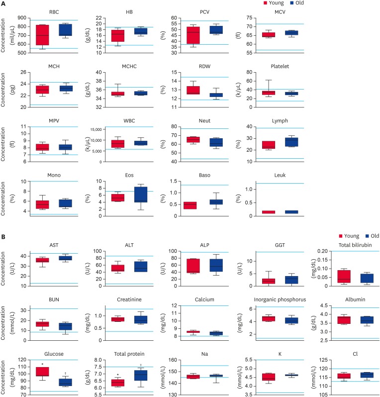
The analysis of the complete blood cell count was performed to assess the differences in the physiological characteristics of large-sized dogs depending on their age (Fig. 2A). The CBC results showed that most of the parameters were within normal ranges. Additionally, there were no significant differences between young and older dogs in the large-sized group for all parameters.
Fig. 2. Evaluation of (A) complete blood cell count and (B) serum biochemistry parameters in large-sized dogs depending on their age. RBC, HB, PCV, MCV, MCH, MCHC, RDW, MPV, WBC, Neut, Lymph, Mono, Eos, Baso, Leuk, AST, ALT, ALP, GGT, BUN, Na, K, and Cl levels were analyzed. Bars represent normal reference intervals. The blood sample has been analyzed for one time per dog.
RBC, red blood cell; HB, hemoglobin; PCV, packed cell volume; MCV, mean cell volume; MCH, mean cell hemoglobin; MCHC, mean corpuscular hemoglobin concentration; RDW, red cell distribution width; MPV, mean platelet volume; WBC, white blood cell; Neut, neutrophil; Lymph, lymphocyte; Mono, monocyte; Eos, eosinophil; Baso, basophil; Leuk, leukocyte; AST, aspartate aminotransferase; ALT, alanine aminotransferase; ALP, alkaline phosphatase; GGT, gamma glutamyltransferase; BUN, blood urea nitrogen; Na, sodium; K, potassium; Cl, chlorine; Young, younger dogs; Old, older dogs.
*,†Within groups, values with different superscript letters are significantly different (p < 0.05).
Analysis of parameters related to serum chemistry in large-sized dogs in different age groups
Serum chemistry analysis was performed in younger and older dogs in the large-sized group (Fig. 2B). All the serum chemistry results were within normal ranges except for the parameters BUN and Na+. There were significant differences between the 2 groups with respect to two factors; glucose and total protein. A significantly increased level of glucose was observed in the younger dogs (100.4 ± 7.0 mg/dL, p < 0.05) compared to the older dogs (87.6 ± 5.7 mg/dL, p < 0.05). Meanwhile, there was a significant lower level of total protein in the young group (6.4 ± 0.2 g/dL, p < 0.05) compared to the old group (6.9 ± 0.5 g/dL, p < 0.05).
ELISA analysis in younger and older dogs depending on their size
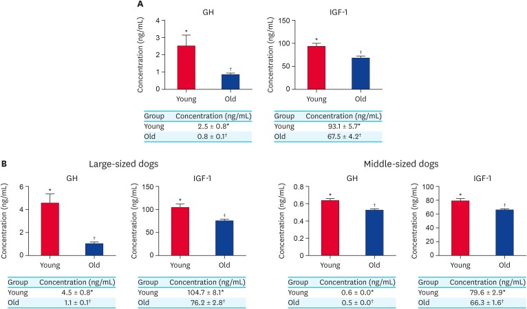
The concentrations of GH and IGF-1 in the serum of each group were analyzed (Fig. 3A and B). First, we analyzed the GH and IGF-1 concentration in the younger and older dogs regardless of their size to evaluate the overall tendency of those factors with ages. As a result, both the GH and IGF-1 levels were significantly increased in the young group (2.5 ± 0.6 ng/mL and 93.1 ± 5.7 ng/mL, respectively, p < 0.05) compared to the old group (0.8 ± 0.1 ng/mL and 67.5 ± 4.2 ng/mL, respectively, p < 0.05) (Fig. 3A). To refine the results whether those decreasing tendencies of GH and IGF-1 factors with ages are influenced depending on their sizes, the groups were subdivided into large and middle-sized dogs depending on their age. The results showed that the levels of GH and IGF-1 significantly increased in younger dogs (4.5 ± 0.8 ng/mL and 104.7 ± 8.1 ng/mL, respectively, p < 0.05) compared to older dogs (1.1 ± 0.1 ng/mL and 76.2 ± 2.8 ng/mL, respectively, p < 0.05) in the large-sized group (Fig. 3B). Moreover, the concentration of GH and IGF-1 was also significantly up-regulated in younger dogs (0.6 ± 0.0 ng/mL and 79.6 ± 2.9 ng/mL, respectively, p < 0.05) compared to the older dogs (0.5 ± 0.0 ng/mL and 66.3 ± 1.6 ng/mL, respectively, p < 0.05) in the middle-sized group (Fig. 3B).
Fig. 3. (A) Concentration of GH and IGF-1 from serum in younger and older dog groups without classification of their body size. (B) Concentration of GH and IGF-1 from serum in younger and older dogs in large-sized group and middle-sized group. At least 3 times replications were performed.
GH, growth hormone; IGF-1, insulin growth factor-1; Young, younger dogs; Old, older dogs.
*,†Within groups, values with different superscript letters are significantly different (p < 0.05).
ELISA analysis in large and middle-sized dogs in different age groups
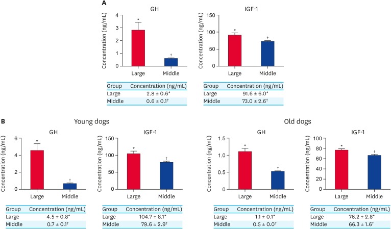
First, the concentration of GH and IGF-1 was assessed in both large and middle-sized dogs regardless of their age to evaluate the overall tendency of those factors with body-size (Fig. 4A). As a result, the levels of GH and IGF-1 were significantly higher in the large-sized dogs (2.8 ± 0.6 ng/mL and 91.5 ± 6.0 ng/mL, respectively, p < 0.05) compared to middle-sized dogs (0.6 ± 0.1 ng/mL and 73.0 ± 2.6 ng/mL, respectively, p < 0.05) (Fig. 4A). Subsequently, these groups were subdivided according to age (i.e. younger and older dogs) to refine the results whether those increasing tendencies of GH and IGF-1 factors with body-size are influenced depending on their ages. As shown in Fig. 4B, there was a significant increase in the levels of GH and IGF-1 in the large-sized group (4.5 ± 0.8 ng/mL and 104.7 ± 8.1 ng/mL, respectively, p < 0.05) compared to the middle-sized group (0.7 ± 0.1 ng/mL and 79.6 ± 2.9 ng/mL, respectively, p < 0.05) among the younger dogs. Moreover, the concentration of both GH and IGF-1 was significantly higher in the large-sized group (1.1 ± 0.1 ng/mL and 76.2 ± 2.8 ng/mL, respectively, p < 0.05) compared to the middle-sized group (0.5 ± 0.0 ng/mL and 66.3 ± 1.6 ng/mL, respectively, p < 0.05) among the older dogs (Fig. 4B).
Fig. 4. (A) Concentration of GH and IGF-1 from serum in large and middle-sized dogs without classification of their ages. (B) Concentration of GH and IGF-1 from serum in larger and middle-sized dogs in younger dogs and older dogs. At least 3 times replications were performed.
GH, growth hormone; IGF-1, insulin growth factor-1; Large, large-sized dogs; Middle, middle-sized dogs.
*,†Within groups, values with different superscript letters are significantly different (p < 0.05).
DISCUSSION
In terms of lifestyle, the dog is the most similar animal to humans in terms of medical care, living in an environment with pollutants and pathogens, and access to physical activity [14,15], which are markedly different compared with other laboratory animals [16]. Since there have been a large number of dogs in the world, the proportion of aging pet dogs is markedly increased, because the quality of veterinary care has been improved. This need to improve the welfare of companion dogs is also useful for the development of aging research in other organisms, namely humans. As such, we need to improve our understanding of the biological effects of aging on screening analysis generally performed in field of veterinary medicine. To this end, our study aimed to identify specific biomarkers in dogs of different ages in comparison to their size as a foundation for the identification of the hematological and biochemical biomarkers that influence the aging process.
Aging is considered to be a continuous ontogenetic process, starting at the birth and ending at the death of the individual [17]. Recently, a large number of cellular and molecular parameters associated with aging, including altered intercellular communication, genomic instability, mitochondrial dysfunction, and epigenetic alterations, were identified in mammals [18]. It has been previously reported that, to investigate the changes in the physiological properties of dogs, CBCs and serum biochemistry profiles are more objective with a greater specificity for the detection of physiological changes than routine physical examinations, wherein hematology involves a simple examination used to monitor and evaluate in experimental dogs to analyze their physiological characteristics [7,19]. Thus, it is common to utilize blood tests in older dogs to identify a specific disease related to aging. A previous retrospective study that investigated the hematological phenotypes in client-owned dogs of various breeds found that the dog is an ideal model for evaluating the blood traits of mammals [19]. However, the authors used a range of dogs without geriatric ages or serum biochemistry parameters. Therefore, the aim of the current study was to identify notable parameters in younger and older dogs of different sizes to represent the process aging. The classification of age in dogs is well-established in the field of veterinary research [1].
According to the CBC and serum biochemical profiles results, significant differences were observed in specific parameters in middle and large-sized dogs. One of the most striking findings of the current study is the evidence of a decline in the levels of total protein and increased levels of glucose in younger dogs, regardless of their body size (Fig. 1A and B). A previous study suggested that the total protein in elderly people with chronic inflammation is composed of higher globulin concentrations and lower levels of albumin [20]. Specifically, increases in serum globulin are closely related to cancer, chronic liver disease, and diabetes mellitus, whereas decreased albumin levels are associated with malnutrition, chronic infection, and nephrotic disease, and is one of the main physiological symptoms for senescence. Also, in another previous study, a decreased ratio of albumin/globulin was reported in older dogs [21,22] due to increased levels of immunoglobulin A [23]. In line with these previous studies, we found that the older dogs in both the middle and large-sized groups showed significantly higher levels of total protein than their younger counterparts, which suggests that the total protein parameters could be used for the identification of the aging process.
Meanwhile, in our present study, a decline in the concentration of glucose was observed in older dogs in both the middle and large-sized groups (Fig. 1A and B). Previous studies have found differing patterns of serum glucose changes with aging. One such study reported that the serum glucose levels increased with age [21], which was attributed to a decreased sensitivity of organ to concentrations changes due to decreases in organ production and increased insulin resistance. Other studies demonstrated that glucose decreases with age [22,24], which was consistent with our results. These studies suggested that the storage competence of glycogen in the liver or functional hepatic mass decreases with age, which was therefore attributed to the decreased concentration of glucose in older individuals [24,25]. Likewise, in our current study, these findings could explain the observed reduction in the serum glucose levels with age.
It has been previously suggested that there is a reciprocal relationship between GH and IGF-1 in age-related physiological actions [9]. A previous study demonstrated that the age-related decline in GH concentration in the plasma is closely related with a reduction of IGF-1 levels. Although well-established biological variation values have existed in the field of human medicine for decades [5,6], the corresponding information in veterinary medicine remains to be elucidated, and size/age-related variations in hematologic and plasma biochemical markers in dogs have not been well documented [26]. Therefore, in this study, we aimed to identify potential biomarkers for physiological change in relation to aging.
Recently, interest in GH as a marker/regulator of aging has increased. For example, plasma GH levels showed significantly age-related down-regulation in rats, suggesting that deceased stimulation of GH-releasing hormone induces age-related effects [10]. In addition, other studies have indicated that there is a decline in GH concentration in age-related processes in mice and humans [27,28]. These findings suggest that a reduction in age-related GH levels characterizes the period of senescence. Also, another potential biomarker for aging, IGF-1, has been demonstrated to act as a physiological predictor for frailty in elderly humans [2]. One previous report indicated that the IGF-1 pathway is closely related with life extension in organisms including nematode worms and mice, and found that IGF-1 signaling is down-regulated in a subset of elderly individuals [29]. Moreover, epidemiological studies have demonstrated that high levels of IGF-1 could reduce the rate of cognitive deterioration and dementia [11], which are closely associated with the progression of aging. Therefore, the pattern of IGF-1 expression is a good candidate for an indicator of lifespan in dogs.
Meanwhile, across animal species, individual lifespan is robustly correlated with body size [30,31]. Moreover, it is well known that the body size is inversely related to longevity in dogs [32,33]. In previous studies, the correlation between lifespan and size in dogs has been characterized in terms of both cellular [34] and metabolic [35] properties. Recently, it was demonstrated that larger breeds of dogs spend a disproportionately higher amount of energy on growth and development at birth compared to smaller breeds, due to their smaller size at birth. Therefore, larger breeds of dogs have comparatively less energy to repair body functions, which reduces their overall lifespan [36]. In addition, it has been reported that a GH/IGF-1 is a trait dependent on adult body size, and which is closely associated with extended life span in rodents [13,37], cats [38], dogs [33,39], horses [40], and humans [33]. This indicates that GH/IGF-1 are one of the main factors for longevity in dogs.
In line with previous findings on the role of GH and IGF-1 in the aging process, in this study, we found that a relationship between GH and IGF-1 develops during the aging process. According to our results, the overall concentration of GH and IGF-1 was markedly increased in younger individuals compared to older individuals in both large and middle-sized dogs, respectively (Fig. 3B). Interestingly, the levels of GH and IGF-1 were also significantly increased in the younger dogs regardless of their body size (Fig. 3A). Moreover, we demonstrated that GH and IGF-1 were significantly high in large-sized dogs compared to middle-sized dogs, with or without classification of their ages (Fig. 4). Therefore, we suggested that the level of age-related biomarkers, GH and IGF-1, are inversely related to age and positively correlated to size in dogs.
In conclusion, our study demonstrates that the concentrations of serum glucose, GH, and IGF-1 were markedly high in younger dogs compared to older dogs regardless of their body size. Furthermore, the biomarkers of GH and IGF-1 were significantly higher in large-sized dogs compared to middle-sized dogs. Finally, the levels of total protein in the serum were found to decline with age in both middle- and large-sized dogs. These results suggest that the 4 representative biomarkers shown to be significantly different with regards to ages or size could be considered as potential biomarkers for regulating the aging process in dogs, which represents invaluable information for the current field of aging research.
Footnotes
Funding: This work was carried out with the support of “Cooperative Research Program of Center for Companion Animal Research (Project No. PJ013958012019)” Rural Development Administration, Republic of Korea. This study was supported by the Research Institute for Veterinary Science and the BK21 plus program.
Conflict of Interest: The authors declare no conflicts of interest.
- Conceptualization: Oh HJ, Lee BC.
- Data curation: Oh HJ, Lee SH.
- Formal analysis: Lee SH.
- Investigation: Lee SH, Kim JW.
- Methodology: Lee SH, Kim JW.
- Project administration: Oh HJ, Lee BC.
- Supervision: Oh HJ, Lee BC.
- Writing - original draft: Lee SH.
- Writing - review & editing: Oh HJ, Lee BC, Kim JW.
References
- 1.Fortney WD. Implementing a successful senior/geriatric health care program for veterinarians, veterinary technicians, and office managers. Vet Clin North Am Small Anim Pract. 2012;42:823–834. viii. doi: 10.1016/j.cvsm.2012.04.011. [DOI] [PubMed] [Google Scholar]
- 2.Walston J. Frailty--the search for underlying causes. Sci Aging Knowledge Environ. 2004;2004:pe4. doi: 10.1126/sageke.2004.4.pe4. [DOI] [PubMed] [Google Scholar]
- 3.Gou X, Wang Z, Li N, Qiu F, Xu Z, Yan D, Yang S, Jia J, Kong X, Wei Z, Lu S, Lian L, Wu C, Wang X, Li G, Ma T, Jiang Q, Zhao X, Yang J, Liu B, Wei D, Li H, Yang J, Yan Y, Zhao G, Dong X, Li M, Deng W, Leng J, Wei C, Wang C, Mao H, Zhang H, Ding G, Li Y. Whole-genome sequencing of six dog breeds from continuous altitudes reveals adaptation to high-altitude hypoxia. Genome Res. 2014;24:1308–1315. doi: 10.1101/gr.171876.113. [DOI] [PMC free article] [PubMed] [Google Scholar]
- 4.Li Y, Wu DD, Boyko AR, Wang GD, Wu SF, Irwin DM, Zhang YP. Population variation revealed high-altitude adaptation of Tibetan mastiffs. Mol Biol Evol. 2014;31:1200–1205. doi: 10.1093/molbev/msu070. [DOI] [PubMed] [Google Scholar]
- 5.Fraser CG. Inherent biological variation and reference values. Clin Chem Lab Med. 2004;42:758–764. doi: 10.1515/CCLM.2004.128. [DOI] [PubMed] [Google Scholar]
- 6.Harris EK. Effects of intra- and interindividual variation on the appropriate use of normal ranges. Clin Chem. 1974;20:1535–1542. [PubMed] [Google Scholar]
- 7.Metzger FL, Rebar AH. Clinical pathology interpretation in geriatric veterinary patients. Vet Clin North Am Small Anim Pract. 2012;42:615–629. v. doi: 10.1016/j.cvsm.2012.04.004. [DOI] [PubMed] [Google Scholar]
- 8.Veldhuis JD, Norman C, Miles JM, Bowers CY. Sex steroids, GHRH, somatostatin, IGF-I, and IGFBP-1 modulate ghrelin's dose-dependent drive of pulsatile GH secretion in healthy older men. J Clin Endocrinol Metab. 2012;97:4753–4760. doi: 10.1210/jc.2012-2567. [DOI] [PMC free article] [PubMed] [Google Scholar]
- 9.Maggio M, Ble A, Ceda GP, Metter EJ. Decline in insulin-like growth factor-I levels across adult life span in two large population studies. J Gerontol A Biol Sci Med Sci. 2006;61:182–183. doi: 10.1093/gerona/61.2.182. [DOI] [PubMed] [Google Scholar]
- 10.Meites J. Neuroendocrine biomarkers of aging in the rat. Exp Gerontol. 1988;23:349–358. doi: 10.1016/0531-5565(88)90037-x. [DOI] [PubMed] [Google Scholar]
- 11.Westwood AJ, Beiser A, Decarli C, Harris TB, Chen TC, He XM, Roubenoff R, Pikula A, Au R, Braverman LE, Wolf PA, Vasan RS, Seshadri S. Insulin-like growth factor-1 and risk of Alzheimer dementia and brain atrophy. Neurology. 2014;82:1613–1619. doi: 10.1212/WNL.0000000000000382. [DOI] [PMC free article] [PubMed] [Google Scholar]
- 12.Laughlin GA, Barrett-Connor E, Criqui MH, Kritz-Silverstein D. The prospective association of serum insulin-like growth factor I (IGF-I) and IGF-binding protein-1 levels with all cause and cardiovascular disease mortality in older adults: the Rancho Bernardo Study. J Clin Endocrinol Metab. 2004;89:114–120. doi: 10.1210/jc.2003-030967. [DOI] [PubMed] [Google Scholar]
- 13.Bartke A, Sun LY, Longo V. Somatotropic signaling: trade-offs between growth, reproductive development, and longevity. Physiol Rev. 2013;93:571–598. doi: 10.1152/physrev.00006.2012. [DOI] [PMC free article] [PubMed] [Google Scholar]
- 14.Tsai KL, Clark LA, Murphy KE. Understanding hereditary diseases using the dog and human as companion model systems. Mamm Genome. 2007;18:444–451. doi: 10.1007/s00335-007-9037-1. [DOI] [PMC free article] [PubMed] [Google Scholar]
- 15.Ostrander EA, Galibert F, Patterson DF. Canine genetics comes of age. Trends Genet. 2000;16:117–124. doi: 10.1016/s0168-9525(99)01958-7. [DOI] [PubMed] [Google Scholar]
- 16.Lorenzini A. How much should we weigh for a long and healthy life span? the need to reconcile caloric restriction versus longevity with body mass index versus mortality data. Front Endocrinol (Lausanne) 2014;5:121. doi: 10.3389/fendo.2014.00121. [DOI] [PMC free article] [PubMed] [Google Scholar]
- 17.Ackermann M, Chao L, Bergstrom CT, Doebeli M. On the evolutionary origin of aging. Aging Cell. 2007;6:235–244. doi: 10.1111/j.1474-9726.2007.00281.x. [DOI] [PMC free article] [PubMed] [Google Scholar]
- 18.López-Otín C, Blasco MA, Partridge L, Serrano M, Kroemer G. The hallmarks of aging. Cell. 2013;153:1194–1217. doi: 10.1016/j.cell.2013.05.039. [DOI] [PMC free article] [PubMed] [Google Scholar]
- 19.Lawrence J, Chang YM, Szladovits B, Davison LJ, Garden OA. Breed-specific hematological phenotypes in the dog: a natural resource for the genetic dissection of hematological parameters in a mammalian species. PLoS One. 2013;8:e81288. doi: 10.1371/journal.pone.0081288. [DOI] [PMC free article] [PubMed] [Google Scholar]
- 20.Maeda S, Takeya Y, Oguro R, Akasaka H, Ryuno H, Kabayama M, Yokoyama S, Nagasawa M, Fujimoto T, Takeda M, Onishi-Takeya M, Itoh N, Takami Y, Yamamoto K, Sugimoto K, Inagaki H, Ogawa M, Nakagawa T, Yasumoto S, Masui Y, Arai Y, Ishizaki T, Ikebe K, Gondo Y, Kamide K, Rakugi H. Serum albumin/globulin ratio is associated with cognitive function in community-dwelling older people: the septuagenarians, octogenarians, nonagenarians investigation with centenarians study. Geriatr Gerontol Int. 2019;19:967–971. doi: 10.1111/ggi.13751. [DOI] [PubMed] [Google Scholar]
- 21.Strasser A, Niedermüller H, Hofecker G, Laber G. The effect of aging on laboratory values in dogs. Zentralbl Veterinarmed A. 1993;40:720–730. doi: 10.1111/j.1439-0442.1993.tb00689.x. [DOI] [PubMed] [Google Scholar]
- 22.Lowseth LA, Gillett NA, Gerlach RF, Muggenburg BA. The effects of aging on hematology and serum chemistry values in the beagle dog. Vet Clin Pathol. 1990;19:13–19. doi: 10.1111/j.1939-165x.1990.tb00535.x. [DOI] [PubMed] [Google Scholar]
- 23.Blount DG, Pritchard DI, Heaton PR. Age-related alterations to immune parameters in Labrador retriever dogs. Vet Immunol Immunopathol. 2005;108:399–407. doi: 10.1016/j.vetimm.2005.06.015. [DOI] [PubMed] [Google Scholar]
- 24.Kaspar LV, Norris WP. Serum chemistry values of normal dogs (beagles): associations with age, sex, and family line. Lab Anim Sci. 1977;27:980–985. [PubMed] [Google Scholar]
- 25.Radakovich LB, Pannone SC, Truelove MP, Olver CS, Santangelo KS. Hematology and biochemistry of aging-evidence of “anemia of the elderly” in old dogs. Vet Clin Pathol. 2017;46:34–45. doi: 10.1111/vcp.12459. [DOI] [PubMed] [Google Scholar]
- 26.Walton RM. Subject-based reference values: biological variation, individuality, and reference change values. Vet Clin Pathol. 2012;41:175–181. doi: 10.1111/j.1939-165X.2012.00414.x. [DOI] [PubMed] [Google Scholar]
- 27.Kuwahara S, Kesuma Sari D, Tsukamoto Y, Tanaka S, Sasaki F. Age-related changes in growth hormone (GH)-releasing hormone and somatostatin neurons in the hypothalamus and in GH cells in the anterior pituitary of female mice. Brain Res. 2004;1025:113–122. doi: 10.1016/j.brainres.2004.08.012. [DOI] [PubMed] [Google Scholar]
- 28.Corpas E, Harman SM, Blackman MR. Human growth hormone and human aging. Endocr Rev. 1993;14:20–39. doi: 10.1210/edrv-14-1-20. [DOI] [PubMed] [Google Scholar]
- 29.Vitale G, Barbieri M, Kamenetskaya M, Paolisso G. GH/IGF-I/insulin system in centenarians. Mech Ageing Dev. 2017;165:107–114. doi: 10.1016/j.mad.2016.12.001. [DOI] [PubMed] [Google Scholar]
- 30.Austad SN. Cats, “rats,” and bats: the comparative biology of aging in the 21st century. Integr Comp Biol. 2010;50:783–792. doi: 10.1093/icb/icq131. [DOI] [PMC free article] [PubMed] [Google Scholar]
- 31.Metcalfe NB, Monaghan P. Growth versus lifespan: perspectives from evolutionary ecology. Exp Gerontol. 2003;38:935–940. doi: 10.1016/s0531-5565(03)00159-1. [DOI] [PubMed] [Google Scholar]
- 32.Kraus C, Pavard S, Promislow DE. The size-life span trade-off decomposed: why large dogs die young. Am Nat. 2013;181:492–505. doi: 10.1086/669665. [DOI] [PubMed] [Google Scholar]
- 33.Patronek GJ, Waters DJ, Glickman LT. Comparative longevity of pet dogs and humans: implications for gerontology research. J Gerontol A Biol Sci Med Sci. 1997;52:B171–178. doi: 10.1093/gerona/52a.3.b171. [DOI] [PubMed] [Google Scholar]
- 34.Li Y, Deeb B, Pendergrass W, Wolf N. Cellular proliferative capacity and life span in small and large dogs. J Gerontol A Biol Sci Med Sci. 1996;51:B403–408. doi: 10.1093/gerona/51a.6.b403. [DOI] [PubMed] [Google Scholar]
- 35.Speakman JR, van Acker A, Harper EJ. Age-related changes in the metabolism and body composition of three dog breeds and their relationship to life expectancy. Aging Cell. 2003;2:265–275. doi: 10.1046/j.1474-9728.2003.00061.x. [DOI] [PubMed] [Google Scholar]
- 36.Fan R, Olbricht G, Baker X, Hou C. Birth mass is the key to understanding the negative correlation between lifespan and body size in dogs. Aging (Albany NY) 2016;8:3209–3222. doi: 10.18632/aging.101081. [DOI] [PMC free article] [PubMed] [Google Scholar]
- 37.Eklund J, Bradford GE. Longeveity and lifetime body weight in mice selected for rapid growth. Nature. 1977;265:48–49. doi: 10.1038/265048b0. [DOI] [PubMed] [Google Scholar]
- 38.Kienzle E, Moik K. A pilot study of the body weight of pure-bred client-owned adult cats. Br J Nutr. 2011;106(Suppl 1):S113–S115. doi: 10.1017/S0007114511001802. [DOI] [PubMed] [Google Scholar]
- 39.Greer KA, Hughes LM, Masternak MM. Connecting serum IGF-1, body size, and age in the domestic dog. Age (Dordr) 2011;33:475–483. doi: 10.1007/s11357-010-9182-4. [DOI] [PMC free article] [PubMed] [Google Scholar]
- 40.Brosnahan MM, Paradis MR. Demographic and clinical characteristics of geriatric horses: 467 cases (1989–1999) J Am Vet Med Assoc. 2003;223:93–98. doi: 10.2460/javma.2003.223.93. [DOI] [PubMed] [Google Scholar]


