Abstract
Background
Microcotyle erythrini van Beneden & Hesse, 1863 (Platyhelminthes: Monogenea) and other closely related species of the genus are often considered as cryptic. Records in hosts other than the type-host with no species confirmation by molecular analyses have contributed to this situation.
Methods
Gill parasites of five sparid fishes, Boops boops (L.), Pagellus erythrinus (L.), P. acarne (Risso), Dentex dentex (L.) and Pagrus pagrus (L.), from the Western Mediterranean off Spain were collected. Specimens of Microcotyle spp. were characterised both molecularly and morphologically. Partial fragments (domains D1-D3) of the 28S rRNA gene and the cytochrome c oxidase subunit 1 (cox1) gene were amplified and used for molecular identification and phylogenetic reconstruction. Principal components analysis was used to look for patterns of morphological separation.
Results
Molecular analyses confirmed the identity of three species: M. erythrini ex P. erythrinus and Pa. pagrus; M. isyebi Bouguerche, Gey, Justine & Tazerouti, 2019 ex B. boops; and a species new to science described herein, M. whittingtoni n. sp. ex D. dentex. The specific morphological traits and confirmed hosts (P. erythrinus and Pa. pagrus) are delimited here in order to avoid misidentifications of M. erythrini (sensu stricto). Microcotyle erythrini (s.s.) is mostly differentiated by the shape of its haptor, which is also longer than in the other congeners. New morphological and molecular data are provided for M. isyebi from the Spanish Mediterranean enlarging the data on its geographical range. Microcotyle whittingtoni n. sp. is described from D. dentex and distinguished from the remaining currently recognised species of the genus by the number and robustness of the clamps.
Conclusions
New diagnostic morphological traits useful to differentiate Microcotyle spp. are suggested: (i) haptor dimensions including lobes; (ii) the thickness of the clamps; (iii) the size and shape of spines of the genital atrium; (iv) the extension of the posterior extremities of vitelline fields; and (v) the shape of egg filaments. The use of new morphological approaches may allow considering these species of Microcotyle as being pseudocryptic. The use of representative undamaged specimens that have been genetically confirmed as conspecific is considered crucial to avoid abnormally wide morphological ranges that prevent species differentiation.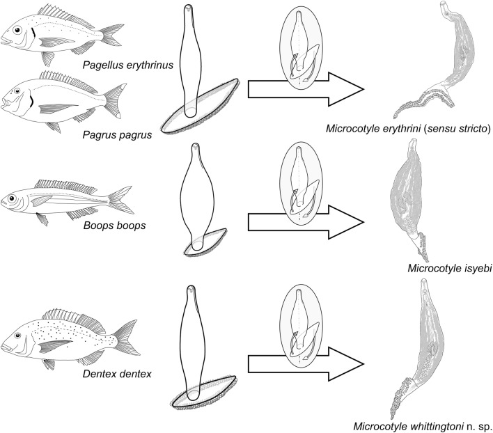
Keywords: Microcotyle erythrini (sensu stricto), M. isyebi, M. whittingtoni n. sp., Haptor morphology, Clamp morphology, Pseudocrypsis
Background
Microcotyle erythrini van Beneden & Hesse, 1863 (Monogenea: Microcotylidae) was originally described from Pagellus erythrinus (L.) (Teleostei: Sparidae) off the coast of Brest (France, North-East Atlantic) and to date it has been listed and considered a valid species [1–3]. Like many of the earliest descriptions of species of Microcotyle, M. erythrini was described briefly, and only differentiated by the number of clamps and testes, and the traits of the genital atrium [4]. Since the original description, many authors have recorded and described new specimens identified as M. erythrini in different sparid species, mostly in the Mediterranean Sea (see Table 1 in Bouguerche et al. [3], for details on the records of M. erythrini). These publications sometimes offered morphological ranges based on a combination of measurements of specimens from different host species (e.g. [5, 6]). Along this process, the morphological ranges of M. erythrini have been enlarged abnormally, which has made it difficult to define a clear and distinguishing morphology. Recently, with the help of molecular tools (cox1 partial fragment), M. erythrini has been split into two species, each in a different sparid host off the Algerian coast: M. erythrini ex P. erythrinus and M. isyebi Bouguerche, Gey, Justine & Tazerouti, 2019 ex Boops boops (L.) [3]. These authors also included the most recent morphometric information on M. erythrini from the type-host P. erythrinus. Bouguerche et al. [2, 3] suggested that morphological and molecular characterization of M. erythrini-like specimens infecting different sparid hosts would reveal higher parasite diversity.
The aim of the present study is a revision of the taxonomy of Microcotyle spp. in sparids from the Western Mediterranean off Spain. The specific objectives of the study are: (i) to describe a new species of Microcotyle parasitic in Dentex dentex (L.); (ii) to redescribe M. erythrini with the support of molecular evidence, define the actual morphological boundaries of the species and indicate the valid historical records; and (iii) to provide new morphological and molecular data useful for the taxonomy of Microcotyle spp. New morphological approaches and classification tools for species discrimination are proposed for these monogeneans which are notoriously difficult to differentiate.
Methods
Sample collection
A total of 150 fishes of four sparid species were examined for microcotylid infections: 40 bogues (Boops boops), 40 common pandoras (Pagellus erythrinus), 40 common dentexes (Dentex dentex) and 30 red porgies (Pagrus pagrus). Additionally, 40 axillary seabreams (P. acarne (Risso)) were also examined. Fishes were caught by commercial bottom trawling vessels during July of 2012 and 2013, off Guardamar del Segura, Alicante, Spain (38°05′N, 0°39′W; Western Mediterranean Sea, FAO fishing subarea 37.1). Fishes were transported on ice to the laboratory, where they were weighed, measured (weight provided in g and standard length in cm, expressed as the range with the mean and standard deviation (SD) in parentheses; only provided for infected hosts in the taxonomic summary) and then dissected for gill examination. Each pair of gills was dissected and inspected for parasites under a stereomicroscope. All parasites were collected and washed in 0.9% saline solution. For Microcotyle spp. specimens, two different protocols were used. Adult and completely mature specimens in optimal conditions (not broken, contracted, stretched, wrinkled or folded) were selected for morphological analyses; these were fixed in 4% formaldehyde solution and preserved for four days, then the specimens were transferred into 70% ethanol. For molecular analyses, fresh specimens were selected; the testes and clamps were counted and photographed and then the specimens were divided into three pieces, storing the anterior and posterior parts as molecular vouchers. The middle pieces were fixed and preserved in molecular-grade ethanol. Prevalence, expressed as a percentage (infected fish and total number of analysed fish in parentheses), and mean intensity, expressed as the mean with standard deviation, in each host, were calculated according to Bush et al. [7].
Sequence generation
Ethanol-preserved specimens of Microcotyle spp. collected from the four fish species were used for genomic DNA isolation. Total genomic DNA was isolated from the excised pieces of the middle part of the worm body which was dried out at 56 °C before DNA isolation. Chelex™100 Resin (BIO-RAD) was used for extraction (see [8] for details).
Mitochondrial cytochrome c oxidase subunit 1 gene (cox1, partial fragment) was amplified using primers JB3 (= COI-ASmit1) (forward: 5′-TTT TTT GGG CAT CCT GAG GTT TAT-3′) and JB4.5 (= ASmit2) (reverse: 5′-AAA GAA AGA ACA TAA TGA AAA TG-3′) [9, 10]. Partial fragment (domains D1-D3) of the 28S rRNA gene was amplified using the primer combination LSU5 (forward: 5′-TAG GTC GAC CCG CTG AAY TTA AGCA-3′) and LSU3′ (reverse: 5′-TAG AAG CTT CCT GAG GGA AAC TTC GG-3′) [11]. Both genes were amplified using puReTaq Ready-To-Go-PCR beads or MiFyTM DNA Polymerase mix (Bioline Inc., Taunton, USA) and PCR amplifications were performed in a total volume of 20 μl containing 8 pmol of each primer and c.50 ng of DNA. The thermocycling profiles consisted of: (i) cox1: initial denaturation at 94 °C for 5 min, followed by 40 cycles of 92 °C for 30 s, 45.5 °C for 45 s, 72 °C for 90 s, and a final extension step at 72 °C for 10 min; (ii) partial 28S rDNA: initial denaturation of 94 °C for 4 min, followed by 30 cycles of 94 °C for 1 min, 50 °C for 30 s, 72 °C for 45 s, followed by a final extension step at 72 °C for 7 min.
PCR amplicons were purified using QIAquick TM PCR Purification Kit (Qiagen Ltd., Hilden, Germany). Sequencing reactions were performed using the PCR primers and two additional internal primers in the case of 28S rRNA gene, i.e. IF15 (forward: 5′-GTC TGT GGC GTA GTG GTA GAC-3′) and IR14 (reverse: 5′-CAT GTT AAA CTC CTT GGT CCG-3′) [12]. Cycle sequencing was carried out at Macrogen Europe Inc. (Amsterdam, the Netherlands).
Alignment and data analyses
Contiguous sequences were assembled in MEGA v.6 [13] and alignments with currently available sequences for Microcotyle spp. in the GenBank database (retrieved on 25th July 2019) were constructed using MAFFT v.7 [14] under default gap parameters on EMBL-EBL bioinformatics web platform (http://www.ebi.ac.uk/Tools/msa/mafft). The outgroup choice was based on previous phylogenies of the group [15–17]. The cox1 alignment (381 nt) comprised a total of 12 newly generated sequences and 20 sequences for 10 species available on GenBank. Bivagina pagrosomi ex Sparus aurata (L.) (GenBank: Z83003) was used as the outgroup. The 28S alignment (823 nt) comprised 4 newly generated sequences and 10 sequences available on GenBank. Bivagina pagrosomi ex S. aurata (GenBank: Z83002) was used as the outgroup. Distance matrices (using the uncorrected p-distance model) were calculated in MEGA v. 6. Neighbour-joining analyses based on Kimura 2-parameter distances were also performed in MEGA v.6 with nodal support estimated using 1000 bootstrap resamplings. Model-based Bayesian inference (BI) and maximum likelihood (ML) analyses were carried out using MrBayes v.3.2.6 on XSEDE at the CIPRES Science Gateway v. 3.3 [18] and PhyML v.3.0 [19] as an online execution on the ATGC bioinformatics platform (http://www.atgc-montpelier.fr/) with a non-parametric bootstrap validation of 1000 pseudoreplicates, respectively. The MCMC chains were run for 10,000,000 generations with trees sampled every 1000 generation. Posterior probability and mean marginal likelihood values were calculated. The first 25% of the sampled trees were discarded as ‛burn-inʼ. Prior to analyses, jModelTest v.2.1.4 [20, 21] was used to select the best-fitting models of nucleotide substitution under the Akaikeʼs information criterion. These were the general time-reversible model with gamma distributed among-site rate variation and estimates of invariant sites (GTR+Г+I) for the cox1 dataset and the Hasegawa-Kishino-Yano model (HKY) for the 28S dataset. Consensus topologies and nodal supports were visualized in FigTree v.1.4.3 [22], posterior probabilities (pp) and bootstrap support (bs) values are summarised on the BI trees (as pp/bs).
Morphological analyses
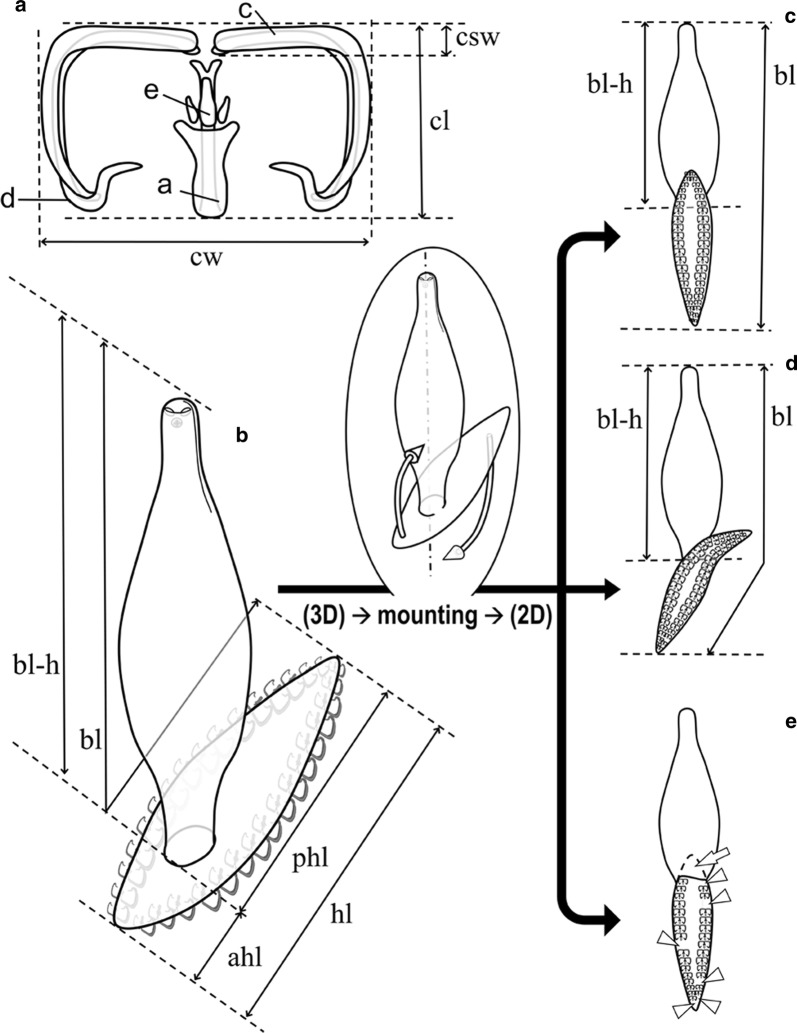
Parasites selected for morphological analyses were stained with iron acetocarmine, dehydrated through an ethanol series, cleared in dimethyl phthalate and prepared as permanent mounts in Canada balsam. After mounting, there was a second selection of specimens suitable for morphological studies, i.e. only specimens in optimal condition (not broken, contracted, stretched, wrinkled or folded). Parasites were examined using a light microscope Nikon Optiphot-2 (Nikon Instruments, Tokyo, Japan) with differential interference contrast at magnifications of 400–1000×. A total of 86 specimens of Microcotyle spp. were selected and drawn (n = 22 ex B. boops; n = 21 ex D. dentex; n = 23 ex P. erythrinus; n = 20 ex Pa. pagrus). Drawings were made with the aid of a drawing tube attached to a light microscope Nikon Optiphot-2. Measurements were taken from digitalized illustrations using ImageJ v.1.48 software [23] and expressed in micrometers as the range followed by the mean in parentheses unless otherwise stated. When characters were visible, a total of 52 morphometric measurements were taken from each specimen. Clamp thickness was estimated as both the maximum width of the distal end of the antero-lateral sclerite (‘c’, see Fig. 1a) and its relation to the clamp length. The type-specimens were deposited in the Collection of the Natural History Museum (NHMUK), London, UK.
Fig. 1.
Schematic drawings for measurements of microcotylid clamps and haptors. a Clamp measurements: ‘a’, ‘c’, ‘d’ and ‘e’, microcotylid sclerites according to Llewellyn [47]. b–e Body outlines, number of clamps and measurements in mounted microcotylids: unmounted specimen in 3D view (b); mounted specimens in 2D view (c–e). Haptor anterior lobe lays on the body in c and e, haptor obliquely mounted in d (anterior lobe not laying on the body); e represents damaged specimen with missing pieces (arrow) and clamps (arrowheads). Abbreviations: ahl, anterior haptor lobe length; bl, body length; bl-h, body length without haptor; cl, clamp length; cw, clamp width; csw, ‘c’ sclerite width; hl, haptor length; phl, posterior haptor lobe length
To look for patterns of separation between Microcotyle spp. specimens from different host species, a principal components analysis (PCA) was applied to a dataset of 86 specimens using morphometrical variables associated with body shape. Prior to the analysis, the data were divided by total body length to account for the effect of body size while visualising possible morphometric differences between species. The specimens were identified as M. erythrini (n = 23 ex P. erythrinus; n = 20 ex Pa. pagrus), M. isyebi (n = 22 ex B. boops) and Microcotyle whittingtoni n. sp. ex D. dentex (n = 21).
Results
Molecular identification
A total of 12 cox1 and four 28S rDNA sequences were generated for the newly collected specimens of Microcotyle spp. from the four fish species from the Western Mediterranean off Spain. Partial cox1 (434 nt) sequences were generated for a total of 12 isolates, i.e. 4 M. isyebi ex B. boops, 6 M. erythrini (4 ex Pa. pagrus and 2 ex P. erythrinus) and 2 M. whittingtoni n. sp. ex D. dentex. Partial 28S rDNA sequences (1238–1527 nt) were generated for a representative subset of the specimens used for cox1 sequence generation; single sequences per species were used for the reconstruction of the 28S rDNA phylogeny. The newly generated sequences for the isolates recovered in the present study were analysed in two separate datasets together with all currently available sequences in the GenBank database for Microcotyle spp. (see Table 1 for details on the ingroup taxa used in the analyses). Posterior probabilities (pp) and bootstrap support (bs) values are summarised on the BI trees (as pp/bs).
Table 1.
Summary data for the isolates of Microcotyle spp. used in the phylogenetic analyses
| Parasite species | Host species | Isolate | FAO Fishing Area | GenBank ID | Source | |
|---|---|---|---|---|---|---|
| cox1 | 28S | |||||
| M. algeriensis Ayadi, Gey, Justine & Tazerouti, 2016 | Scorpaena notata Rafinesque | MO-01 | WM | KX926443 | Ayadi et al. [24] | |
| Scorpaena notata | MO-02 | WM | KX926444 | Ayadi et al. [24] | ||
| Scorpaena notata | MO-03 | WM | KX926445 | Ayadi et al. [24] | ||
| M. archosargi MacCallum, 1913 | Archosargus rhomboidalis (L.) | 81 | WCA | MG586867 | Mendoza-Franco et al. [25] | |
| M. arripis Sandars, 1945 | Arripis georgianus (Valenciennes) | SA | GU263830 | Catalano et al. [26] | ||
| M. caudata Goto, 1894 | Sebastes inermis Cuvier | MC06 | NWP | LC472527 | Kamio & Ono (unpublished data) | |
| “Sebastes inermis species complex” | MC12 | NWP | LC472528 | Kamio & Ono (unpublished data) | ||
| “Sebastes inermis species complex” | MC18 | NWP | LC472529 | Kamio & Ono. (unpublished data) | ||
| “Sebastes inermis species complex” | MC20 | NWP | LC472530 | Kamio & Ono (unpublished data) | ||
| “Sebastes inermis species complex” | MC24 | NWP | LC472531 | Kamio & Ono (unpublished data) | ||
| M. erythrini van Beneden & Hesse, 1863 | Pagellus erythrinus (L.) | MePe1 | WM | MN814848 | Present study | |
| Pagellus erythrinus | MePe2 | WM | MN816012 | Present study | ||
| Pagellus erythrinus | MePe3 | WM | MN816013 | Present study | ||
| Pagellus erythrinus | WM | AY009159 | Jovelin & Justine [15] | |||
| Pagellus erythrinus | WM | AM157221 | Badets et al. [12] | |||
| Pagrus pagrus (L.) | MePp1 | WM | MN816014 | MN814849 | Present study | |
| Pagrus pagrus | MePp2 | WM | MN816015 | Present study | ||
| Pagrus pagrus | MePp3 | WM | MN816016 | Present study | ||
| Pagrus pagrus | MePp4 | WM | MN816017 | Present study | ||
| M. isyebi Bouguerche, Gey, Justine & Tazerouti, 2019 | Boops boops (L.) | MiBb1 | WM | MN816018 | MN814850 | Present study |
| Boops boops | MiBb2 | WM | MN816019 | Present study | ||
| Boops boops | MiBb3 | WM | MN816020 | Present study | ||
| Boops boops | MiBb4 | WM | MN816021 | Present study | ||
| Boops boops | MO01 | WM | MK317922 | Bouguerche et al. [3] | ||
| M. sebastis Goto, 1894 | Sebastes sp. | NSP | AF382051 | Olson & Littlewood [16] | ||
| Microcotyle sp. AKV-2016 | Nemipterus japonicas (Bloch) | VII37_12 | EAS | KU926692 | Verma & Agrawal (unpublished data) | |
| Microcotyle sp. DG-2016 | Helicolenus dactylopterus (Delaroche) | MO-04 | WM | KX926446 | Ayadi et al. [24] | |
| Helicolenus dactylopterus | MO-06 | WM | KX926447 | Ayadi et al. [24] | ||
| Sebastes schlegelii Hilgendorf | NWP | DQ412044 | Park et al. [27] | |||
| Microcotyle sp. YK-2019 | Sebastiscus marmoratus (Cuvier) | MK02 | NWP | LC472525 | Kamio & Ono (unpublished data) | |
| Microcotyle sp. YK-2019 | Sebastiscus marmoratus | MK01 | NWP | LC472526 | Kamio & Ono (unpublished data) | |
| Microcotyle sp. 1 SC-2018 | – | NWP | MH700256 | Chou (unpublished data) | ||
| Microcotyle sp. 2 SC-2018 | – | NWP | MH700266 | Chou (unpublished data) | ||
| Microcotylidae sp. M10 | Sebastes sp. | M10 | NWA | EF653385 | Aiken et al. [28] | |
| Microcotylidae sp. M11 | Argyrosomus japonicus | M11 | SA | EF653386 | Aiken et al. [28] | |
| M. visa Bouguerche, Gey, Justine & Tazerouti, 2019 | Pagrus caeruleostictus (Valenciennes) | PacoerMO01 | WM | MK275652 | Bouguerche et al. [2] | |
| Pagrus caeruleostictus | PacoerMO02 | WM | MK275653 | Bouguerche et al. [2] | ||
| Pagrus caeruleostictus | PacoerMO03 | WM | MK275654 | Bouguerche et al. [2] | ||
| M. whittingtoni n. sp. | Dentex dentex (L.) | MwDd1 | WM | MN816010 | MN814847 | Present study |
| Dentex dentex | MwDd2 | WM | MN816011 | Present study | ||
| “Paramicrocotyle” sp. FAS-2014a | Pinguipes chilensis (Valenciennes) | SWP | KJ794215 | Oliva et al. [29] | ||
| Outgroup | ||||||
| Bivagina pagrosomi (Murray, 1931) | Sparus aurata L.b | SWP | Z83003 | Z83002 | Littlewood et al. [10] | |
aGenus synonymized with Microcotyle [2, 30]
bAs Sparus auratus in Littlewood et al. [10]
Note: The newly generated sequences are indicated in bold
Abbreviations: CPS, Central-South-East Pacific; EAS, Eastern Arabian Sea; NS, North Sea; NWA, North-West Atlantic; NWP, North-West Pacific; SA, Southern Australia; SWP, South-West Pacific; WCA Western-Central Atlantic; WM, Western Mediterranean, –, not specified
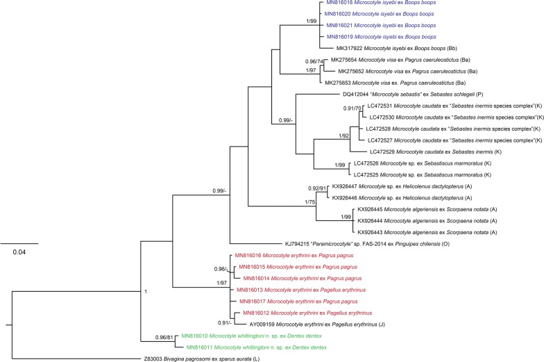
The newly generated cox1 sequences were analysed together with 19 published sequences for Microcotyle spp. (Table 1). Phylogenetic analysis revealed that the newly sequenced isolates belonged to 3 species: M. erythrini ex P. erythrinus and Pa. pagrus; M. isyebi ex B. boops; and M. whittingtoni n. sp. ex D. dentex. The tree from the BI analysis is provided in Fig. 2 together with the statistical support from the ML analysis. The four isolates recovered from B. boops clustered together with an isolate of M. isyebi from the same host species reported from the southern coast of the Western Mediterranean off Algeria [3]. The sequences for the isolates recovered from Pa. pagrus and P. erythrinus clustered together with the published sequences for M. erythrini ex P. erythrinus from the Western Mediterranean off France [15]. The two sequences for M. whittingtoni n. sp. ex D. dentex clustered together in a basal clade to the remaining representatives of Microcotyle spp. All of the above clades were strongly supported in both BI and ML analyses. Overall, the cox1 phylogeny (Fig. 2) recovered three groups of sister species within the Microcotyle although with poor support: (i) M. isyebi and M. visa Bouguerche, Gey, Justine & Tazerouti, 2019; (ii) M. caudata Goto, 1894 and an unidentified Microcotyle sp. ex Sebastiscus marmoratus (Cuvier) from the North-West Pacific off Japan; and (iii) M. algeriensis Ayadi, Gey, Justine & Tazerouti, 2016 ex Scorpaena notata Rafinesque and Microcotyle sp. ex Helicolenus dactylopterus (Delaroche) (syn. M. sebastis sensu Radujković & Euzet, (1989) [31]) (both reported from the Western Mediterranean off Algeria). The single sequence for ‘Microcotyle sebastis’ was close to the M. caudata-Microcotyle sp. from off Japan, and an isolate originally identified as “Paramicrocotyle sp.” (genus synonymised with Microcotyle [30]) ex Pinguipes chilensis Velenciennes from the South-East Pacific off Chile, was recovered as sister species to the major clade comprising the previously reported representatives from the Mediterranean, North-East Atlantic, Indian Ocean and the North-West Pacific. Microcotyle erythrini was recovered apart from the above-mentioned main multi-taxon clade albeit with low nodal support.
Fig. 2.
Bayesian inference (BI) phylogram based on the mitochondrial cox1 dataset for Microcotyle spp. Bivagina pagrosomi was used as the outgroup. Posterior probabilities and bootstrap support values are shown at the nodes; only values > 0.90 (BI) and 75% (ML) are shown. The scale-bar indicates the expected number of substitutions per site. Sequence identification is as in GenBank, followed by a letter: A, Ayadi et al. [24]; Ba, Bouguerche et al. [2]; Bb, Bouguerche et al. [3]; J, Jovelin & Justine [15]; K, Kamio & Ono (unpublished); O, Oliva et al. [29]; P, Park et al. [27]
The intraspecific sequence divergence (see Additional file 1: Table S1) within the newly generated cox1 sequences ranged between 0.2–1.4% (1–6 nt difference) for M. erythrini (ex P. erythrinus and Pa. pagrus); 0.2–0.5% (1–2 nt difference) for M. isyebi and 1.4% (6 nt difference) for M. whittingtoni n. sp. ex D. dentex. The newly generated sequences for the isolates of M. isyebi from off Spain differed by 1.4–1.7% (4–5 nt) from M. isyebi from off Algeria; these for M. erythrini differed by 2.1–2.8% (6–8 nt) from the published isolate from off Corsica (GenBank: AY009159); and the two isolates of M. whittingtoni n. sp. ex D. dentex differed substantially from both M. isyebi and M. erythrini, i.e. by 14.4–15.2% (43–56 nt) and by 10.8–13.5% (41–44 nt), respectively. The overall sequence divergence among the species of Microcotyle ranged between 4.5–18.5% (17–62 nt difference).
Both, ML and BI analyses for the 28S dataset yielded congruent tree topologies (Fig. 3) and high nodal support for most of the clades. Most of the species of Microcotyle clustered in a single multi-taxon clade with the unpublished sequence for an isolate identified as Microcotyle sp. ex Nemipterus japonicus (Bloch) from the Indian Ocean as a distinct, basal species. The newly generated sequences for M. erythrini ex P. erythrinus and Pa. pagrus, clustered in a strongly supported clade together with a previously published sequence for M. erythrini ex P. erythrinus off Corsica, France and a sequence for “Microcotylidae sp.” M11 ex Argyrosomus japonicus (Temminck & Schlegel) from off Australia. Microcotyle whittingtoni n. sp. and M. isyebi clustered together as close relatives of M. erythrini + “Microcotylidae sp.” M11. Microcotyle arripis Sandars, 1945 from the South-West Pacific off Australia and an isolate provisionally identified as Microcotyle sp. 2 from off China clustered together in a strongly supported subclade.
Fig. 3.
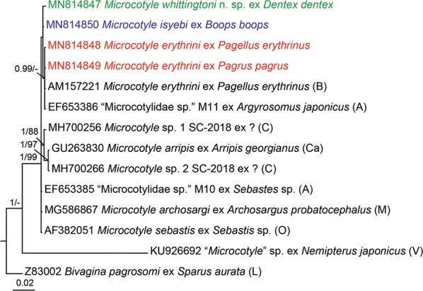
Bayesian inference (BI) phylogram based on the partial 28S rDNA sequences (domains D1-D3) for Microcotyle spp. Bivagina pagrosomi was used as the outgroup. Posterior probabilities and bootstrap support values are shown at the nodes; only values > 0.90 (BI) and 75% (ML) are shown. The scale-bar indicates the expected number of substitutions per site. Sequence identification is as in GenBank, followed by a letter: A, Aiken et al. [28]; B, Badets et al. [12]; C, Chou (unpublished data); Ca, Catalano et al. [26]; L, Littlewood et al. [10]; M, Mendoza Franco et al. [25]; O, Olson & Littlewood [16]; V, Verma & Agrawal (unpublished)
The novel 28S sequences for M. erythrini recovered from the two fish host species differed by a single base. The novel sequence for M. erythrini from P. erythrinus was identical with the published sequence ex P. erythrinus (GenBank: AM157221) in the Western Mediterranean (see Additional file 2: Table S2). The 28S rDNA sequences for M. isyebi and M. whittingtoni n. sp. differed from the M. erythrini isolates by 1 and 3 nt (0.1 and 0.4%), respectively, and by 2 nt (0.2%) between themselves. Microcotyle sp. AKV-2016 (KU926692) differed substantially from the remaining Microcotyle spp., i.e. by 80–116 nt (12.8–14.7%) corresponding to intergeneric-level differences. The overall sequence divergence among the species of Microcotyle ranged between 1–8 nt (0.1–1.0%).
Morphological data
Microcotyle erythrini van Beneden & Hesse 1863 (sensu stricto)
Hosts: Pagellus erythrinus (L.) (type-host), common pandora [weight: 98.9–160.0 g (123 ± 17.3 g); standard length: 15.8–23 cm (17.5 ± 1 cm)] off Guardamar del Segura, Spain; Pagrus pagrus (L.), red porgy [weight: 84.2–289.3 g (175.0 ± 44.5 g); standard length: 13.4–20.5 cm (17.12 ± 1.5 cm)], off Guardamar del Segura, Spain (both Perciformes: Sparidae).
Locality: Off Guardamar del Segura, Western Mediterranean off Spain. Other localities with valid records: off Brest, France (type-locality); Boka Kotorska Bay and off Montenegro coast, Montenegro; off Sète, France.
Voucher material: Specimens from P. erythrinus (n = 3) and Pa. pagrus (n = 3) from off Guardamar del Segura are deposited in the Natural History Museum, London, UK (NHMUK.2019.12.10.6-8 and NHMUK.2019.12.10.9-11, respectively); the remaining material from Guardamar del Segura is deposited in the Parasitological Collection of the Cavanilles Institute of Biodiversity and Evolutionary Biology, University of Valencia, Spain.
Infection parameters: P. erythrinus (n = 40): prevalence, 51% (21 out of 40); mean intensity, 2.2 ± 3.8; Pa. pagrus (n = 30); prevalence, 45% (14 out of 30); mean intensity, 2.2 ± 1.7.
Site on host: Gill filaments.
Representative DNA sequences: GenBank accession numbers: MN816012 and MN816013 ex P. erythrinus; MN816014, MN816015, MN816016 and MN816017 ex Pa. pagrus (cox1); MN814848 ex P. erythrinus and MN814849 ex Pa. pagrus (28S).
Description
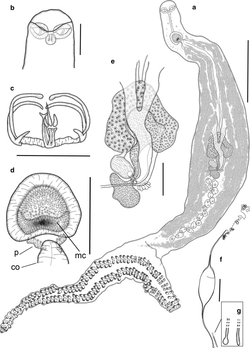
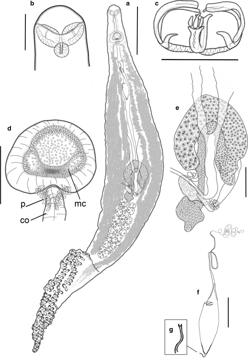
[Based on 43 mature adults (23 ex P. erythrinus and 20 ex Pa. pagrus), except where otherwise indicated; data in the description are reported as mean ± SD for specimens ex P. erythrinus [mean ± SD ex Pa. pagrus]; ranges are provided in Table 2; Fig. 4]. Body fusiform, elongate, slender, 3532 ± 918 [3630 ± 835] long, 182 ± 38 [182 ± 37] wide at level of genital atrium and 249 ± 92 [258 ± 57] wide at level of testes, tapered anteriorly up to 566 ± 107 (n = 18) [513 ± 141 (n = 13)] from anterior extremity of body; body laterally narrowed at posterior end of anterior tapered region, 204 ± 55 (n = 18) [190 ± 39 (n = 13)] wide, often posteriorly delimited by lateral notches. Haptor dorsoventrally bi-lobed, elongated (haptor length/total body length ratio: 38–62% (44%) [35–60% (42%)]), well differentiated from body, sometimes with peduncle 448 ± 103 (n = 7) [414 ± 127 (n = 4)] long, with minimum width 174 ± 36 (n = 7) [158 ± 47 (n = 4)]; haptor laterally symmetric, ventrally projected in anterior (ventral) lobe and longer posterior (dorsal) lobe (anterior/posterior haptoral lobe length ratio 60–90% (72%) (n = 20) [52–81% (66%) (n = 19)]. Haptor armed with two rows of sessile clamps, 90–124 in number for specimens from both host species, in two lateral frills, joining at anterior and posterior extremities of haptor; with slightly smaller clamps at anterior and posterior margins of haptor. Clamps of “microcotylid” type, slender, “c” sclerite maximum width 3 ± 1 for specimens from both host species and 0.084 ± 0.015 (n = 25) [0.068 ± 0.019 (n = 25)] corrected by clamp length; with trident-shaped accessory sclerite (‘e’, see Fig. 1a) formed by thick central bar reaching to distal tips of antero-lateral sclerites ‘c’ and two thin short sclerites directly branched from basis of ‘e’.
Table 2.
Metrical ranges for Microcotyle erythrini (sensu stricto), M. isyebi and M. whittingtoni n. sp. described in this study based on collections from off Guardamar del Segura, Spain, Western Mediterranean
| Parasite species | M. erythrini (s.s.) | M. isyebi | M. whittingtoni n. sp. | |
|---|---|---|---|---|
| Host species | P. erythrinus | Pa. pagrus | B. boops | D. dentex |
| Sample size | (n = 23) | (n = 20) | (n = 22) | (n = 21) |
| Body length | 1998–6215 | 2042–6183 | 2355–6401 | 2719–4569 |
| Body length without haptor | 1376–3760 | 1520–5307 | 1757–4970 | 1916–3591 |
| Maximum body width | 194–647 | 189–610 | 322–966 | 314–605 |
| Body width at level of buccal suckers | 95–179 | 68–153 | 95–185 | 110–173 |
| Body width at level of genital atrium | 102–251 | 109–270 | 157–322 | 159–260 |
| Body width at level of testes | 156–574 | 150–357 | 237–790 | 225–468 |
| Length of anterior tapered region | 359–749 | 132–687 | 288–922 | 457–730 |
| Width of anterior tapered region | 135–378 | 124–242 | 180–388 | 147–354 |
| Haptor length | 1126–1840 | 761–1590 | 702–1436 | 862–1264 |
| Anterior haptoral lobe length | 353–735 | 307–690 | 164–339 | 187–370 |
| Posterior haptoral lobe length | 758–1,163 | 608–991 | 474–1,226 | 632–1,038 |
| Peduncle length | 339–615 | 263–574 | 178–651 | 199–476 |
| Width of peduncle at connection with haptor | 120–246 | 72–227 | 92–414 | 100–369 |
| Minimum peduncle width | 120–276 | 67–227 | 91–414 | 81–368 |
| No. of clamps | 90–124 | 90–124 | 80–110 | 60–78 |
| Clamp length | 20–50 | 22–42 | 21–40 | 22–47 |
| Clamp width | 55–86 | 40–64 | 46–68 | 52–75 |
| Sclerite ‘c’ width | 2–4 | 2–3 | 1–3 | 3–6 |
| Buccal sucker length | 47–80 | 38–78 | 50–82 | 57–90 |
| Buccal sucker width | 34–59 | 19–50 | 33–63 | 39–59 |
| Pharynx length | 29–47 | 20–42 | 29–55 | 27–41 |
| Pharynx width | 23–41 | 18–34 | 23–54 | 25–43 |
| Oesophagus length | 159–307 | 217–343 | 150–360 | 209–367 |
| Testes to anterior extremity distance | 982–2223 | 1055–4018 | 1215–3288 | 1305–2342 |
| No. of testes | 12–20 | 14–22 | 19–26 | 16–27 |
| No. of testis rows | 1–2 | 1–2 | 2–3 | 1–3 |
| Testes length | 32–115 | 38–82 | 32–162 | 39–112 |
| Testes width | 51–116 | 42–75 | 52–132 | 36–88 |
| Testicular area length | 298–1007 | 364–1250 | 88–301 | 68–717 |
| Testicular area width | 41–193 | 56–155 | 408–1558 | 393–1023 |
| Genital atrium to anterior extremity distance | 108–277 | 206–424 | 128–355 | 183–300 |
| Genital atrium length | 71–151 | 71–165 | 73–177 | 105–184 |
| Genital atrium width | 88–168 | 54–201 | 109–255 | 115–173 |
| No. of spines in the main chamber of the genital atrium | 216–408 | 275–363 | 253–356 | 272–391 |
| Length of spines in the main chamber of the genital atrium | 4–6 | 4–6 | 4–7 | 4–7 |
| No. of spines in the “pockets” of the genital atrium | 20–33 | 21–41 | 19–49 | 34–47 |
| Length of spines in the “pockets” of the genital atrium | 6–8 | 6–9 | 7–11 | 7–13 |
| Copulatory organ length | 43–100 | 47–70 | 52–86 | 46–107 |
| Copulatory organ width | 54–100 | 33–100 | 54–118 | 37–93 |
| Germarium to anterior extremity distance | 1093–2149 | 1381–2460 | 1141–3240 | 1240–2240 |
| Vagina to anterior extremity distance | 321–550 | 380–585 | 328–425 | 380–550 |
| Germarium length | 566–1428 | 890–1428 | 896–1792 | 730–1199 |
| Germarium maximum width | 37–127 | 83–96 | 47–134 | 38–86 |
| Seminal receptacle length | 144–171 | 140–185 | 289–316 | 182–208 |
| Seminal receptacle width | 89–105 | 80–99 | 96–112 | 70–87 |
| Vitellarium to anterior extremity distance | 204–560 | 319–613 | 244–578 | 320–566 |
| Length of vitellarium within peduncle | 120–450 | 138–589 | 18–530 | 94–621 |
| Length of vitellarium within haptor | 10–111 | 19–42 | 0–0 | 17–404 |
| Distance between vitellarium posterior | 0–149 | 0–152 | 0–462 | 129–498 |
| Left efferent vitelline duct length | 87–287 | 124–444 | 127–858 | 216–467 |
| Right efferent vitelline duct length | 114–328 | 153–377 | 150–584 | 202–442 |
| Different vitelline duct length | 143–400 | 224–367 | 205–456 | 121–300 |
| Egg length (without filaments) | 166–223 | 145–201 | 189–230 | 184–264 |
| Egg width (without filaments) | 57–91 | 57–90 | 51–88 | 66–84 |
| Abopercular filament length | 95–151 | 98–147 | 110–208 | 40–111 |
Fig. 4.
Microcotyle erythrini van Beneden & Hesse (1863) (sensu stricto) ex Pagellus erythrinus (L.) from off Guardamar del Segura, Spain. All drawings from the same voucher specimen. a Whole mount. b Anterior body end. c Clamp. d Genital atrium, including copulatory organ. e Germarium. f Egg. g Detail of abopercular egg filament end. Abbreviations: co, copulatory organ; mc, main chamber of the genital atrium; p, small posterior chambers (“pockets” sensu Mamaev [44]). Scale-bars: a, 500 µm; b, d–f, 100 µm; c, 50 µm
Mouth subventral, within conical vestibule with pair of septate buccal suckers. Pharynx subspherical; oesophagus short; intestinal bifurcation posterior to genital atrium, sometimes at level of atrium. Caeca extend into haptor or peduncle, with inner and intricate external lateral ramifications.
Testes numerous, 12–20 [14–22] in number, dorso-ventrally flattened, subelliptical to irregular, most anterior located at 1536 ± 360 [1922 ± 585] from anterior extremity, post-germarial and pre-haptoral, partially extending into haptor peduncle, arranged in clusters of 1 or 2 rows, with some testes dorso-ventrally overlapped. Vas deferens relatively straight, dorsal to uterus; copulatory organ muscular, 68 ± 22 (n = 12) [62 ± 7 (n = 8)], located in posterior part of genital atrium. Genital atrium at 216 ± 40 [267 ± 63] from anterior extremity of body, with wide medial muscular chamber, armed with small conical spines, 216–408 [275–363] in number, communicated with 2 lateral posterior small chambers (“pockets” sensu Mamaev, 1989 [44]) armed with longer spines, 20–33 [21–41] in number.
Germarium at 1503 ± 292 (n = 8) [1758 ± 268 (n = 6)] from anterior extremity of body; 867 ± 251 (n = 8) [913 ± 150 (n = 6)] long, question mark-shaped, with proximal globular germinal area, 71 ± 25 × 110 ± 20 (n = 8) [76 ± 9 × 121 ± 43 (n = 6)], connected with narrow straight section, 278 ± 104 × 35 ± 20 (n = 8) [292 ± 92 × 45 ± 14 (n = 6)], widening in long distal globular region, 599 ± 50 (n = 8) [621 ± 51 (n = 6)] long, with proximal arched section directed dextro-sinistrally, connected to wide arched section directed sinistro-dextrally; maximum width at distal section, 71 ± 28 (n = 8) [90 ± 10 (n = 6)]. Oviduct slightly sinuous, including elongated seminal receptacle, 99 ± 7 × 160 ± 12 (n = 4) [93 ± 8.3 × 163 ± 13 (n = 2)] directed postero-sinistrally; ending in oötype; Mehlis’ gland well developed.
Vaginal pore mid-dorsal, often imperceptible, at 465 ± 64 (n = 11) [473 ± 59 (n = 8)] from anterior extremity. Vitelline follicles dispersed, starting at 356 ± 81 [437 ± 82] from anterior body extremity, in 2 lateral fields surrounding caecal ramifications; vitelline follicles extending within haptor or peduncle in all specimens. Posterior extremities of vitelline fields asymmetrical in 52% [95%] of specimens, distance between fields usually short, 83 ± 54 [89 ± 46]; right field longer in 56% [61%] of specimens with asymmetrical fields; posterior extremities of vitelline fields often joined (83% [45%] of specimens with symmetrical fields). Vitelline ducts Y-shaped (Fig. 4e), with 2 separate efferent ducts, right 204 ± 67 [224 ± 91] long, left 178 ± 77 [304 ± 119] long, joining in common different duct 214 ± 79 [289 ± 67] long, ventral, at germarium level. Eggs fusiform (Fig. 4f), with 2 filaments; opercular filament long, thin, slightly thickened at posterior end; abopercular filament shorter with solid tip, capitate or pointed (Fig. 4g). Opercular end of egg narrowed to connect abruptly with tubular hollow section (1/3–1/7 of total egg length, not including filaments, for specimens for both host species) leading to opercular filament.
Remarks
Microcotyle erythrini was described by van Beneden & Hesse [4] and mostly characterized by its specific host, P. erythrinus, as authors provided limited morphological information (mostly at the generic level) and with no supporting drawing. Parona & Perugia [5] redescribed this species; however the description is unreliable as these authors provided pooled morphological information from material ex B. boops, host of M. isyebi (see [3] and present study) and ex P. acarne, a host not confirmed for M. erythrini. Morphological data with pooled information form specimens collected from more than one host species or parasites collected in fish species different from the type-host or other confirmed hosts should not be considered as suitable (see also Additional file 3: Table S3). Several new geographical records of M. erythrini ex P. erythrinus, exclusively, have been published by other authors since 1863 (see Table 1 in [3]). Among these records, only Radujković & Euzet [31] and Bouguerche et al. [3] provided morphological and morphometric data for specimens off Montenegro and Séte, respectively (see also Additional file 3: Table S3). Here, we provide metrical data (Table 2) for newly collected specimens ex P. erythrini and Pa. pagrus (new host record) from the Spanish Western Mediterranean. Specimens from these two hosts collected in the present study are genetically and morphologically indistinguishable.
Only considering the specimens reported ex P. erythrinus and Pa. pagrus by van Beneden & Hesse [4], Radujković & Euzet [31], Bouguerche et al. [3] and the present study [from here onwards M. erythrini (sensu stricto)], the diagnostic characters of M. erythrini (s.s.) agree and measurements mostly overlap but wide ranges for some features are still observed (see also Additional file 3: Table S3), which hampers the differentiation from other congeneric species. Paying attention to the characters traditionally used in the taxonomy of Microcotyle, the number of clamps (82–132) and the number of testes (9–24) of M. erythrini (s.s.), combining the information from all descriptions in confirmed hosts (see Table 2 and Additional file 3: Table S3), resemble or overlap with those of several species reported in the Mediterranean (M. donavini van Beneden & Hesse, 1863 and M. pomatomi Goto, 1899) and in other sparid hosts (M. isyebi and M. visa). Regarding the traits more recently used to differentiate the species of Microcotyle, such as the genital atrium armature and combining the information from all descriptions in confirmed hosts (see Table 2 and Additional file 3: Table S3), M. erythrini (s.s.) resembles other species with large number of spines in the main chamber (201–408) and “pockets” of the genital atrium (20–41), overlapping with M. isyebi, M. pomatomi, M. visa, M. whittingtoni n. sp. and Microcotyle sp. ex H. dactylopterus (see [24, 31]; numbers estimated from the drawing for M. pomatomi) (see Additional file 3: Table S3).
According to the combination of the characters listed above, M. isyebi, M. pomatomi and M. visa appear most similar morphologically to M. erythrini (s.s.). Microcotyle pomatomi, the only species described and reported from a non-sparid host (Pomatomus saltatrix (L.); Pomatomidae), is difficult to differentiate due to the numerous circumglobal records and descriptions which have increased abnormally the ranges for the metrical data of this species. Moreover, the only Mediterranean description of M. pomatomi (off Turkey, Sezen & Price, 1967 in [32]) is particularly similar to M. erythrini (s.s.). Detailed morphological and molecular studies are needed to differentiate the two species. The other two species, both sparid parasites, were described as hardly morphologically distinguishable from M. erythrini. Microcotyle visa was differentiated from M. erythrini by the smaller clamp size, larger pharynx and greater number of testes; however, these differences are not completely sufficient to differentiate species as all they overlap (even with those of M. erythrini (s.s.)) [2]. No diagnostic morphological differences were provided by Bouguerche et al. [3] to distinguish M. isyebi from M. erythrini, other than body size, different hosts and large genetic divergence based on cox1 data. New evidence reported in the present study allows characterizing M. erythrini (s.s.) based on the size and shape of the haptor which is relatively longer in relation to body length (35–62% vs 27–34% in M. visa and 21–32% in M. isyebi) and the greater ratio of anterior/posterior haptoral lobe length (52–90% vs 34–50% in M. visa and 17–52% in M. isyebi) (data for M. visa estimated from figure 3A in Bouguerche et al. [2]; those for M. isyebi from the present study). The anterior/posterior haptoral lobe length ratio range for M. erythrini (s.s.) is very close to the upper range limits for these two species; however, the ratio was > 60% in some of the M. erythrini (s.s.) specimens examined here (11 out of 21 ex P. erythrinus and 5 out 18 ex Pa. pagrus). Additionally, vitelline fields always extend within the haptor in M. erythrini (s.s.). Bougherche et al. [3] reported that the left caecum-vitellarium branch of M. isyebi extends into haptor; however, both vitellarium fields of the M. isyebi specimens analysed in the present study are always prehaptoral. Finally, in the new material from the Spanish Mediterranean, the tips of the abopercular filaments of the eggs are solid (capitated or pointed) in M. erythrini (s.s.) vs half cup-shaped to bifid in M. isyebi. No information on this trait is available for M. visa.
Microcotyle isyebi Bouguerche, Gey, Justine & Tazerouti, 2019
Host: Boops boops (L.) (type-host) (Teleostei: Sparidae), bogue [weight: 112.9–216.7 g (157.2 ± 22.3 g); standard length: 19.8–24.0 cm (21.7 ± 1 cm)], off Guardamar del Segura, Spain.
Locality: Off Guardamar del Segura, Western Mediterranean off Spain. Other localities with valid records: off Bouharoun, Algeria (type-locality) and off Granada, Spain.
Voucher material: Three specimens from off Guardamar del Segura are deposited in the Natural History Museum, London, UK (NHMUK.2019.12.10.12-14); the remaining material from Guardamar del Segura is deposited in the Parasitological Collection of the Cavanilles Institute of Biodiversity and Evolutionary Biology, University of Valencia, Spain.
Infection parameters: Prevalence: 70% (28 out of 40); mean intensity, 4.96 ± 4.46 (n = 40).
Site on host: Gill filaments.
Representative DNA sequences: GenBank accession numbers: MN816018, MN816019, MN816020 and MN816021 (cox1); MN814850 (28S).
Description
[Based on 22 mature adults (paragenophores sensu [3]); data in the description are reported as mean ± SD, ranges are provided in Table 2; Fig. 5]. Body fusiform, stout to elongate. Anterior region tapered, 585 ± 147 long, posteriorly delimited by lateral notches, which narrow body to 271 ± 57 wide. Body width 221.0 ± 45 at level of genital atrium, 411 ± 130 at level of testes. Haptor relatively short [haptor length/total body length ratio 21–32% (26%)], dorsoventrally bi-lobed, well differentiated from body by peduncle 402 ± 149 long, with minimum width 238 ± 72 (n = 19); laterally symmetric, divided into anteriorly projected very short ventral lobe and longer posterior lobe (dorsal) [anterior haptoral lobe/posterior haptoral lobe length ratio 17–52% (33%)]. Haptor armed with two rows of sessile clamps, 80–110 in number, in two lateral frills, joining at anterior and posterior extremities of haptor. Clamps at anterior and posterior extremities of haptor slightly smaller. Clamps of “microcotylid” type, slender, “c” sclerite maximum width 2 ± 1 and 0.057 ± 0.021, corrected by clamp length (n = 25); with trident-shaped accessory sclerite (‘e’, see Fig. 1) formed by long thick central bar reaching to distal tips of antero-lateral sclerites ‘c’ and 2 thin branches directly ramified from basis of ‘e’.
Fig. 5.
Microcotyle isyebi Bouguerche, Gey, Justine & Tazerouti, 2019 ex Boops boops (L.) from off Guardamar del Segura, Spain. All drawings are from the same voucher specimen, except for the egg. a Whole mount. b Anterior end. c Clamp. d Genital atrium, including copulatory organ. e Germarium. f Egg. g Detail of abopercular egg filament. Abbreviations: co, copulatory organ; mc, main chamber of the genital atrium; p, small posterior chambers (“pockets” sensu Mamaev [44]). Scale-bars: a, 500 µm; b, d–f, 100 µm; c 50 µm
Mouth subventral, within funnel-shaped vestibule with pair of septate buccal suckers. Pharynx subspherical; oesophagus short, connected to intestinal bifurcation at posterior margin of genital atrium, or more posterior. Caeca extend up to haptor peduncle, with inner and external lateral ramifications (external more profuse).
Testes numerous, 19–26 in number, dorso-ventrally flattened, sub-elliptical to irregular, grouped in testicular fields, most anterior located at 2016 ± 476 from anterior extremity of body, post-germarial and pre-haptoral (partially extending into haptor peduncle); testes arranged in clusters of 2 or 3 rows, with some testes overlapped dorso-ventrally. Vas deferens wide, straight, dorsal to uterus, terminating in short muscular copulatory organ, 67 ± 13 × 81 ± 21 (n = 10), located in posterior part of genital atrium. Genital atrium at 233 ± 45 from anterior extremity of body, formed by main wide medial muscular chamber, armed with small conical spines, 253–356 in number, followed by 2 small chambers (“pockets”) located posteriorly, at both sides of copulatory organ; “pockets” armed with conical spines, 19–49 in number, longer than spines in main chamber (see Table 2).
Germarium 1274.7 ± 199 long (n = 10), question mark-shaped, at 1674 ± 472 (n = 10) from anterior extremity of body; proximal globular germinal area 75 ± 28 × 135 ± 38 (n = 10), connected to straight, narrow section 398 ± 104 × 43 ± 14 (n = 10), widening in tubular region 877.1 ± 231 long (n = 10) with proximal arched dextro-sinistral section, connected to wider distal arch directed sinistro-dextrally, with maximum distal width 83 ± 28 (n = 10). Oviduct directed postero-sinistrally ending in oötype, sinistral to germarium, with short sinuous proximal section connected with wide elongated chamber filled with sperm (oviducal seminal receptacle 106 ± 7 × 304 ± 112 (n = 3). Mehlis’ gland well developed.
Vaginal pore medial, dorsal, unarmed, often unobserved, at 372.8 ± 62 (n = 8) from anterior extremity. Vitelline follicles dispersed, extending from 391 ± 82 from anterior extremity of body, extended in 2 lateral fields together with caeca and surrounding testes, usually pre-haptoral but partly extending within peduncle. Posterior extremities of vitelline fields mostly different in length (79% of specimens with asymmetrical fields), always unjoined; distance between fields, 0–462, right field longer in 70% of the specimens. Vitelline ducts Y-shaped, with 2 unjoined ducts 339 ± 150 and 357 ± 213 long (right and left, respectively) (n = 10), joining ventral to germarium in slightly sinuous defferent duct, 337 ± 84 (n = 10) long. Egg fusiform (Fig. 5f), with 2 filaments; opercular filament long, thin, often with thickened final tip; abopercular filament short, ending in thickened tip, half cup-shaped to bifid (Fig. 5g). Opercular extremity of egg narrowing abruptly in tubular hollow section (1/3–1/6 of total egg length, not including filaments) leading to opercular filament.
Remarks
Both morphological and molecular data reported in the present paper agree with the original description of M. isyebi based on material from B. boops off Algeria [3] and from the Spanish Mediterranean [33]. Parona & Perugia [5] and Akmirza [6] also provided morphological data from specimens identified as M. erythrini ex B. boops but these were not considered as species diagnostic in the present study as they represent pooled information for parasites ex B. boops and another host, P. acarne; Microcotyle spp. in sparids are highly host species-specific (see [2, 3] and the present study). In the present study, no specimens of Microcotyle spp. were found in P. acarne.
Some comments on the original diagnosis of the species can be added in light of the data from the description of López-Román & Guevara Pozo [33] and the present study. The range for the number of clamps seems too wide in the original description of M. isyebi based on material collected off Algeria (54–102) compared with that reported by López-Román & Guevara Pozo [33] (90–100) and the present study (80–110) based on material collected off Spain. These numbers should be re-examined, especially because this trait is particularly differential among the species of Microcotyle and, as previously reported, it could help differentiate M. isyebi from other similar species such as Microcotyle sp. ex H. dactylopterus [24] and M. whittingtoni n. sp. (see [3] and Remarks to the new species below). Particular attention must be paid to the lower range for clamp number as a small number of clamps is often related to young or damaged specimens. The number of spines in the main chamber of the genital atrium of M. isyebi is also clearly lower in the original description than in the present material (136–230 vs 253–356) (see [3] and Table 2), thus enlarging the range for M. isyebi and making this trait almost useless in characterizing this species as it overlaps with most of the species except for M. donavini and M. omanae Machkewskyi, Dimitrieva, Al-Jufaili & Al-Mazrooei, 2013 (with lower and higher number of spines respectively, see Additional file 4: Table S4). The presence of posterior small chambers of the genital atrium (“pockets”) was also reported as diagnostic in the original description of M. isyebi; however, this feature requires a further comment. According to Bouguerche et al. [3], “pockets” are absent in M. archosargi, M. lichiae Ariola, 1899 and M. pomatomi; however, this difference seems to be valid only for M. lichiae, as these small chambers exist in M. archosargi and M. pomatomi according to the drawings in [25] and [32], respectively.
In the original description of the species, M. isyebi was differentiated from M. pomatomi and from Microcotyle sp. ex H. dactylopterus [24] by traits with overlapping ranges (the number of clamps and spines of the genital atrium for Microcotyle sp. ex H. dactylopterus) or almost overlapping ranges (the number of clamps and testes for M. pomatomi). Microcotyle pomatomi and Microcotyle sp. ex H. dactylopterus [24] require further taxonomic research; M. pomatomi has numerous descriptions and synonyms worldwide which have expanded extremely the ranges for morphological features (see [32]; also the only Mediterranean record by Sezen & Price (1967) in [32]), and the morphology Microcotyle sp. ex H. dactylopterus has been only briefly described [24, 31].
Bouguerche et al. [3] reported that M. isyebi is almost indistinguishable from M. erythrini. As mentioned above, examination of mature, entire, uncontracted, unstretched and unfolded specimens of this species would be helpful to define or shorten some of the descriptive morphological ranges. Other morphological traits suggested in the present study reveal additional differences. Thus, M. isyebi differs from M. erythrini (s.s.) in having a shorter haptor in relation to body length (21–32 vs 35–62%) and a shorter anterior haptoral lobe in relation to posterior haptoral lobe length (17–52 vs 52–90%) and from M. whittingtoni n. sp. in the possession of slender clamps (ratio “c” sclerite maximum width/total clamp length, 0.027–0.88 vs 0.100–0.146; see the Remarks for M. whittingtoni n. sp. below).
Microcotyle whittingtoni n. sp.
Synonym: Microcotyle erythrini van Beneden & Hesse, 1863 of González González (2005) [36].
Type-host: Dentex dentex (L.) (Teleostei: Sparidae), common dentex [weight: 204.0–296.2 g (227.5 ± 24 g); standard length: 22.3–20.0 cm (20.8 ± 0.7 cm)], off Guardamar del Segura, Spain).
Type-locality: Off Guardamar del Segura, Western Mediterranean off Spain. Other locality with a valid record: off Balearic Islands, Spain.
Type-material: The holotype (NHMUK.2019.12.10.1) and 3 paratypes (NHMUK.2019.12.10.2-5) from off Guardamar del Segura are deposited in the Natural History Museum, London, UK; the remaining material from off Guardamar del Segura is deposited in the Parasitological Collection of the Cavanilles Institute of Biodiversity and Evolutionary Biology, University of Valencia, Spain.
Infection parameters: Prevalence, 58% (23 out of 40); mean intensity, 4.36 ± 5.18 (n = 40).
Site on host: Gill filaments.
Representative DNA sequences: GenBank accession numbers: MN816010 and MN816011 (cox1); MN814847 (28S).
ZooBank registration: To comply with the regulations set out in article 8.5 of the emended 2012 version of the International Code of Zoological Nomenclature (ICZN, 2012) details of the new species have been submitted to ZooBank. The life Science Identifer (LSID) for Microcotyle whittingtoni n. sp. is urn:lsid:zoobank.org:act:5E369A8A-0EA2-4ED2-A3C6-0D6E4CC5A390.
Etymology: The new species is named in honour of the late Dr Ian David Whittington, eminent researcher on monogenean biology and taxonomy. His comprehensive, meticulous and brilliant studies have inspired and encouraged fish parasitologists worldwide.
Description
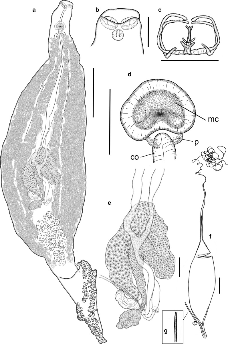
[Based on 21 mature adults, except when otherwise indicated; data in the description are reported as mean ± SD; ranges are provided in Table 2; Fig. 6]. Body fusiform, elongate, occasionally slender, 3509 ± 507 long, tapered anteriorly at 563 ± 79 (n = 20) from anterior extremity of body; anterior tapered region posteriorly delimited by lateral notches, which narrow body to 253 ± 54 (n = 20) wide. Body 212 ± 27 wide at level of genital atrium and 316 ± 69 wide at testes level. Haptor dorsoventrally bi-lobed, relatively long [haptor length/total body length ratio 24–35% (30%)], well differentiated, sometimes with peduncle [peduncle 309 ± 104 long, with minimum width 204 ± 77 (n = 6)]; laterally symmetric, with short ventral lobe projected anteriorly and longer posterior (dorsal) lobe [anterior/posterior haptoral lobe length ratio 21–52% (36%)]. Haptor armed with sessile clamps, 60–78 in number, in two rows in lateral frills, joining at anterior and posterior extremities of haptor; clamps slightly smaller at anterior and posterior extremities of haptor. Clamps robust, “c” sclerite maximum width 5 ± 1 and 0.120 ± 0.017 corrected by clamp length (n = 25); “microcotylid” type with trident-shaped accessory sclerite (‘e’, see Fig. 1) with long thick central bar reaching to distal tips of antero-lateral sclerites ‘c’ and 2 delicate branches ramified from basis of ‘e’.
Fig. 6.
Microcotyle whittingtoni n. sp. ex Dentex dentex (L.) from off Guardamar del Segura, Spain. Holotype. a Whole mount. b Anterior end. c Clamp. d Genital atrium, including copulatory organ. e Germarium. f Egg. g Detail of abopercular egg filament. Abbreviations: co, copulatory organ; mc, main chamber of the genital atrium; p, small posterior chambers (“pockets” sensu Mamaev [44]). Scale-bars: a, 500 µm; b, d–f, 100 µm; c, 50 µm
Mouth subterminal, ventral, with 2 septate buccal suckers within funnel-shaped vestibule; oesophagus short; intestinal bifurcation at level of posterior margin of genital atrium or just proterior. Caeca with inner and profuse external lateral ramifications extending into haptor.
Testes numerous, 16–27 in number, dorso-ventrally flattened, subelliptical to irregular, arranged in clusters of 1–3 rows, with some testes overlapping dorso-ventrally; testicular field at 1807 ± 304 from anterior extremity of body, post-germarial and pre-haptoral, partially extending into haptor peduncle. Vas deferens wide, coursing dorsal to uterus, straight up to short muscular copulatory organ, 80 ± 17 × 60 ± 19 (n = 10), opening into posterior part of genital atrium. Genital atrium at 240 ± 34 from anterior extremity of body, with muscular wall, formed by wide main medial chamber, covered with tiny conical spines (272–391 in number), connected with 2 postero-lateral small chambers (“pockets”) armed with longer curved spines (34–47 in number), flanking copulatory organ.
Germarium elongated 953 ± 331 long (n = 11), question mark-shaped, at 1336 ± 167 (n = 11) from anterior extremity of body; proximal globular germinal area 58 ± 13 × 119 ± 44 (n = 11) followed by straight narrow section 311 ± 59 × 37 ± 8 (n = 11), connected with wide tubular region 642 ± 180 (n = 11) formed by 2 arches crossing first dextro-sinistrally and then sinistro-dextrally, gradually widening up to maximum width of 60 ± 16 (n = 11) at distal section. Oviduct directed postero-sinistrally ending in oötype; connected to elongated oviducal seminal receptacle 83 ± 7 × 197 ± 12 (n = 3) by sinuous narrow section. Mehlis’ gland well developed.
Vaginal pore mid-dorsal, unarmed, inconspicuous, at 487 ± 59 from anterior extremity of body (n = 10). Vitelline follicles small, scattered from 423 ± 59 from anterior extremity of haptor, with lateral fields accompanying caecal ramifications and surrounding testes; vitellarium spread along peduncle and within haptor in all specimens. Posterior extremities of vitelline fields unjoined, always different in length (distance between fields, 308 ± 113; right field longer in 86% of the individuals). Vitelline ducts Y-shaped; efferent ducts 316 ± 81 and 331 ± 79 (right and left, respectively) long (n = 10) separated up to germarium and ventrally joined in slightly sinuous defferent duct, 228 ± 55 long (n = 10). Eggs fusiform (Fig. 6f), with 2 filaments; opercular filament very long, thin, with slightly thicker final tip; abopercular filament shorter, ending in half cup-shaped to bifid tip (Fig. 6g). Opercular end of egg narrowing gradually to connect through conical hollow section (1/6–1/8 of total egg length, not including filaments) leading to opercular filament.
Remarks
Microcotyle whittingtoni n. sp. differs from M. erythrini (s.s.) by the number of clamps (60–78 vs 82–132), the haptor length/total body length ratio (24–35 vs 35–62%), the anterior/posterior haptoral lobe length ratio (21–52 vs 52–90%) and the shape of the tip of the abopercular egg filament (half cup-shaped to bifid vs capitate or pointed). The number of clamps of M. whittingtoni n. sp. is particularly low (60–78), similar to other species reported in non-sparid hosts (M. lichiae and Microcotyle sp. ex H. dactylopterus [24]). The ranges for this trait also overlap with those in the original descriptions of two species described from sparids: M. isyebi ex B. boops (54–102 clamps; see [3]) and M. visa ex Pa. caeruleosticus (59–126 clamps; see [2]). These ranges are abnormally wide and should be reviewed (see also the Remarks for M. isyebi above). The number of testes of M. whittingtoni n. sp. (16–27) is a less defining character as the range overlaps the ranges for most Microcotyle spp. (see e.g. [34] and Additional file 3: Table S3). Considering the species reported in the Mediterranean or in sparid hosts, this trait is only useful for differentiating the new species from M. omanae (34–55 testes) and from Microcotyle sp. ex H. dactylopterus [24], a species with lower but slightly overlapping number of testes (10–17). Regarding the genital atrium armature, the number of spines in the main chamber (272–391) in the new species overlaps with the ranges for M. erythrini (s.s.), M. isyebi ([3]; present study), M. pomatomi and Microcotyle sp. ex H. dactylopterus [24]. The spines in the “pockets” of the genital atrium in M. whittingtoni n. sp. appear to be longer and more curved than those of the other species examined in the present study (M. erythrini (s.s.) and M. isyebi). The number of spines in the “pockets” of the genital atrium in M. whittingtoni n. sp. (34–47) overlaps with the ranges for M. erythrini (s.s.), M. isyebi, M. omanae and Microcotyle sp. ex H. dactylopterus [24].
The combination of characters for M. whittingtoni n. sp. previously mentioned in the remarks is also present in M. isyebi, M. lichiae and Microcotyle sp. ex H. dactylopterus [24]. Ariola [35] differentiated M. lichiae from other Microcotyle spp. predominantly by its large body size, asymmetrical haptor and concentric arrangement of the spines in the genital atrium. Additionally, M. whittingtoni n. sp. differs from M. lichiae by the shape of the haptor and the genital atrium; in fact Ariola [35] described a genital atrium with five rings of concentric spines in M. lichiae, unique among Microcotyle spp. Moreover, M. lichiae parasitizes a non-sparid host (Carangidae). More specimens of M. lichiae must be examined in order to determine the taxonomic status of this species. Regarding the outstandingly greater body length of M. lichiae (8000 vs 2719–4569 µm), this must be considered with caution as the size of polyopisthocotylean monogeneans is strongly related to host size, and thus not a reliable character in the taxonomy of polyopisthocotyleans [32]. Regarding M. isyebi and Microcotyle sp. ex H. dactylopterus [24], it is difficult to depict differential features for M. whittingtoni n. sp. other than parasitism in different hosts. The only clear differential morphological trait of M. whittingtoni n. sp. is the possession of more robust clamps, distinctly different from those in M. isyebi (ratio “c” sclerite maximum width/total clamp length, 0.100–0.146 vs 0.027–0.88). Another difference can be found in the posterior extremities of vitelline fields, always asymmetrical in M. whittingtoni n. sp., while M. isyebi includes some specimens with symmetrical fileds (21% according to the present study, see species description above). The data on Microcotyle sp. ex H. dactylopterus is limited, and some characters, such as clamp or egg morphology are not reported, therefore, until more specimens are analysed, the only evidence to differentiate these species is parasitism in different host species and molecular delineation ([24]; present study).
The specimens recorded by González González [36] in D. dentex from the Balearic Islands (identified as M. erythrini) belong to M. whittingtoni n. sp. due to the congruent morphology, host, and geographical distribution. The description by González González [36] agrees well with the description of the new species, except for the greater number of clamps (110–120 vs 60–78). However, the number of clamps in the specimen of the drawing and photograph in González González [36] has 60 clamps (figures 6 and 8 in [36]) in agreement with the description of the new species.
Multivariate morphometric analysis
The PCA using seven morphometric variables associated with body shape produced a plot of the 86 specimens (one extreme outlier was removed prior to analyses) in the first plane of the PCA showing the morphological variability between the species of Microcotyle from the Spanish Western Mediterranean (Fig. 7). The first two axes of the PCA explained 73.81% of the variation in the dataset. The first axis explained 55.32% of the variation and showed a separation between M. erythrini (s.s.) and M. isyebi, while M. whittingtoni n. sp. overlapped with the other two species. The specimens of M. erythrini ex P. erythrinus and Pa. pagrus showed a wider variation, whereas, for M. whittingtoni n. sp. ex D. dentex and M. isyebi ex B. boops the variation was lower. The first axis was positively correlated with the maximum body width (0.844), body length without the haptor (0.736), body width at level of the genital atrium (0.726) and body width at testis area (0.777), and negatively correlated with the length of the anterior haptor lobe (−0.856) and haptor length (−0.803). The second axis which was negatively correlated with body width at the level of the buccal suckers (−0.838) showed intraspecific separation between the specimens of M. erythrini ex P. erythrinus and ex Pa. pagrus.
Fig. 7.
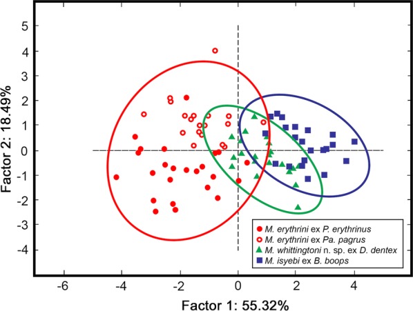
Plot of 87 specimens of Microcotyle spp. in the first plane of the PCA. Ellipses indicate 95% confidence intervals
Discussion
No type-species was selected for the genus Microcotyle in the original definition by van Beneden & Hesse [4], which included the descriptions of two species, M. donavini and M. erythrini (also M. canthari and M. labracis, but these species currently belong to the genera Neobivagina and Serranicotyle, respectively). Later, Sproston [37] selected M. donavini as the type-species for the genus but at the time of the erection of the genus, these two species were the first morphological references. First descriptions of new Microcotyle species were based on vague morphological differences (mostly the number of clamps and testes [5, 38, 39]). Many more species have been described since then worldwide and several genera of the subfamily Microcotylinae have been erected, and M. erythrini has continued being considered valid [2, 3]. Recently, Bouguerche et al. [2] and Bouguerche et al. [3] provided molecular evidence that despite the validity of this species, several Microcotyle spp. from different host species have been wrongly identified as M. erythrini because of their morphological homogeneity. These authors referred to a M. erythrini complex of cryptic species and suggested that a molecular re-evaluation may reveal additional parasite diversity [3]. Caution must be taken in order to select representative specimens in perfect conditions of maturity, completeness and constitution. Whittington [40] stated that to separate monogeneans of a species complex with high levels of diversity “it is vital to ensure that there is a useful trail of high quality parasite material” for taxonomic studies, also stressing the importance of supporting the results with molecular genetic analyses. The present study shows that morphological differences between M. erythrini (s.s.) and similar species can be found: a new species of Microcotyle is described in D. dentex, together with the redescription of M. erythrini (s.s.) (including a new host record, Pa. pagrus) and a new geographical record of M. isyebi with additional morphological information, all supported by molecular evidence.
Molecular analyses of the cox1 gene showed clear differences between Microcotyle spp. distinctly separating the three species described here. Previous studies have suggested levels of intraspecific variation lower than 5% for species of mazocraeids and microcotylids (up to 5.6% and up to 4.5%, respectively, Yan et al. [41]; Mladineo et al. [42]). Based on cox1 sequences, M. whittingtoni n. sp. appears markedly distinct, since the genetic distance from the remaining congeners was higher than 10.8%; specifically, the two isolates ex D. dentex differed from M. erythrini (s.s.) by 10.8–13.5% and from M. isyebi by 14.4–15.2%. The available 28S rDNA sequences for Microcotyle spp. are scarce as this region is not commonly used as a marker for interspecific differences.
We have delimited the valid morphological ranges of M. erythrini (s.s.) and similar species in sparids; in addition, we suggest the use of new diagnostic characters and morphological tools for assessment of multivariate patterns (e.g. PCA). One of the issues in defining the differential traits of similar species of microcotylids is to avoid abnormally wide morphological ranges by selecting only representative specimens: (i) not including specimens potentially belonging to other species (e.g. morphologically similar parasites from other host species not confirmed by molecular analysis); (ii) selecting morphologically optimal specimens (mature, unbroken, uncontracted, unstretched, not wrinkled and unfolded); and (iii) characterizing these specimens accurately to ensure that the diagnostic species-specific characters are properly described. More than 150 years after the original description of M. erythrini, numerous descriptions of this species from P. erythrinus and other hosts have provided extremely wide ranges of morphological information for this species, making it almost impossible to find differentiating features. By defining M. erythrini (sensu stricto) here, we aimed to characterize the species by much narrower morphological ranges only considering valid genetically tested specimens from confirmed hosts. Regarding the optimal specimen selection, only completely mature adults should be representative for standardized taxonomic descriptions. Worms with fully developed both male and female reproductive systems must be selected, as testes in young adults are early functional and vas deferens is full of sperm while no developed oocytes exist in the germarium. Also, the preservation and completeness of the specimens must be ensured.
Knowledge of the three-dimensional structure of the monogeneans in fresh preparations is crucial to understand the morphology of the specimens mounted in Canada balsam as under the coverslide they are represented in a two-dimensional view; knowledge of the natural shapes allows detecting possible folds and missing parts. For example, the haptor of Microcotyle spp. has a dorsal lobe and a ventral lobe (sometimes notched anteriorly), both with clamps; when the specimens are mounted (usually in ventral view), dorsal and ventral projections are folded and often the ventral lobe overlaps the haptor peduncle and/or the posterior end of the body (Fig. 1c). When measuring the haptor, this morphology must be considered in order to measure the body length and the haptor dimensions (see Fig. 1c, d). Moreover, when clamps are counted, possible gaps in the sequence of clamp frills (Fig. 1e, arrowhead) or possible missing pieces of haptor (Fig. 1e, arrow) must be considered, taking into account that the most distal clamps at the ends of the haptoral lobes are smaller.
In general, we recommend the revision and adequate counting of some discrete characters as the number of clamps or testes in the descriptions of several previously described species of Microcotyle, as some ranges are often abnormally high (e.g. 59–126 clamps for M. visa or 9–24 testes for M. erythrini, see [2, 3]). We must be particularly rigorous with this consideration as these traits are key in the species diagnoses of polyopisthocotyleans. For example, the number of clamps herein reported for M. whittingtoni n. sp. is 60–78, as the much higher range reported for M. erythrini of González González [36] in Dentex dentex (110–120) was not confirmed and the drawing and the photomicrograph show that their specimens had 60 clamps [36]. Regarding testes, it should be considered that they are flattened and stacked in at least two dorsoventral levels, so they must be detected and counted at different depth levels.
Some traditionally used morphological traits are intrinsically highly variable, and must be considered with extreme caution when used for taxonomy. Total length has been considered to characterize species such as M. archosargi and M. lichiae which are, in general, much larger than M. erythrini (s.s.) and similar species; however this trait is uncertain as monogenean sizes are known to be highly dependent on host size [32, 43]; e.g. Mendoza-Franco et al. [25] described smaller specimens of M. archosargi, lowering the range of body length to numbers that overlap with most of the similar species in sparid fishes (see Additional file 3: Table S3). In the case of M. lichiae, the original description was based on a single specimen and there are no data for its intraspecific variability. The number of spines in the genital atrium has been used more recently to differentiate species of Microcotyle; this character is often highly variable (e.g. for M. isyebi Bouguerche et al. [3] reported 154–267 vs 272–395 in the present study) and can depend on the condition of the specimen (e.g. incorrect fixation or genital atrium more or less evaginated) or discordances related to the observers. The “pockets” of the genital atrium (sensu Mamaev [44]; posterior small chambers), typical of the genus, have also provided taxonomic information. Mamaev [44] already indicated that the presence or absence of spines in these “pockets” was a good diagnostic character. For example, Bouguerche et al. [3] also stated that M. isyebi shared the presence of genital atrium “pockets” with the other Microcotyle species parasitic in sparid fishes (i.e. M. archosargi, M. erythrini, M. isyebi and M. visa) and not in species parasitic in fishes of other families (i.e. M. donavini and M. lichiae; these authors also listed M. pomatomi and Microcotyle sp. ex H. dactylopterus [24] but “pockets” are present in these species, see Remarks to M. isyebi above). “Pockets” are often not described and sometimes not clearly drawn (e.g. M. pomatomi [32]), as sometimes they can be unarmed or armed with a few spines [44]. Moreover, when the genital atrium is evaginated, chambers often become indistinguishable in ventral view. Their absence implies a different general structure of the genital atrium, a feature used for differentiation at the generic level within the subfamily Microcotylinae [30, 45]. Our last considerations of the traditionally used diagnostic traits refer to the dimensions of the soft muscular organs such as the pharynx or the genital atrium, both contractile and highly variable depending on the specimen, often mentioned in species descriptions (e.g. [2, 43]). All these soft organs can entail diagnostic evidence, but reliable differences should be outstanding, mostly referred to their volume or area, and if possible, relative to the specimen size.
The use of the correct tools and procedures can allow that the currently genetically differentiated species (M. erythrini, M. isyebi and M. whittingtoni n. sp.) become pseudocryptic with defining diagnostic characters or combinations of characters. When the morphometric data of individual worms was integrated in the PCA, the resulting components could not be useful to separate species but provided useful information on specimen groupings based on their shape. The results of the PCA in the present study illustrated that additional diagnostic information can be extracted from the general form of the worms, particularly regarding the relative dimensions and arrangement of the haptor and the remaining of the body. In view of this evidence, we suggest new diagnostic characters revealing previously unnoticed morphological differences: (i) haptor dimensions including anterior and posterior lobes (the larger values for haptor length to body length ratio and for anterior/posterior haptoral lobe length ratio differentiate M. erythrini (s.s.) from M. isyebi and M. whittingtoni n. sp.); (ii) thickness of the clamps (the higher ratio between “c” sclerite maximum width/total clamp length differentiates M. whittingtoni n. sp. from M. isyebi and M. erythrini (s.s.)); (iii) relative size and shape of spines of the “pockets” of the genital atrium (spines of the “pockets” in M. whittingtoni n. sp. appear to be longer and more curved than those of M. isyebi and M. erythrini (s.s.)); (iv) extension and symmetry of the posterior extremities of vitelline fields (posterior extremities of vitelline fields always asymmetrical in M. whittingtoni n. sp. vs occasionally symmetrical in M. isyebi and M. erythrini (s.s.)); and (v) shape of the tip of the abopercular filament of the egg; the solid (capitated or pointed) tips of the abopercular filaments differentiate M. erythrini (s.s.) from M. isyebi and M. whittingtoni n. sp. We propose that the region that can provide more taxonomic information is the haptor, taking into account its three-dimensional structure as an oval to fusiform (when pointed at both ends) “foot” holding a body perpendicularly inserted, directly or through a peduncle (Fig. 1c–e). In this way the total and relative haptor dimensions must include both lobes (anterior and posterior), and one of them is often unnoticed in mounted specimens because they fold over the body (see Fig. 1c, d). In fact, some authors have described the haptor of some Microcotyle species as triangular (e.g. [3, 26, 33, 34]) only referring to the lobe not folded over the body. In this way, M. erythrini (s.s.) can be defined by its relatively longer ventral lobe, the one that is usually unnoticed as it is adhered to the body in permanent mounts. As a note of caution, we must stress the need of examination of adult specimens only, as the relative dimensions of the haptor are known to change significantly during the development (see, for example Machkewskyi et al. [43]). The shape and size of the clamps also provides useful taxonomic information. These structures are usually described only as Microcotyle-type, and the width and length are provided (sometimes wrongly addressed, see Additional file 3: Table S3, Additional file 4: Table S4 and Fig. 1a for correct measuring). However, within this morphological description, some variations can be found. A more detailed study of clamp features can provide further taxonomic information. For example, the accessory sclerite (‘e’) is herein described as trifid or trident-shaped for all three species analysed, but it is mostly not described and not drawn, and the few authors drawing the sclerite represent it as single or lancet-shaped (e.g. [24, 46]). We also suggest that more attention should be paid to the thickness of the clamps: among the three species herein analysed, M. whittingtoni n. sp. shows noticeably thicker clamps; we explored this attribute through the width of the antero-lateral sclerite (‘c’) in absolute value and in relation to clamp length, as this region of the clamp appeared to be constant in all the specimens observed. The number of the spines in the “pockets” of the genital atrium is sometimes reported separately, but no specific information on the shape of these spines is usually found, except sometimes detailing that they are equal to those in the main chamber of the genital atrium [2, 3, 24]. Interestingly, these spines were observed to be longer than the spines of the main chamber in all three species herein described, and those in M. whittingtoni n. sp. were distinctly curved; the lack of information from other species prevented us to reach to further taxonomic conclusions, but we encourage the authors to provide specific information on the spines in the “pockets” in their descriptions of species of Microcotyle.
In the specimens of Microcotyle from the Spanish Western Mediterranean we observed some differences in the extension of the posterior extremities of vitelline fields (also including the extension of the caeca, as they accompany the vitellarium): extending into the haptor or peduncle in M. erythrini (s.s.) and into the haptor in M. whittingtoni n. sp. and prehaptoral in M. isyebi. However, this trait was not here suggested to characterize M. isyebi as according to the original description the posterior extension of the left caecum (and consequently the accompanying vitelline fields) extends into haptor “for a short distance” of the specimens from off Algeria [3]. This character may be dependent on the degree of contraction of the specimen, and therefore all specimens should be fixed and mounted in a similar way to be comparable. Other aspect related to the posterior extension of the vitelline fields of the vitellarium is their symmetry. We observed that the posterior extensions of the vitelline fields were always asymmetrical in M. whittingtoni n. sp., while in the other two species we found both specimens with symmetric and asymmetric vitelline fields. Gill polyopisthocotyleans show more or less distinct asymmetry related with the side of the gill filament they attach to [47, 48]; interestingly Bouguerche et al. [3] reported that left caecum (and consequently the accompanying vitelline fields) was longer in M. isyebi, while in all the species herein observed included specimens with both dextral or sinistral asymmetry.
Mamaev [44] described the eggs of Microcotyle spp. as two-filamented, with usually long opercular and shorter abopercular filament, but no further morphological details are normally provided in the species descriptions. The examination of the new specimens from the Spanish Western Mediterranean also revealed differential details regarding the eggs such as the different shapes of the end of the abopercular filament: solid (pointed or capitate) in M. erythrini (s.s.) (Fig. 4f) and hollow (bifid or cup-shaped) in M. isyebi and M. whittingtoni n. sp. (Figs. 5f, 6f). Other possible differential details were observed such as the type of connection between the egg and the opercular filament: abruptly connected in M. erythini (s.s.) and M. isyebi (Figs. 4f, 5f) and inserted through a gradual transition in M. whittingtoni n. sp. (Fig. 6f). This trait is not used for diagnosis in the present study as it requires a more standardized description. More detailed descriptions are recommended as this trait can be taxonomically useful and other authors, e.g. Sproston [37], have already reported interspecific differences regarding the egg shape. The information on this trait can be limited as the egg shape varies depending on the condition and presence of uterine eggs.
Conclusions
The present study suggests new diagnostic morphological traits to differentiate Microcotyle spp. in Mediterranean sparids and shed light on the case of M. erythrini species complex changing its previously considered cryptic status. More detailed descriptions are recommended, including molecular data, preferably of more informative gene markers regarding the interspecific differences in the polyopisthocotyleans such as cox1 [41, 42, 49], but also 28S rDNA sequences as they can provide useful complementary information. This study also shows that M. erythrini (s.s.) is not species-specific (even not genus-specific) to its hosts, as it parasitizes Pa. pagrus in addition to the type-host, P. erythrini; therefore, although the host species must continue as referential in the taxonomy of Microcotyle spp., a new host record does not necessarily mean a new species. However, further studies are needed in order to establish the morphological traits defining the microcotylids, especially for genera such as Microcotyle, with numerous species reported worldwide.
Supplementary information
Additional file 1: Table S1. Mean genetic divergence (uncorrected p-distance in % and number of pairwise nucleotide differences in parentheses) estimated for the partial cox1 sequence pairs within- (along the diagonal, emboldened) and among species of Microcotyle (below the diagonal).
Additional file 2: Table S2. Pairwise nucleotide differences among species of Microcotyle for the partial 28S rDNA sequences, including Bivagina pagrosomi.
Additional file 3: Table S3. Metrical data for Microcotyle erythrini (sensu stricto) and other species of Microcotyle in sparid fishes in the Mediterranean Sea and North-East Atlantic. Measurements are in micrometres expressed as ranges, except where a single value was provided.
Additional file 4: Table S4. Metrical data from descriptions of Microcotyle spp. similar to M. erythrini (sensu stricto) from Mediterranean non-sparid or non-Mediterranean fishes. Measurements are in micrometres expressed as ranges, except where a single value was provided.
Acknowledgements
The authors thank Hermanos Narejo S.L., and particularly Francisco Piedecausa, for their collaboration providing the fish smaples. We are indebted to Professor Aneta Kostadinova (Bulgarian Academy of Sciences) for her generous advice and comments. We are indebted to Rachel V. Pool (University of Valencia) for revising the English and to the anonymous reviewers for their helpful comments and suggestions. MV-M, APO and FEM also thank Dr Natalia Fraija (AZTI) for her guidance in applying molecular thechniques and analyses at the initial stage of the study.
Abbreviations
- AHL
anterior haptor lobe length
- BL
body length
- BL-H
body length without haptor
- CL
clamp length
- CO
copulatory organ
- CPS
Central South-East Pacific
- CSW
‘c’ sclerite width
- CW
clamp width
- EAS
Eastern Arabian Sea
- HL
haptor length
- MC
main chamber of the genital atrium
- NS
North Sea
- NWA
North-West Atlantic
- NWP
North-West Pacific
- P
small posterior chambers (“pockets” sensu Mamaev [44])
- PHL
posterior haptor lobe length
- SA
Southern Australia
- SWP
South-West Pacific
- WCA
Western-Central Atlantic
- WM
Western Mediterranean
Authorsʼ contributions
MV-M conceived the study, obtained the samples, undertook the morphological characterisation and drawings. APO co-designed and planed the project, carried out the multivariate morphometric analysis and helped draft the manuscript. SG and MV-M carried out the sequencing, performed the phylogenetic analyses, contributed to the taxonomic discussion and drafted the corresponding parts. JAR took part in the preparation of the manuscript and discussed the results. FEM coordinated and co-designed the project, drafted the manuscript and defined the general structure. All authors read and approved the final manuscript.
Funding
This study was supported by the projects AGL2015-68405-R (MINECO/FEDER, Spanish Government/UE) and Prometeo/2015/018, Revidpaqua ISIC/2012/003 and GV/2019/143 (Valencian Regional Government, Spain). SG benefited from a postdoctoral fellowship ‘Juan de la Cierva-Formación’ of the MICINN (FJCI-2016-29535), Spain.
Availability of data and materials
All data generated or analyzed during this study are included in this published article. The newly generated sequences were submitted to the GenBank database under the accession numbers MN816010-MN816021 (cox1) and MN814847-MN814850 (28S). The holotype and paratypes of M. whittingtoni n. sp., and vouchers of M. erythrini (s.s.) and M. isyebi were deposited in the Natural History Museum, London, UK (NHMUK.2019.12.10.1-NHMUK.2019.12.10.14); the remaining voucher material is deposited in the Parasitological Collection of the Cavanilles Institute of Biodiversity and Evolutionary Biology, University of Valencia, Spain (Acc. Nos. ICBiBE/PeMe2019, ICBiBE/PpMe2019, ICBiBE/BbMi2019 and ICBiBE/DdMw2019).
Ethics approval and consent to participate
All applicable institutional, national and international guidelines for the care and use of animals were followed.
Consent for publication
Not applicable.
Competing interests
The authors declare that they have no competing interests.
Footnotes
Publisher's Note
Springer Nature remains neutral with regard to jurisdictional claims in published maps and institutional affiliations.
Contributor Information
María Víllora-Montero, Email: Maria.Villora@uv.es.
Ana Pérez-del-Olmo, Email: Ana.perez-olmo@uv.es.
Simona Georgieva, Email: simona.georgieva@gmail.com.
Juan Antonio Raga, Email: Raga@uv.es.
Francisco Esteban Montero, Email: Francisco.E.Montero@uv.es.
Supplementary information
Supplementary information accompanies this paper at 10.1186/s13071-020-3878-9.
References
- 1.World Register of Marine Species Database. WoRMS Editorial Board; 2019. http://www.marinespecies.org/index.php. Accessed 21 June 2019.
- 2.Bouguerche C, Gey D, Justine JL, Tazerouti F. Microcotyle visa n. sp. (Monogenea: Microcotylidae), a gill parasite of Pagrus caeruleostictus (Valenciennes) (Teleostei: Sparidae) off the Algerian coast, Western Mediterranean. Syst Parasitol. 2019;96:131–147. doi: 10.1007/s11230-019-09842-2. [DOI] [PubMed] [Google Scholar]
- 3.Bouguerche C, Gey D, Justine JL, Tazerouti F. Towards the resolution of the Microcotyle erythrini species complex: description of Microcotyle isyebi n. sp. (Monogenea, Microcotylidae) from Boops boops (Teleostei, Sparidae) off the Algerian coast. Parasitol Res. 2019;118:1417–1428. doi: 10.1007/s00436-019-06293-y. [DOI] [PubMed] [Google Scholar]
- 4.van Beneden P-J, Hesse C-E. Recherches sur les bdellodes ou hirudinés et les trématodes marins. Brussels: Mém Acad Roy Sc Belgique; 1863. pp. 112–116. [Google Scholar]
- 5.Parona C, Perugia A. Res ligusticae, XIV, Contribuzione per una monografía del genere Microcotyle. Ann Museo Civico Storia Nat Giacomo Doria Genoa, Ser. 2a. 1890;10:173–220. [Google Scholar]
- 6.Akmirza A. Monogeneans of fish near Gökçeada, Turkey. Turk J Zool. 2013;37:441–448. [Google Scholar]
- 7.Bush A, Lafferty K, Lotz J, Shostak A. Parasitology meets ecology on its own terms: Margolis et al. revisited. J Parasitol. 1997;88:575–583. doi: 10.2307/3284227. [DOI] [PubMed] [Google Scholar]
- 8.Georgieva S, Selbach C, Faltýnková A, Soldánová M, Sures B, Skírnisson K, et al. New cryptic species of the ‘revolutum’ group of Echinostoma (Digenea: Echinostomatidae) revealed by molecular and morphological data. Parasit Vectors. 2013;6:64. doi: 10.1186/1756-3305-6-64. [DOI] [PMC free article] [PubMed] [Google Scholar]
- 9.Bowles J, Blair D, McManus DP. A molecular phylogeny of the human schistosomes. Mol Phylogenet Evol. 1995;4:103–109. doi: 10.1006/mpev.1995.1011. [DOI] [PubMed] [Google Scholar]
- 10.Littlewood D, Rohde K, Clough K. Parasite speciation within or between host species? Phylogenetic evidence from site-specific polystome monogeneans. Int J Parasitol. 1997;27:1289–1297. doi: 10.1016/S0020-7519(97)00086-6. [DOI] [PubMed] [Google Scholar]
- 11.Littlewood D, Johnston D. Molecular phylogenetics of the four Schistosoma species groups determined with partial 28S ribosomal RNA gene sequences. Parasitology. 1995;111:167–175. doi: 10.1017/S003118200006491X. [DOI] [PubMed] [Google Scholar]
- 12.Badets M, Whittington I, Lalubin F, Allienne J-F, Maspimby J-L, Bentz S, et al. Correlating early evolution of parasitic platyhelminths to Gondwana breakup. Syst Biol. 2011;60:762–781. doi: 10.1093/sysbio/syr078. [DOI] [PubMed] [Google Scholar]
- 13.Tamura K, Stecher G, Peterson D, Filipski A, Kumar S. MEGA6: molecular evolutionary genetics analysis version 6.0. Mol Biol Evol. 2013;30:2725–2729. doi: 10.1093/molbev/mst197. [DOI] [PMC free article] [PubMed] [Google Scholar]
- 14.Katoh K, Standley DM. MAFFT multiple sequence alignment software version 7: improvements in performance and usability. Mol Biol Evol. 2013;30:772–780. doi: 10.1093/molbev/mst010. [DOI] [PMC free article] [PubMed] [Google Scholar]
- 15.Jovelin R, Justine J-L. Phylogenetic relationships within the polyopisthocotylean monogeneans (Platyhelminthes) inferred from partial 28S rDNA sequences. Int J Parasitol. 2001;31:393–401. doi: 10.1016/S0020-7519(01)00114-X. [DOI] [PubMed] [Google Scholar]
- 16.Olson P, Littlewood D. Phylogenetics of the Monogenea—evidence from a medley of molecules. Int J Parasitol. 2002;32:233–244. doi: 10.1016/S0020-7519(01)00328-9. [DOI] [PubMed] [Google Scholar]
- 17.Littlewood D, Rohde K, Bray R, Herniou E. Phylogeny of the Platyhelminthes and the evolution of parasitism. Biol J Linn Soc. 1999;68:257–287. doi: 10.1111/j.1095-8312.1999.tb01169.x. [DOI] [Google Scholar]
- 18.Miller MA, Pfeiffer W, Schwartz T. Creating the CIPRES Science Gateway for inference of large phylogenetic trees. In: 2010 Gateway Computing Environments Workshop (GCE); 2010. p. 1–8.
- 19.Guindon S, Dufayard JF, Lefort V, Anisimova M, Hordijk W, Gascuel O. New algorithms and methods to estimate maximum-likelihood phylogenies: assessing the performance of PhyML 3.0. Syst Biol. 2010;59:307–321. doi: 10.1093/sysbio/syq010. [DOI] [PubMed] [Google Scholar]
- 20.Guindon S, Gascuel O. A simple, fast, and accurate algorithm to estimate large phylogenies by maximum likelihood. Syst Biol. 2003;52:696–704. doi: 10.1080/10635150390235520. [DOI] [PubMed] [Google Scholar]
- 21.Darriba D, Taboada GL, Doallo R, Posada D. jModelTest 2: more models, new heuristics and parallel computing. Nat Methods. 2012;9:772. doi: 10.1038/nmeth.2109. [DOI] [PMC free article] [PubMed] [Google Scholar]
- 22.Rambaut A, Drummond A. FigTree version 1.4.0; 2012. http://tree.bio.ed.ac.uk/software/figtree/
- 23.Schneider CA, Rasband WS, Eliceiri KW. NIH Image to ImageJ: 25 years of image analysis. Nat Methods. 2012;9:671–675. doi: 10.1038/nmeth.2089. [DOI] [PMC free article] [PubMed] [Google Scholar]
- 24.Ayadi ZEM, Gey D, Justine JL, Tazerouti F. A new species of Microcotyle (Monogenea: Microcotylidae) from Scorpaena notata (Teleostei: Scorpaenidae) in the Mediterranean Sea. Parasitol Int. 2017;66:37–42. doi: 10.1016/j.parint.2016.11.004. [DOI] [PubMed] [Google Scholar]
- 25.Mendoza-Franco EF, Tun MCR, Anchevida AJD, del Río Rodríguez RE. Morphological and molecular (28S rRNA) data of monogeneans (Platyhelminthes) infecting the gill lamellae of marine fishes in the Campeche Bank, southwest Gulf of Mexico. ZooKeys. 2018;783:125. doi: 10.3897/zookeys.783.26218. [DOI] [PMC free article] [PubMed] [Google Scholar]
- 26.Catalano SR, Hutson KS, Ratcliff RM, Whittington ID. Redescriptions of two species of microcotylid monogeneans from three arripid hosts in southern Australian waters. Syst Parasitol. 2010;76:211–222. doi: 10.1007/s11230-010-9247-x. [DOI] [PubMed] [Google Scholar]
- 27.Park J-K, Kim K-H, Kang S, Kim W, Eom KS, Littlewood D. A common origin of complex life cycles in parasitic flatworms: evidence from the complete mitochondrial genome of Microcotyle sebastis (Monogenea: Platyhelminthes) BMC Evol Biol. 2007;7:11. doi: 10.1186/1471-2148-7-11. [DOI] [PMC free article] [PubMed] [Google Scholar]
- 28.Aiken HM, Bott NJ, Mladineo I, Montero FE, Nowak BF, Hayward CJ. Molecular evidence for cosmopolitan distribution of platyhelminth parasites of tunas (Thunnus spp.) Fish Fish. 2007;8:167–180. doi: 10.1111/j.1467-2679.2007.00248.x. [DOI] [Google Scholar]
- 29.Oliva ME, Sepulveda FA, González MT. Parapedocotyle prolatili gen. n. et sp. n., of a new subfamily of the Diclidophoridae (Monogenea), a gill parasite of Prolatilus jugularis (Teleostei: Pinguipedidae) from Chile. Folia Parasitol. 2014;61:543. doi: 10.14411/fp.2014.067. [DOI] [PubMed] [Google Scholar]
- 30.Mamaev YuL. The taxonomical composition of the family Microcotylidae Taschenberg, 1879 (Monogenea) Folia Parasitol. 1986;33:199–206. [Google Scholar]
- 31.Radujković BM, Euzet L. Faune des parasites de poissons marins du Montenegro (Adriatique Sud): Monogenes. Acta Adriat. 1989;30:51–135. [Google Scholar]
- 32.Williams A. Monogeneans of the families Microcotylidae Taschenberg, 1879 and Heteraxinidae Price, 1962 from Western Australia, including the description of Polylabris sandarsae n. sp. (Microcotylidae) Syst Parasitol. 1991;18:17–43. doi: 10.1007/BF00012221. [DOI] [Google Scholar]
- 33.López-Román R, Guevara Pozo D. Especies de la familia Microcotylidae (Monogenea) halladas en teleosteos marinos de la costa de Granada. Rev Ibérica Parasitol. 1973;33:197–233. [Google Scholar]
- 34.Yamaguti S. Systema Helminthum. Monogenea and Aspidocotylea. New York: Interscience Publishers, Wiley; 1963. [Google Scholar]
- 35.Di Ariola V. alcuni trematodi di pesci marini. Boll Mus Zool Anat Comp Genova. 1899;4:1–10. [Google Scholar]
- 36.González González P. Parasitofauna branquial de Dentex dentex (Lineo, 1758) (Pisces; Sparidae). PhD thesis, University of Valencia; 2005. http://roderic.uv.es/handle/10550/15041. Accessed 26 Dec 2019.
- 37.Sproston NG. A synopsis of the monogenetic trematodes. Trans Zool Soc Lond. 1946;25:185–600. [Google Scholar]
- 38.Goto S. Studies on the ectoparasitic trematodes of Japan. Tokyo: Imperial University; 1894. [Google Scholar]
- 39.MacCallum G. Further notes on the genus Microcotyle. Zool Jahrb. 1913;35:389–402. [Google Scholar]
- 40.Whittington ID. The Capsalidae (Monogenea: Monopisthocotylea): a review of diversity, classification and phylogeny with a note about species complexes. Folia Parasitol. 2004;51:109. doi: 10.14411/fp.2004.016. [DOI] [PubMed] [Google Scholar]
- 41.Yan S, Wang M, Yang C-P, Zhi T-T, Brown CL, Yang T-B. Comparative phylogeography of two monogenean species (Mazocraeidae) on the host of chub mackerel, Scomber japonicus, along the coast of China. Parasitology. 2016;143:594–605. doi: 10.1017/S0031182016000160. [DOI] [PubMed] [Google Scholar]
- 42.Mladineo I, Šegvić T, Grubišić L. Molecular evidence for the lack of transmission of the monogenean Sparicotyle chrysophrii (Monogenea, Polyopisthocotylea) and isopod Ceratothoa oestroides (Crustacea, Cymothoidae) between wild bogue (Boops boops) and cage-reared sea bream (Sparus aurata) and sea bass (Dicentrarchus labrax) Aquaculture. 2009;295:160–167. doi: 10.1016/j.aquaculture.2009.07.017. [DOI] [Google Scholar]
- 43.Machkewskyi VK, Dmitrieva EV, Al-Jufaili S, Al-Mazrooei NA. Microcotyle omanae n. sp. (Monogenea: Microcotylidae), a parasite of Cheimerius nufar (Valenciennes) (Sparidae) from the Arabian Sea. Syst Parasitol. 2013;86:153–163. doi: 10.1007/s11230-013-9444-5. [DOI] [PubMed] [Google Scholar]
- 44.Mamaev YL. On species composition and morphological features of the Microcotyle genus (Microcotylidae, Monogenoidea) In: Lebedev BI, editor. Investigations in parasitology. Collection of papers. Vladivostok: DBNTs AN SSSR; 1989. pp. 32–38. [Google Scholar]
- 45.Mamaev YL. The composition of the genera Atriaster and Atrispinum (Microcotylidae, Monogenea) and some peculiarities of their morphology. Parazitologiya. 1984;18:204–208. [Google Scholar]
- 46.Dillon WA, Hargis WJ, Harrises AE. Monogeneans from the southern Pacific Ocean: Polyopisthocotyleids from the Australian fishes. Subfamilies Polylabrinae (Genus Polylabroides) and Microcotylinae (Genus Neobivagina) Parazitol Sbornik. 1985;33:83–87. [Google Scholar]
- 47.Llewellyn J. The host-specificity, micro-ecology, adhesive attitudes, and comparative morphology of some trematode gill parasites. J Mar Biol Assoc UK. 1956;35:113–127. doi: 10.1017/S0025315400009000. [DOI] [Google Scholar]
- 48.Kearn G. Some aspects of the biology of monogenean (Platyhelminth) parasites of marine and freshwater fishes. Oceanography. 2014;2:1–8. [Google Scholar]
- 49.Shi S-F, Li M, Yan S, Wang M, Yang C-P, Lun Z-R, Brown CL, Yang T-B. Phylogeography and demographic history of Gotocotyla sawara (Monogenea: Gotocotylidae) on Japanese Spanish mackerel (Scomberomorus niphonius) along the coast of China. J Parasitol. 2014;100:85–93. doi: 10.1645/13-235.1. [DOI] [PubMed] [Google Scholar]
Associated Data
This section collects any data citations, data availability statements, or supplementary materials included in this article.
Supplementary Materials
Additional file 1: Table S1. Mean genetic divergence (uncorrected p-distance in % and number of pairwise nucleotide differences in parentheses) estimated for the partial cox1 sequence pairs within- (along the diagonal, emboldened) and among species of Microcotyle (below the diagonal).
Additional file 2: Table S2. Pairwise nucleotide differences among species of Microcotyle for the partial 28S rDNA sequences, including Bivagina pagrosomi.
Additional file 3: Table S3. Metrical data for Microcotyle erythrini (sensu stricto) and other species of Microcotyle in sparid fishes in the Mediterranean Sea and North-East Atlantic. Measurements are in micrometres expressed as ranges, except where a single value was provided.
Additional file 4: Table S4. Metrical data from descriptions of Microcotyle spp. similar to M. erythrini (sensu stricto) from Mediterranean non-sparid or non-Mediterranean fishes. Measurements are in micrometres expressed as ranges, except where a single value was provided.
Data Availability Statement
All data generated or analyzed during this study are included in this published article. The newly generated sequences were submitted to the GenBank database under the accession numbers MN816010-MN816021 (cox1) and MN814847-MN814850 (28S). The holotype and paratypes of M. whittingtoni n. sp., and vouchers of M. erythrini (s.s.) and M. isyebi were deposited in the Natural History Museum, London, UK (NHMUK.2019.12.10.1-NHMUK.2019.12.10.14); the remaining voucher material is deposited in the Parasitological Collection of the Cavanilles Institute of Biodiversity and Evolutionary Biology, University of Valencia, Spain (Acc. Nos. ICBiBE/PeMe2019, ICBiBE/PpMe2019, ICBiBE/BbMi2019 and ICBiBE/DdMw2019).


