Abstract
Background:
One-visit apexification is a treatment of choice in necrotic immature open apex teeth. Calcium silicate base materials are suitable for this method. The purpose of this study was to investigate and compare the sealing efficiency of Biodentine, mineral trioxide aggregate (MTA) ProRoot, and calcium-enriched mixture (CEM) cement orthograde apical plug using bacterial leakage method.
Materials and Methods:
In this in vitro study a total of 70 extracted maxillary incisors were cleaned and shaped. A 1.1-mm standardized artificially open apex was created in all samples. The teeth were randomly divided into three experimental groups of 20, and two negative and positive control groups of 5. In experimental groups, 4-mm thick apical plugs of ProRoot MTA, CEM cement, or Biodentine were placed in an orthograde manner. Negative control samples were completely filled with MTA while positive control samples were left unfilled. Sealing efficiency was measured by bacterial leakage method, and results were analyzed by Kaplan–Meier and Chi-square tests. The level of significance was set at 0.05.
Results:
The highest number of turbidity was recorded for ProRoot MTA samples, while the lowest for Biodentine. There was a significant difference in the number of turbidity between ProRoot MTA and Biodentine groups (P < 0.001), but there was no significant difference between CEM cement and Biodentine (P = 0.133) and ProRoot MTA (P = 0.055).
Conclusion:
Within the limitation of this in vitro study, Biodentine showed promising results as a substance with good-sealing efficiency.
Keywords: Apexification, biocompatible materials, calcium-enriched mixture cement, dental leakage, mineral trioxide aggregate
INTRODUCTION
Necrotic pulps in immature permanent teeth lead to incomplete root development and apical closure. In such cases, common root canal treatments are not able to provide an adequate apical seal.[1,2,3] One of the treatment options for necrotic teeth is apexification, induction of apical closure, to produce more favorable conditions for conventional root canal filling. Although apexification with calcium hydroxide (CH) has been a proven approach and successfully performed over a long time, disadvantages of long-term CH therapy justified the search for alternative therapies such as one-visit apexification technique.[4] Nowadays, one-visit apexification method using osteoconductive materials such as mineral trioxide aggregate (MTA) is becoming more popular.[5] MTA is a biocompatible powder consisting of small low-solubility hydrophilic particles, which is able to set in a humid environment.[4] On the other hand, MTA has been demonstrated to have some disadvantages, including long setting time, high cost, potential discoloration, and lack of good handling characteristics.[6] The recently introduced new biomaterials such as calcium-enriched mixture cement (CEM cement) and Biodentine have shown considerable clinical success and have overcome some MTA-related disadvantages. CEM cement is a hydrophilic tooth-colored cement that releases CH during and after setting.[7] The physical properties of this material are favorable. It also possesses the ability to set in aqueous environments with shorter setting time than MTA and a sealing ability comparable to MTA. Due to higher flow, its thickness is lower than MTA.[8]
In an in vitro study on the sealing efficiency of MTA and CEM-cement apical plugs using different obturation techniques, Tabrizizade et al. showed no significant differences between the sealing efficiency of both materials.[9] Biodentine, a new calcium silicate-based restorative cement with dentin-like mechanical properties, has been available since 2009.[10,11] Biodentine was used not only in restorative-endodontic treatments such as repair of perforations and resorptive lesions but also in endodontic treatments such as pulp capping, pulpotomy, and root end filling.[12] Biodentine has a good-sealing efficiency and mechanical properties.[13] Its setting time is 12 min, and it does not cause discoloration.[14] An important factor for successful endodontic treatment in open-apex teeth is the sealing efficiency of apical plug. At the present time, only two in vivo studies by Bani et al.[15] and Cechella et al.[16] using Biodentine as an apical barrier are available for us. Based on the results of these studies, the apical sealing efficiency of Biodentine was comparable to that of and lower than MTA, respectively.
Regarding few studies on Biodentine as an apical barrier for one-visit apexification treatments and comparing it with other materials, the present study was conducted to evaluate and compare the apical microleakage of Biodentine, ProRoot MTA, and CEM cement orthograde apical plugs.
MATERIALS AND METHODS
In this in vitro study, a total of 70 human single-root maxillary incisors were selected among freshly extracted teeth due to periodontal reasons. The inclusion criteria were intact single root teeth with straight canals, absence of fracture or microcracks, and root resorption or intracanal calcification. The specimens were decontaminated by immersing in 5.25% sodium hypochlorite (NaOCl) (Golrang, Tehran, Iran) for 1 h and then cleaned under tap water by a brush to remove the residual debris. The samples were kept in normal saline (0.9% NaCl, Darupakhsh, Tehran, Iran) until they were used.
The teeth were decoronated by a 0.3 diamond disc (Tizkavan, Tehran, Iran) so that approximately 18 mm of roots remained. Then, about 3 mm of the apical tip of the roots were resected by a fissure bur (Tizkavan, Tehran, Iran) to eliminate apical deltas and ramifications.
The standardized 15 mm root lengths were navigated by inserting a #20 K-File (Mani, Tehran, Iran) into the canals. The working length was determined visually 0.5 mm shorter than the apex. The apical portion of the canals was prepared by hand instrumentation up to #40 k-File, and the coronal two-thirds of the canals were shaped by #1, 2, and 3 Gates Glidden burs (Mani, Tehran, Iran). To simulate the clinical situation of an open apex, apical foramina were prepared by #1, 2, and 3 Peeso Reamers (Mani, Tehran, Iran) in a retrograde manner, which resulted in a diameter of 1.1 mm.[17] Throughout preparation, by replacing each instrument, the canals were irrigated by 2 ml of 2.5% NaOCl. To remove the smear layer, canals were filled with 17% ethylenediaminetetraacetic acid (Dentonics, INC. USA) for 1 min, followed by 5.25% NaOCl for 3 min. Final irrigation was done with 5 ml of normal saline. The samples were then randomly divided into three experimental groups of ProRoot MTA (Dentsply, Germany), Biodentine (Septodont, France), and CEM cement (YektazistDandan, Iran) of 20 teeth each, and two control groups of 5.
To simulate the periapical soft tissues during plug placement, the teeth were compressed into a wet sponge. Canals were dried with paper point (DiaDent, South Korea). The materials, prepared according to the manufacturer's instructions, were carried into the canals in an orthograde manner by MTA carrier (Juya, Tehran, Iran), guided by the tip of a #80 paper point and finally compressed by an appropriate hand plugger (Juya, Tehran, Iran) to a thickness of 4 mm.
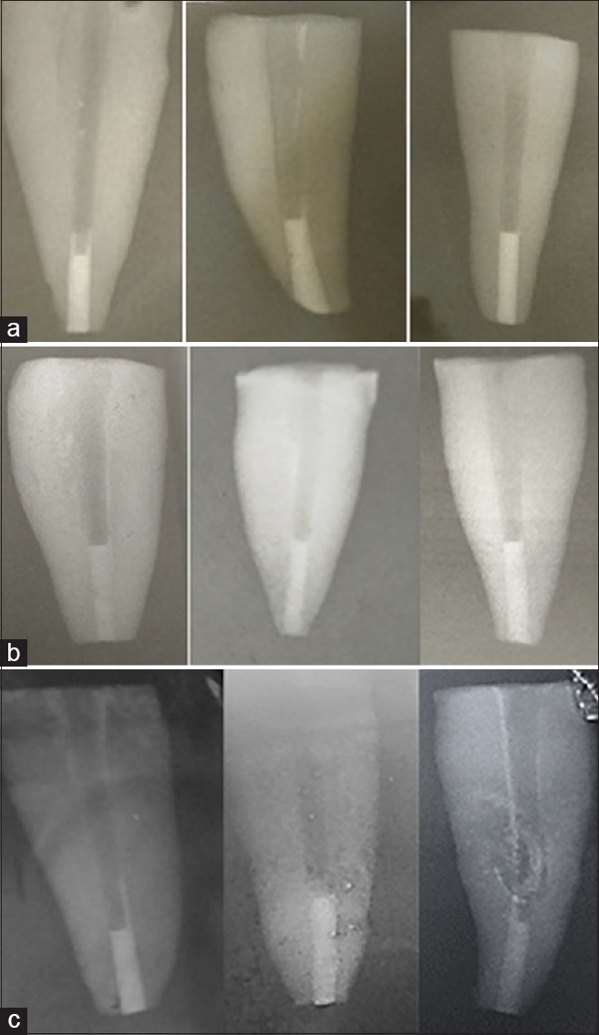
Radiographs were taken to confirm the density and proper placement of the 4-mm thick plug [Figure 1]. The rest of the canals were left unfilled. A wet cotton pellet was placed on the MTA and CEM cement samples. All the canals were sealed with temporary restorative material (Coltosol, Ariadent, Tehran, Iran) and then stored at 37°C and 100% humidity for 72 h.
Figure 1.

Radiographs confirm the density and proper placement of the three tested material. (a) Samples of ProRoot mineral trioxide aggregate group. (b) Samples of Biodentine group. (c) Samples of calcium-enriched mixture cement group.
The root canals of the positive and negative control groups were cleaned and shaped like the test groups. Negative control samples were filled with ProRoot MTA completely; but in positive samples, no apical plugs were used to fill the apical third.
To prevent bacterial leakage from the accessory canals and cemental tears on the root surface, external root surfaces, but apical foramens and access cavities, of experimental and positive control samples, were covered with two layers of nail polish (My, Ariankimiatak, Iran). In negative control samples, the entire root surfaces, including the apical foramens, but not the access cavities, were covered with sticky wax.
Bacterial leakage test
To evaluate the bacterial leakage, the teeth were transferred into a system with two upper and lower chambers. The upper chamber was assembled by a plastic Eppendorf cylinder (Padtan Teb, Tehran, Iran) after cutting off 5 mm from its end. The sample was passed through the tube so that about 2 mm of the apical part of the root was left outside. The gaps between the sample and the inner side of cylinder were completely sealed with cyanoacrylate adhesive and sticky wax. This tightly fitted model of the tube and tooth was sterilized with ethylene oxide in an 8-h cycle.
A volume of 8–10 ml sterile brain heart infusion broth (BHI-broth) (HiMedia, Germany) was introduced into the sterile disposable culture tubes (Padtan Teb, Tehran, Iran), as a lower chamber of the leakage apparatus. The previously assembled upper chamber with teeth was then inserted in the culture tubes under aseptic conditions so that a minimum of 2–3 mm of the apical part of each root was immersed in BHI-broth. The junction area between the upper and lower chambers was sealed using Parafilm tapes. To ensure sterilization, the whole system was incubated at 37°C for 3 days. The samples showing the evidence of turbidity in BHI-broth were excluded from the study, in which none of the samples showed turbidity.
Two ml of BHI-broth was inoculated with 9 × 10⁸ colony-forming unit/ml (McFarland no. 3) of Enterococcus faecalis (ATCC 29212) to form a bacterial suspension that was added to the upper chamber every 2 days. Bacterial leakage was evaluated by the severity of turbidity in culture media in the lower chamber. The culture tubes were observed daily for 75 days. The date of the early signs of turbidity was recorded, and the sample was discarded. To confirm the purity of E. faecalis in BHI-broth, a sample was taken from the culture tube and cultivated.
Statistical analysis of the data was accomplished using Chi-square and Kaplan–Meier survival analysis tests in SPSS 22 environment (IBM, Chicago, IL, USA). The level of significance was set at 0.05.
RESULTS
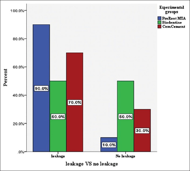
During the entire observational period, all positive samples exhibited complete leakage within 24 h, while the samples in negative control groups did not show any bacterial leakage. Furthermore, 18 samples in ProRoot MTA group (90%), 10 samples in Biodentine group (50%), and 14 samples in CEM cement group (70%) showed turbidity throughout the experiment.
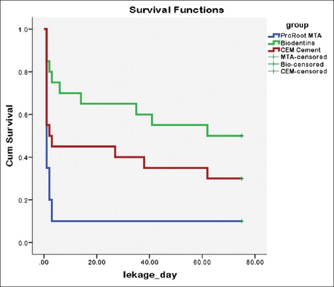
According to Kaplan–Meier survival analysis, there was a significant difference in mean survival time of study between ProRoot MTA and Biodentine groups (P < 0.001), but the distributions of survival rates were the same in ProRoot MTA and CEM cement groups (P = 0.055) as well as in Biodentine and CEM cement groups (P = 0.133) [Figure 2].
Figure 2.

Data distribution of survival test in experimental groups.
Similar to Kaplan–Meier survival analysis, Chi-square test showed statistically significant difference between ProRoot MTA and Biodentine experimental groups (P = 0.006). Moreover, there were no significant differences between ProRoot MTA and CEM cement groups (P = 0.235) as well as between Biodentine and CEM cement groups (P = 0.197) [Figure 3].
Figure 3.

Percentage of Enterococcus faecalis leakage and no leakage among all experimental groups.
DISCUSSION
Calcium silicate-based materials, one of the materials used in single-visit apexification therapeutic approach, were first introduced with the advent of MTA and recently used in CEM cement, tracal, bioaggregate, EndoSequence bioceramic, and Biodentine.[5,18] These new biocompatible materials, while maintaining the desired properties of MTA, are designed to overcome the disadvantages of MTA, including long setting time, tooth discoloration, high price, and poor handling characteristics.[5,7,15] Root canal filling materials are not fixed, inert, and impenetrable, and potentially have the gaps which large amounts of bacteria, ions, and molecules can pass through.[19] This clinically invisible microleakage is a major factor jeopardizing the long-term success of endodontic treatments.[19] Dye penetration method is one of the oldest ones to evaluate the sealing efficiency of apical plugs. Due to the chemical properties, PH, and dye molecules size, which affect the penetration rate, as well as the potential of decoloration of the materials such as MTA, CEM cement, and CH, it is logical to use alternative methods to evaluate microleakage.[20,21] The use of bacteria to assess sealing efficiency is a more reliable method and resembles clinical conditions.[22] The present data suggested that the type of microorganism and the time of microleakage testing directly affected the results of bacterial microleakage evaluations.[23] In the present study, bacterial leakage method and E. faecalis were recruited to simulate oral conditions. E. faecalis, a Gram-positive, anaerobic, and the most common species isolated from a failed root canal therapy, is in a normal oral bacterial flora which has the ability to withstand adverse environmental conditions and penetrate in the dentinal tubules.[24] The studies investigated that the antibacterial activity of MTA, CEM cement, and Biodentine have indicated that although these materials have excellent anti-bactericidal activities due to high pH, all three were unable to completely eliminate E. faecalis.[25,26,27] All dental specimens were identical in terms of the length and diameter of the apical entries before placing the plugs. Furthermore, 4-mm thick plugs were used, which was considered within the guidelines.[28] The results of positive and negative control groups confirmed the accuracy of the study.
The present study focused on comparing the sealing efficiency of ProRoot MTA, CEM cement, and Biodentine as an apical barrier, which was absent in previous studies. The lowest and highest rates of microleakage belonged to Biodentine and MTA, respectively. Biodentine as an apical barrier produced a better seal in 75 days compared to ProRoot MTA and CEM cement; however, the difference in microleakage volume was statistically significant only between Biodentine and ProRoot MTA. Al-kahtani et al.'s[29] study on MTA as an orthograde apical barrier using bacterial penetration method did not show any microleakage. Perhaps, the thickness of plugs and the type of bacterial species (Actinomyces viscosus in) have yielded different findings. In a comparative study of the antibacterial activity of materials such as MTA and CEM cement on most pulpal and periapical pathogenic species, A. viscosus and E. faecalis had the lowest and highest resistance rates, respectively.[30] The results of this study are in disagreement with those of Al- Hezaimi et al. They compared two types of white and gray MTA.[31] The samples did not show any microleakage during the 42-day trial period, and this could be due to the thickness of plugs and shorter duration of the study.[32]
The present research found no significant difference between sealing efficiency of MTA and CEM cement. This result is consistent with results of the two recent studies which were conducted by Moradi et al.[24] and Tabrizizade et al.[9] who compared the sealing ability of ProRoot MTA and CEM cement with bacterial penetration and fluid filtration methods, respectively. Both of them showed that these two materials had similar sealing properties.
The feasible reasons for lower microleakage of CEM cement compared to MTA are higher antibacterial activity of CEM cement, similar to CH (although this ability is identical in both materials in the presence of dentin powder);[33] good setting expansion; better marginal adaptation; shorter setting time;[34] and finally, ability of CEM cement in saline environments to produce similar to standard hydroxyapatite crystals on its surface.[35] Only two studies have examined and compared the Biodentine's sealing efficiency as an apical barrier with ProRoot MTA, which contradicts the results of the current study. Bani et al. compared the microleakage of 1–4-mm thick MTA and Biodentine materials by fluid filtration method and showed the same results in both groups.[15] However, the microleakage was decreased by increasing the thickness of the material; but, in the same thicknesses, the differences were not significant. Cechella et al.[16] analyzed the sealing ability of these two biomaterials, with or without phosphate-buffered saline intracanal dressing, using a glucose leakage method after 2 months. They showed that the Biodentine had lower sealing efficiency than MTA. So far, no study has been conducted to compare the microleakage of Biodentine and CEM cement as orthograde apical barriers.
In this study, Biodentine showed lower microleakage compared to ProRoot MTA and CEM cement. The reasons for this difference are as follows:
Biodentine in contact with dentine forms tag-like structures along the contact area called “mineral penetration zone.” This creates adhesion to dentin.[36] Han and Okiji showed that calcium and silicon, untaken into dentine, form tag-like structures stronger in Biodentine than in Pro-Root MTA.[13] Furthermore, the shear bond strength of Biodentine was similar to that of glass ionomer to dentin, which is higher than the shear bond strength of Pro-Root MTA.[37] In addition, it has been shown that Biodentine has a more uniform structure and is less porous than MTA.[38] By adding a softener and setting time accelerator in a single-dose capsule, the modified Biodentine powder has been reported to produce better physical and functional properties and adaptability.[39]
All of these factors can affect the volume of microleakage of this material and decrease it more compared with MTA. Furthermore, a study evaluating Biodentine, MTA, and glass ionomer as root end-filling materials showed a better marginal adaptability for Biodentine than MTA.[40]
It should be noted that in this study, all of the microleakages of Pro-Root MTA samples happened in the early days of the experiment, while no microleakage was observed in all the remaining days until the end of study. This was not true for Biodentine and CEM cement groups. Despite some leakage in the early days, the samples in these two groups showed a sporadic pattern of microleakage during the study. Because of the association of the presence or absence of microleakage with tooth survival in clinical conditions in long term, and considering the limited time and sample size of this study, evaluating the sealing ability of these materials over a longer period and with more samples may lead to a real and more comparable results in clinical conditions.
CONCLUSION
Based on our findings, Biodentine can predictably prevent bacterial leakage as an apical barrier in permanent necrotic teeth with undeveloped open apices.
Financial support and sponsorship
Nil.
Conflicts of interest
The authors of this manuscript declare that they have no conflicts of interest, real or perceived, financial or nonfinancial in this article.
Acknowledgments
This study was a part of thesis and research project which was supported and funded by Islamic Azad University of Isfahan (Khorasgan).
REFERENCES
- 1.Kalaskar RR, Kalaskar AR. Maturogenesis of non-vital immature permanent teeth. Contemp Clin Dent. 2013;4:268–70. doi: 10.4103/0976-237X.114891. [DOI] [PMC free article] [PubMed] [Google Scholar]
- 2.Kim DS, Park HJ, Yeom JH, Seo JS, Ryu GJ, Park KH, et al. Long-term follow-ups of revascularized immature necrotic teeth: Three case reports. Int J Oral Sci. 2012;4:109–13. doi: 10.1038/ijos.2012.23. [DOI] [PMC free article] [PubMed] [Google Scholar]
- 3.Shabahang S. Treatment options: Apexogenesis and apexification. J Endod. 2013;39:S26–9. doi: 10.1016/j.joen.2012.11.046. [DOI] [PubMed] [Google Scholar]
- 4.Rafter M. Apexification: A review. Dent Traumatol. 2005;21:1–8. doi: 10.1111/j.1600-9657.2004.00284.x. [DOI] [PubMed] [Google Scholar]
- 5.Hargreaves Kenneth M, Berman Louis H. Cohen's Pathways of thePulp. 11th ed. 24. St Louis: Mosby Elsevier; 2016. pp. e35–9. [Google Scholar]
- 6.Parirokh M, Torabinejad M. Mineral trioxide aggregate: A comprehensive literature review – Part III: Clinical applications, drawbacks, and mechanism of action. J Endod. 2010;36:400–13. doi: 10.1016/j.joen.2009.09.009. [DOI] [PubMed] [Google Scholar]
- 7.Asgary S, Eghbal MJ, Parirokh M. Sealing ability of a novel endodontic cement as a root-end filling material. J Biomed Mater Res A. 2008;87:706–9. doi: 10.1002/jbm.a.31678. [DOI] [PubMed] [Google Scholar]
- 8.Adel M, Nima MM, Shivaie Kojoori S, Norooz Oliaie H, Naghavi N, Asgary S. Comparison of endodontic biomaterials as apical barriers in simulated open apices. ISRN Dent. 2012;2012:359873. doi: 10.5402/2012/359873. [DOI] [PMC free article] [PubMed] [Google Scholar]
- 9.Tabrizizade M, Asadi Y, Sooratgar A, Moradi S, Sooratgar H, Ayatollahi F. Sealing ability of mineral trioxide aggregate and calcium-enriched mixture cement as apical barriers with different obturation techniques. Iran Endod J. 2014;9:261–5. [PMC free article] [PubMed] [Google Scholar]
- 10.Grech L, Mallia B, Camilleri J. Characterization of set intermediate restorative material, biodentine, bioaggregate and a prototype calcium silicate cement for use as root-end filling materials. Int Endod J. 2013;46:632–41. doi: 10.1111/iej.12039. [DOI] [PubMed] [Google Scholar]
- 11.Nowicka A, Lipski M, Parafiniuk M, Sporniak-Tutak K, Lichota D, Kosierkiewicz A, et al. Response of human dental pulp capped with biodentine and mineral trioxide aggregate. J Endod. 2013;39:743–7. doi: 10.1016/j.joen.2013.01.005. [DOI] [PubMed] [Google Scholar]
- 12.Rajasekharan S, Martens LC, Cauwels RG, Verbeeck RM. Biodentine™ material characteristics and clinical applications: A review of the literature. Eur Arch Paediatr Dent. 2014;15:147–58. doi: 10.1007/s40368-014-0114-3. [DOI] [PubMed] [Google Scholar]
- 13.Han L, Okiji T. Bioactivity evaluation of three calcium silicate-based endodontic materials. Int Endod J. 2013;46:808–14. doi: 10.1111/iej.12062. [DOI] [PubMed] [Google Scholar]
- 14.Shayegan A, Jurysta C, Atash R, Petein M, Abbeele AV. Biodentine used as a pulp-capping agent in primary pig teeth. Pediatr Dent. 2012;34:e202–8. [PubMed] [Google Scholar]
- 15.Bani M, Sungurtekin-Ekçi E, Odabaş ME. Efficacy of biodentine as an apical plug in nonvital permanent teeth with open apices: An in vitro study. Biomed Res Int. 2015;2015:359275. doi: 10.1155/2015/359275. [DOI] [PMC free article] [PubMed] [Google Scholar]
- 16.Cechella B, de Almeida J, Kuntze M, Felippe W. Analysis of sealing ability of endodontic cements apical plugs. J Clin Exp Dent. 2018;10:e146–e150. doi: 10.4317/jced.54186. [DOI] [PMC free article] [PubMed] [Google Scholar]
- 17.Andreasen Jens O, Andreasen Frances M, Andersson Lars. Text Book and Color Atlas of Traumatic Injuries to the Teeth. 4th ed. 23. Oxford, UK: Blackwell Munksgaard; 2007. p. 662. [Google Scholar]
- 18.Torabinejad M, Parirokh M, Dummer PM. Mineral trioxide aggregate and other bioactive endodontic cements: An updated overview – Part II: Other clinical applications and complications. Int Endod J. 2018;51:284–317. doi: 10.1111/iej.12843. [DOI] [PubMed] [Google Scholar]
- 19.Muliyar S, Shameem KA, Thankachan RP, Francis PG, Jayapalan CS, Hafiz KA. Microleakage in endodontics. J Int Oral Health. 2014;6:99–104. [PMC free article] [PubMed] [Google Scholar]
- 20.Veríssimo DM, do Vale MS. Methodologies for assessment of apical and coronal leakage of endodontic filling materials: A critical review. J Oral Sci. 2006;48:93–8. doi: 10.2334/josnusd.48.93. [DOI] [PubMed] [Google Scholar]
- 21.Wu MK, Kontakiotis EG, Wesselink PR. Decoloration of 1% methylene blue solution in contact with dental filling materials. J Dent. 1998;26:585–9. doi: 10.1016/s0300-5712(97)00040-7. [DOI] [PubMed] [Google Scholar]
- 22.Timpawat S, Amornchat C, Trisuwan WR. Bacterial coronal leakage after obturation with three root canal sealers. J Endod. 2001;27:36–9. doi: 10.1097/00004770-200101000-00011. [DOI] [PubMed] [Google Scholar]
- 23.Torabinejad M, Parirokh M. Mineral trioxide aggregate: A comprehensive literature review – Part II: Leakage and biocompatibility investigations. J Endod. 2010;36:190–202. doi: 10.1016/j.joen.2009.09.010. [DOI] [PubMed] [Google Scholar]
- 24.Moradi S, Disfani R, Ghazvini K, Lomee M. Sealing ability of orthograde MTA and CEM cement in apically resected roots using bacterial leakage method. Iran Endod J. 2013;8:109–13. [PMC free article] [PubMed] [Google Scholar]
- 25.Estrela C, Bammann LL, Estrela CR, Silva RS, Pécora JD. Antimicrobial and chemical study of MTA, Portland cement, calcium hydroxide paste, sealapex and dycal. Braz Dent J. 2000;11:3–9. [PubMed] [Google Scholar]
- 26.Asgary S, Akbari Kamrani F, Taheri S. Evaluation of antimicrobial effect of MTA, calcium hydroxide, and CEM cement. Iran Endod J. 2007;2:105–9. [PMC free article] [PubMed] [Google Scholar]
- 27.Bhavana V, Chaitanya KP, Gandi P, Patil J, Dola B, Reddy RB. Evaluation of antibacterial and antifungal activity of new calcium-based cement (Biodentine) compared to MTA and glass ionomer cement. J Conserv Dent. 2015;18:44–6. doi: 10.4103/0972-0707.148892. [DOI] [PMC free article] [PubMed] [Google Scholar]
- 28.Hachmeister DR, Schindler WG, Walker WA, 3rd, Thomas DD. The sealing ability and retention characteristics of mineral trioxide aggregate in a model of apexification. J Endod. 2002;28:386–90. doi: 10.1097/00004770-200205000-00010. [DOI] [PubMed] [Google Scholar]
- 29.Al-Kahtani A, Shostad S, Schifferle R, Bhambhani S. In vitro evaluation of microleakage of an orthograde apical plug of mineral trioxide aggregate in permanent teeth with simulated immature apices. J Endod. 2005;31:117–9. doi: 10.1097/01.don.0000136204.14140.81. [DOI] [PubMed] [Google Scholar]
- 30.Hasan Zarrabi M, Javidi M, Naderinasab M, Gharechahi M. Comparative evaluation of antimicrobial activity of three cements: New endodontic cement (NEC), mineral trioxide aggregate (MTA) and Portland. J Oral Sci. 2009;51:437–42. doi: 10.2334/josnusd.51.437. [DOI] [PubMed] [Google Scholar]
- 31.Al-Hezaimi K, Naghshbandi J, Oglesby S, Simon JH, Rotstein I. Human saliva penetration of root canals obturated with two types of mineral trioxide aggregate cements. J Endod. 2005;31:453–6. doi: 10.1097/01.don.0000145429.04231.e2. [DOI] [PubMed] [Google Scholar]
- 32.Coneglian PZ, Orosco FA, Bramante CM, de Moraes IG, Garcia RB, Bernardineli N, et al. In vitro sealing ability of white and gray mineral trioxide aggregate (MTA) and white Portland cement used as apical plugs. J Appl Oral Sci. 2007;15:181–5. doi: 10.1590/S1678-77572007000300006. [DOI] [PMC free article] [PubMed] [Google Scholar]
- 33.Asgary S, Kamrani FA. Antibacterial effects of five different root canal sealing materials. J Oral Sci. 2008;50:469–74. doi: 10.2334/josnusd.50.469. [DOI] [PubMed] [Google Scholar]
- 34.Asgary S, Shahabi S, Jafarzadeh T, Amini S, Kheirieh S. The properties of a new endodontic material. J Endod. 2008;34:990–3. doi: 10.1016/j.joen.2008.05.006. [DOI] [PubMed] [Google Scholar]
- 35.Asgary S, Eghbal MJ, Parirokh M, Ghoddusi J. Effect of two storage solutions on surface topography of two root-end fillings. Aust Endod J. 2009;35:147–52. doi: 10.1111/j.1747-4477.2008.00137.x. [DOI] [PubMed] [Google Scholar]
- 36.Raskin A, Eschrich G, Dejou J, About I. In vitro microleakage of biodentine as a dentin substitute compared to Fuji II LC in cervical lining restorations. J Adhes Dent. 2012;14:535–42. doi: 10.3290/j.jad.a25690. [DOI] [PubMed] [Google Scholar]
- 37.Kaup M, Dammann CH, Schäfer E, Dammaschke T. Shear bond strength of biodentine, ProRoot MTA, glass ionomer cement and composite resin on human dentine ex vivo. Head Face Med. 2015;11:14. doi: 10.1186/s13005-015-0071-z. [DOI] [PMC free article] [PubMed] [Google Scholar]
- 38.Camilleri J, Grech L, Galea K, Keir D, Fenech M, Formosa L, et al. Porosity and root dentine to material interface assessment of calcium silicate-based root-end filling materials. Clin Oral Investig. 2014;18:1437–46. doi: 10.1007/s00784-013-1124-y. [DOI] [PubMed] [Google Scholar]
- 39.Atmeh AR, Chong EZ, Richard G, Festy F, Watson TF. Dentin-cement interfacial interaction: Calcium silicates and polyalkenoates. J Dent Res. 2012;91:454–9. doi: 10.1177/0022034512443068. [DOI] [PMC free article] [PubMed] [Google Scholar]
- 40.Ravichandra PV, Vemisetty H, Deepthi K, Jayaprada Reddy S, Ramkiran D, Jaya Nagendra Krishna MJ, et al. Comparative evaluation of marginal adaptation of biodentine (TM) and other commonly used root end filling materials-an in vitro study. J Clin Diagn Res. 2014;8:243–5. doi: 10.7860/JCDR/2014/7834.4174. [DOI] [PMC free article] [PubMed] [Google Scholar]


