



Abstract
Allergy is a hypersensitivity reaction when exposed to certain environmental substances. It shows high relation between immunoglobulin E (IgE) binding to a specific receptor (FcεRI), pro-inflammatory cytokines, and mediators with allergic inflammation responses. Curcumin is a yellow pigment isolated from the turmeric. Curcumin possesses antioxidant and anti-inflammatory properties as well as exhibits significant chemopreventive activity. This study was aimed to investigate the in vitro assessment of the regulation of curcumin on allergic inflammatory responses on rat basophil leukemia (RBL)-2H3 and human pre-basophils (KU812) cell lines. Curcumin showed the activity against histamine and β-hexosaminidase releases from both IgE-mediated and A23187-induced cells degranulation. The morphological observation also confirmed that curcumin inhibits cells degranulation. IgE-mediated allergic responses and significantly induced mast cells intracellular reactive oxygen species (ROS) production. Curcumin reduced ROS production from IgE-mediated or A23187-induced cells degranulation. Curcumin also successfully reduced FcεRI expressions and some pro-inflammatory cytokines, such as interleukin (IL)-4 and IL-13. Furthermore, curcumin inhibited protein kinase C (PKC)-δ translocation from cytosolic to particulate. These results suggested that curcumin can alleviate both the IgE-mediated and calcium ionosphere A23187-stimulated allergic responses through reducing the release of the allergic mediators.
Keywords: Allergic responses, Curcumin, Degranulation, Immunoglobulin E, Protein kinase C
Introduction
Allergy is a hypersensitivity reaction occur when exposed to certain environmental substances. It can target some organs of the body, include the nose, lung, skin, and gastrointestinal tract (Juckmeta et al. 2014). The prevalence of allergic diseases, such as allergic rhinitis (hay fever), atopic dermatitis (eczema), and asthma was increased recently and considered as a serious health problem among worldwide (Galli et al. 2008; Hong et al. 2012; Rosenwasser 2011). Both immunoglobulin E (IgE)-mediated and non-IgE-mediated components are involved in allergic diseases (Juckmeta et al. 2014). The allergic reaction is stimulated by the binding of immunoglobulin E (IgE) secreted by B cell to a specific receptor (FcεRI) on the surface of mast cells and basophils (Metzger et al. 1986).
The rat basophilic leukemia (RBL)-2H3 cells are mucosal mast cells with functions identic to normal basophils and primary mast cells (Chen et al. 2010). This cell line has been widely used for a long-term to evaluate allergic inflammation (Choi et al. 2012; Kobayashi and Tanabe 2006). Whereas, the human pre-basophil cell lines (KU812) cells derived from human leukemia have the potential to differentiate into basophils. The KU812 cells have been studied for allergic inflammation reaction based on the FcεRI expression and histamine production (Hara et al. 1998; Shim et al. 2002). Therefore, both RBL2H3 and KU812 cells are considered as pertinent models for allergic inflammation study. Monoclonal anti-dinitrophenol immunoglobulin E (anti-DNP IgE) and DNP-bovine serum albumin (DNP-BSA) have been used for triggering the release of histamine release from the mast cells (Bohn and König 1982, 1985). Whereas, a calcium ionosphere (A23187) has been used to upregulate the calcium (Ca2+) expression for mast cells degranulation and protein kinase C (PKC) activation (Shim et al. 2009). Histamine is known as a hallmark of an allergic reaction which secreted by mast cell during an allergic reaction (Galli et al. 2008). The β-hexosaminidase is also released along with histamine upon the cell degranulation (Matsuda et al. 2004).
The natural products and functional foods have been studied for their ability to prevent or to inhibit the allergic response on mast cells, such as ethanol extract from Geranium sibiricum (Shim et al. 2009), Boesenbergia pandurate rhizome (Choi et al. 2012), alkaloids compound from Tiliacora triandra, and flavonoids extracted from Ficus racemose (Juckmeta et al. 2014). Curcumin is an active ingredient which can be extracted from turmeric (Curcuma longa). Curcumin has been widely used as a food ingredient and also used for the treatment of various diseases. Curcumin has been emerged as favorable functional foods by its potent antioxidant activity and observed in experimental models for some diseases, such as hepatic and pancreatic diseases, arteriosclerosis, diabetic, and colitis disease model (Chainani-Wu 2003; Hatcher et al. 2008; Kao et al. 2016; Pan et al. 2018). Various previous studies reported that curcumin possesses the anticarcinogenic activity and suppress the nuclear factor kappa B (NF-κB), cyclooxygenase (COX)-2, and matrix metalloproteinase (MMP)-9 expressions (Kurup and Barrios 2008; Soudamini and Kuttan 1989). Additionally, curcumin suppressed TNF-α and interleukin (IL)-4 as well as Syk kinase level in vitro study (Lee et al. 2008). According to these previous studies, curcumin showed some potential biological functions includes anti-inflammatory properties. However, the study of protein kinase C delta (PKC-δ) regulation-mediated allergic reactions has not been reported. Therefore, this study is the first investigation of the role of curcumin against PKC-δ translocation in allergic reactions. As such, this study was aimed to evaluate the modulatory effects of curcumin on the allergic responses in RBL2H3 and KU812 cells especially in the case of IgE receptors and protein kinase C delta regulation.
Materials and methods
Materials
Curcumin (Diferuloylmethane, molecular weight 368.38 g/mol, purity 97%) and phthaldialdehyde (OPA) were purchased from Fluka, Japan. Dimethyl sulfoxide (DMSO), sodium pyruvate, Dulbecco’s phosphate buffered saline (PBS), 3-(4,5-dimethylthiazol-2-yl)-2,5-diphenyl tetrazolium bromide (MTT), anti-mouse DNP IgE, piperazine-N,N′-bis(2-ethanesulfonic acid) (PIPES), p-nitrophenyl-N-acetyl-b-d-glucosaminidase, 2′,7′-Dichlorofluorescin diacetate (DCFH-DA), phenylmethylsulfonyl fluoride (PMSF), and phenol were purchased from Sigma Aldrich (St. Louis, USA). Minimum essential medium (MEM), RPMI medium 1640, fetal calf serum (FCS), and trypsin–EDTA were purchased from Gibco Life Technologies (New York, USA). Oligo (dT)15 prime, dNTP Mix, ImProm-II™ Reverse Transcriptase, and ethidium bromide were purchased from Promega Corporation (Wisconsin, USA). The anti-rat Protein Kinase C isotype delta (PKC-δ) antibody was purchased from BD Transduction Laboratories (California, USA). Purified anti-human FcεRIα and anti-mouse IgG-AP antibodies were purchased from eBioscience Inc. (California, USA).
Cells culture and viability assay
The rat basophilic leukemia (RBL-2H3) cells (mast cell model, CRL-2256) was obtained from Professor Tsong-Long Hwang Laboratory (Graduate Institute of Natural Products, Chang Kung University, Taiwan). Whereas, human pre-basophil cell lines (KU812) was purchased from American Type Culture Collection (ATCC, Virginia, USA). The RBL-2HE cells were cultured in MEM and supplemented with 15% FCS. Whereas, the KU812 cells were cultured in RPMI 1640 and supplemented with 10% FCS. Each of the cells was incubated at 37 °C and 5% CO2.
Cellular viability was performed by using a 3-[4,5-dimethylthiazol-2-yl]-2,5 diphenyl tetrazolium bromide (MTT)-based colorimetric assay. Briefly, RBL-2H3 (2 × 10⁵ cells/mL) or KU812 (5 × 10⁵ cells/mL) were seed in 96-wells microplate. For the RBL-2H3 cells, the cell was co-cultured with serial concentrations of curcumin (0.2–200 µg/mL) for short-time (4 h and 6 h), whereas KU812 cells were co-cultured with curcumin (0.2–100 µg/mL) for 24 h. The curcumin was dissolved by using the complete cell culture medium (medium and serum) of each cell according to the previous publications (Pan et al. 1999; Wang et al. 1997). Each of the cell cultures solution (100 µL/well) was incubated at 37 °C and 5% CO2. After incubation times, the culture supernatant was removed and cells were washed with PBS. After this, 100 µL of MTT solution (1 mg/mL) was added to cell wells and incubated at 37 °C for 4 h. Later, cells were dissolved with 100 µL of DMSO and shaken at dark-room temperature for 15 min and immediately recorded the absorbance at 570 nm.
β-hexosaminidase release assay
Anti-DNP IgE/DNP-BSA as a stimulant
The β-hexosaminidase release was analyzed by following Matsuda et al. (2004) method. Briefly, the RBL-2H3 cells (2 × 10⁵ cells/mL) were cultured in 24-wells plate with anti-DNP IgE (500 ng/mL) for 24 h. The cells were washed with PIPES buffer (140 mM NaCl, 5 mM KCl, 0.6 mM MgCl2, 10 mM PIPES, pH 7.4) supplemented with 5.5 mM glucose, 0.1% BSA (w/v), and 1.0 mM CaCl2 (incubation buffer). After that, curcumin solution was added to each well, followed by the addition of antigen (DNP-BSA, 500 ng/mL) for 1 h. The reaction was stopped by cooling in an ice bath. The supernatant was transferred into 96-wells plate and incubated with equal volume of substrate (p-nitrophenyl-N-acetyl-β-d-glucosaminidase) in 0.2 M citrate buffer (pH 4.5) at 37 °C for 1 h. The reaction was stopped by adding stop solution (0.1 M Na2CO3/NaHCO3, pH 10.0). Cell disruption with 0.2% (v/v) Triton X-100 to obtain pellet β-hexosaminidase release. The absorbance was measured with a microplate reader at 405 nm.
Ca2+ ionophore A23187 as a stimulant
The RBL-2H3 cells was cultured in 24-wells plate (2 × 10⁵ cells/mL). The cells were washed with PIPES buffer and then incubated in incubation buffer. After that, curcumin solution was added to each well, followed by an addition of A23187 (1 μM) for 30 min. The reaction was stopped by cooling in an ice bath. The supernatant was taken and measured with the same condition with supernatant from antigen as a stimulant.
Histamine release assay
Anti-DNP IgE/DNP-BSA as a stimulant
The histamine release was measured by using the properties of histamine and ortho-phthalaldehyde (OPA), becoming a fluorescent substances in an alkaline environment according to a previous method (Zhao et al. 2001). Briefly, the RBL-2H3 cells in 24-well plates (2 × 10⁶ cells/mL) were sensitized with 1 μg/mL anti-DNP IgE overnight. The cells were washed with PIPES buffer and then incubated in incubation buffer. After that, curcumin solution was added to each well, followed by the addition of antigen (DNP-BSA, 500 ng/mL) for 1 h. The reaction was stopped by cooling in an ice bath. The 100 μL of supernatant was mixed separately with 1 N NaOH and OPA at room temperature for 4 min. The reaction was stopped by adding 3 M HCl and the supernatant was transferred into 96-wells plate. The absorbance was measured with fluorescence analyzer at excitation at 360 nm and emission at 450 nm. Cell disruption with 0.2% (v/v) Triton X-100 to obtain total histamine release.
Ca2+ ionophore A23187 as a stimulant
The RBL-2H3 cells (2 × 10⁶ cells/mL) were seeded in 24-well plates. The cells were washed with PIPES buffer and then incubated in incubation buffer. After that, curcumin solution was added to each well, followed by the addition of A23187 (2 μM) for 30 min. The reaction was stopped by cooling in an ice bath. The supernatant was taken and measured with the same condition with supernatant from antigen as a stimulant.
Intracellular reactive oxygen species (ROS) production assay
Anti-DNP IgE/DNP-BSA as a stimulant
The intracellular reactive oxygen species (ROS) production was analyzed according to Suzuki et al. (2003) method. Briefly, the RBL-2H3 cells (5 × 10⁵ cells/mL) were seeded in 24-wells plate and sensitized with 1 μg/mL of anti-DNP IgE for 24 h. The cells were harvested by trypsin–EDTA and washed with PIPES buffer and then incubated in incubation buffer. After that, curcumin solution was added to each well, followed by the addition of DCFH-DA at a final concentration of 5 μM for 30 min at 37 °C, washed twice, and resuspended in PIPES buffer. After that, antigen (DNP-BSA, 500 ng/mL) was added to each well. The cells were incubated for 13 min and the resultant oxidized DCF was analyzed at 40-s intervals using a microplate fluorometer with excitation at 485 nm and emission at 527 nm.
Ca2+ ionophore A23187 as a stimulant
The RBL-2H3 cells (5 × 10⁵ cells/mL) were treated with curcumin for 30 min. After that, DCFH-DA was added at a final concentration of 5 μM for 30 min at 37 °C, washed twice, followed by the addition of A23187 (2 μM). The cells were incubated for 13 min, and the production of intracellular ROS was analyzed at 40-s intervals using a microplate fluorometer with excitation at 485 nm and emission at 527 nm. The assay was performed according to the previous method (Matsui et al. 2000).
Conventional reverse transcription polymerase chain reaction (RT-PCR)
The KU812 cells (5 × 10⁵ cells/mL) were incubated with curcumin for 12 h and 24 h. The total RNA was extracted and quantified. Whereas, cDNA was prepared using ImProm-II™ Reverse Transcription System according to the manufacturer’s protocols. PCRs were performed in a 30 μL reaction volume containing 3 μL of cDNA, 10× Taq buffer (containing 15 mM MgCl2), Taq DNA polymerase (5 U/μL) and dNTP mixture (10 mM) in order. PCR primers specific to each gene was described in Tables 1 and 2. Whereas, G3PDH and α-actin as an internal control. 2% agarose gel was prepared and each well was loaded to DNA of sample and DNA marker which mixed with tracking dye. After electrophoresis, the gel was stained with Ethidium bromide (EtBr). After dyeing, the gel was observed by UV light and photographed. The image was quantified and compared with the marker to determine the molecular weight. The data of gene quantification obtained by computer analysis was divided by internal control, then used as a result.
Table 1.
The primer sequence and products size for PCR related to FcεRI
| mRNA | Primer sequence | Length (bp) |
|---|---|---|
| FcεRI α chain | ||
| Sense | 5′-CTTAGGATGTGGGTTCAGAAGT-3′ | 495 |
| Anti-sense | 5′-GACAGTGGAGAATACAAATGTCA-3′ | |
| FcεRI γ chain | ||
| Sense | 5′-TAGGGCCAGCTGGTGTTAATGGCA-3′ | 364 |
| Anti-sense | 5′-GATGATTCCAGCAGTGGTCTTGCT-3′ | |
| G3PDH | ||
| Sense | 5′-GCTCAGACACCATGGGGAAGGT-3′ | 404 |
| Anti-sense | 5′-GTGGTGCAGGAGGCATTGCTGA-3′ | |
Table 2.
The primer sequence and products size for PCR related to Th2 cytokines
| mRNA | Primer sequence | Length (bp) |
|---|---|---|
| IL-4 | ||
| Sense | ATGGGTCTCACCTCCCAACTGCT | 455 |
| Anti-sense | CGAACACTTTGAATATTTCTCTCTCAT | |
| IL-13 | ||
| Sense | CCACGGTCATTGCTCTCACTTGCC | 263 |
| Anti-sense | CCTTGTGCGGGCAGAATCCGCTCA | |
| α-actin | ||
| Sense | GTGGGGCGCCCCAGGCACCA | 496 |
| Anti-sense | GTCCTTAATGTCACGCACGATTTC | |
Western blot analysis
The RBL-2H3 cells and KU812 cells (5 × 10⁵ cells/mL) were incubated with different concentrations of curcumin. The total protein lysates were extracted and quantified according to Lowry methods (Lowry et al. 1951). The equal proteins of cell lysates were resolved on SDS-PAGE and transferred to immobilon polyvinyl difluoride (PVDF) membranes. The blots were blocked with blocking buffer (containing 5% non-fat skim milk) for 1 h at room temperature and then probed with the primary antibodies against PKC monoclonal antibodies and anti-FcεRI α chain overnight at 4 °C. The blots were subsequently incubated with the secondary goat anti-rabbit antibodies conjugated with horseradish peroxidase for 1 h at room temperature.
Statistical analysis
All data were expressed as the mean ± standard deviation (SD). Multiple comparisons of different groups were analyzed by Duncan’s test the value of P < 0.05 significant level using SPSS 22.0 program.
Results
Effects of curcumin on RBL-2H3 and KU812 cells viability
Curcumin showed no significant toxicity on RBL-2H3 cells after treated for 4 h and 6 h (Fig. 1a). The cell viability higher than 90% on 30 µM of curcumin. Figure 1b shows that the cell viability of KU812 cells reduced by 60% on 50 µM of curcumin and more cell death was observed in 100 µM after treated with curcumin for 24 h. The inhibitory concentration (IC50) of curcumin about 36.8 µM.
Fig. 1.

Effects of curcumin on cells viability. a 2 × 10⁵ cells/mL of RBL-2H3 cells were co-cultured with curcumin for short-term; b 5 × 10⁵ cells/mL of KU812 cells were co-cultured with curcumin for 24 h. Data are shown as the mean ± standard deviation (SD) of three independent experiments
Effects curcumin on morphological cells degranulation
Under microscope observation, normal RBL-2H3 cells were spindle-shaped and cell type were intact (Fig. 2a). Whereas the sensitized cells were stimulated by anti-DNP IgE (500 ng/mL) and DNP-BSA (500 ng/mL), the cell type changed immediately, and the cells showed degranulation after antigen stimulation (Fig. 2b). The cells degranulation inhibited by pretreatment with 5 μM and 30 μM curcumin (Fig. 2c, d, respectively).
Fig. 2.

Morphological observation of curcumin suppressed IgE-mediated degranulation on RBL-2H3 cells. Normal, unstimulated group
In the same condition with anti-DNP IgE/DNP-BSA stimulation, under microscopic observation, the RBL-2H3 also changed immediately after A23178-induced cell degranulation as shown in Fig. 3. However, both low- and high-dose of curcumin successfully inhibited the cells degranulation, whereas the high-dose (30 µM) stronger when compared to low-dose (10 µM).
Fig. 3.

Morphological observation of curcumin suppressed A23187-induced degranulation on RBL-2H3 cells. Normal, unstimulated group
Effects of curcumin on histamine releases on RBL-2H3 cells
The histamine release in untreated groups (positive control, PC) were significantly higher after treated with both anti-DNP IgE/DNP-BSA and A23187 when compared to unstimulated groups (normal group, N) as shown in Fig. 4a, b, respectively. After treated with various concentration of curcumin, the histamine release significantly decreased in a dose-depended manner.
Fig. 4.

Effects of curcumin on histamine release of RBL-2H3 cells. a IgE-mediated and b A23186-induced histamine releases. Data are shown as the mean ± standard deviation (SD) of three independent experiments. N, normal (unstimulated) group; PC; positive control (untreated, 0 µM) groups
Effects curcumin on β-hexosaminidase releases on RBL-2H3 cells
The RBL-2H3 cells were specifically treated with anti-dinitrophenol immunoglobulin E (anti-DNP IgE) and DNP-bovine serum albumin (DNP-BSA) to find the optimal concentrations for cells degranulation. We measured the β-hexosaminidase release as its indicator (Fig. 5a) and then treated with curcumin as shown in Fig. 5b. Figure 3a shows that the β-hexosaminidase release increased after treated with 500 ng/mL of anti-DNP IgE and 500 ng/mL of DNP-BSA. Whereas, after treated with different concentrations of curcumin, the β-hexosaminidase release significantly decreased in a dose-dependent manner (Fig. 5b).
Fig. 5.

The β-hexosaminidase releases stimulated by anti-DNP IgE/DNP-BSA on RBL-2H3 cells. a Optimal concentration curve; b after treated by curcumin. Data are shown as the mean ± standard deviation (SD) of three independent experiments. N, normal (unstimulated) group; PC; positive control (untreated, 0 µM) groups
The RBL-2H3 cells also were treated with calcium (Ca2+) ionophore A23187 to find the optimal concentration for cells degranulation (Fig. 6a). The results showed that the β-hexosaminidase release significantly increased after treated by A23187 in RBL-2H3 cells. However, in the case of A23187 (1, 2, and 3 µM), there is no significant difference between the groups. The β-hexosaminidase release significantly decreased after treated with curcumin in a dose-depended manner (Fig. 6b).
Fig. 6.

The β-hexosaminidase releases stimulated by A23187 on RBL-2H3 cells. a The optimal concentration of A23187; b after treated by curcumin. Data are shown as the mean ± standard deviation (SD) of three independent experiments. N, normal (unstimulated) group; PC; positive control (untreated, 0 µM) groups
Effects of curcumin on reactive oxygen species (ROS) and catalase activity in RBL-2H3 cells
Both anti-DNP IgE/DNP-BSA- and A23187-mediated reactive oxygen species (ROS) release was measured. The ROS levels significantly higher in untreated groups when compared to Control groups as shown in Fig. 7a, b, respectively. After treated with curcumin, the ROS levels significantly decreased in a doses-depended manner. Additionally, the treatment significantly increased the catalase (CAT) enzymatic antioxidant activity when compared to untreated (0 µM) group (Fig. 7c).
Fig. 7.

Effect of curcumin on reactive oxygen species (ROS) production in RBL-2H3 cells. a Anti-DNP IgE/DNP-BSA-triggered and b A23187-triggered ROS production; c catalase activity. Data are shown as the mean ± standard deviation (SD) of three independent experiments. N, normal (unstimulated) group
Effects of curcumin on the gene expression of IL-4 and IL-13 mRNAs in KU812 cells
Figure 8a shows the RT-PCR bands of interleukin (IL)-4 and IL-13 expressions. Whereas, Fig. 8b, c showed the quantitative expression of IL-4 and IL-13, respectively. These figures showed that high expression of IL-4 and IL-13 in both untreated (0 µM) and treated with low-doses of curcumin (1 and 5 µM). Whereas, treatment with high-dose of curcumin (10, 25, and 50 µM) significantly reduced the IL-4 and IL-13 expressions.
Fig. 8.

Effects of curcumin on the gene expression of IL-4 and IL-13 mRNAs in KU812 cells. a RT-PCR bands; b IL-4 expression; and c IL-13 expression. Different letter (A, B) and (a–d) indicate the significant difference (P < 0.0001). N, Normal (unstimulated) group; PC, positive control (untreated, 0 µM) group
Effects of curcumin on FcεRI mRNAs expression in KU812 cells
The relative intensity of FcεRI-α and FcεRI-γ mRNAs expressions reduced after treated with curcumin (Fig. 9a, b, respectively). High-dose of curcumin (50 µm) significantly inhibited FcεRI expressions after treated for 12 h and 24 h. Especially in the case of FcεRI-γ, the high-dose of curcumin strongly inhibited its expression. Figure 9c shows that the curcumin inhibited the cellular expression of FcεRI-α chain protein by both 10 µM and 50 µM treatments as analyzed by immunoblotting technique. Whereas, treated for 24 h more effectively to inhibit the FcεRI-α expression.
Fig. 9.

Effects of curcumin on the expression of FcεRI mRNAs in KU812 cells. a Treated for 12 h and b 24 h; c immunoblot analysis of the expression the cellular FcεRI α chain protein. Different letter (A, B) and (a–d) indicate the significant deference at P < 0.005 and P < 0.0001, respectively. Data are shown as the mean ± standard deviation (SD) of three independent experiments. Control, untreated (0 µM) group
Effect of curcumin on PKC-δ translocation in RBL-2H3 Cells
Figure 10 shows that the high portion of protein kinase C (PKC)-δ in particulate with antigen-stimulated on untreated condition (0 µM). However, treated with curcumin (5, 10, and 30 µM) successfully inhibited the PKC-δ translocation from cytosolic to particulate form. As shown in Fig. 10, this study reported that curcumin no effect on the total of PKC delta. However, it only significantly affects to PKC translocation.
Fig. 10.
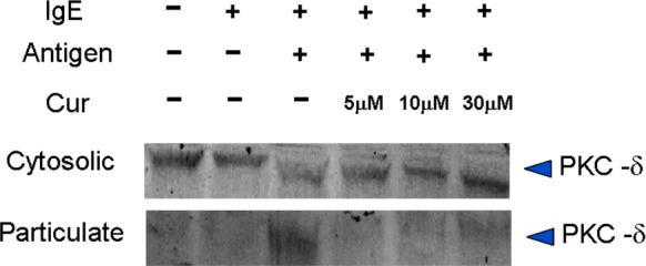
Effect of curcumin on PKC-δ translocation induced by antigen in RBL-2H3 cells
Discussion
This study investigated the ability of curcumin (Diferuloylmethane) against allergic inflammation response in vitro model by using rat basophil leukemia (RBL)-2H3 cells and human pre-basophils (KU812) cell lines. Curcuminoids have been approved as “Generally Recognized as Safe” (GRAS) by the US Food and Drug Administration (FDA) (Gupta et al. 2012). Additionally, curcuminoids also safety profiles and good tolerability have been shown by clinical trials, even at doses between 4000 and 8000 mg/day (Basnet and Skalko-Basnet 2011).
Figure 1a shows that the curcumin concentrations show the IC50 for cell viability more than 200 µM when incubated for short-terms. The figure also indicated that more than 90% cell viability when treated with 30 µM. Whereas, KU812 cells with IC50 about 36.8 µM when incubated for 24 h (Fig. 1b). As such, we used 30 µM of curcumin for next treatments both RBL-2H3 and KU812 cells. Previously study reported that 2-benzylidene cyclopentanone analogs of curcumin showed the IC50 of RBL-2H3 cell viability more than 100 µM when incubated for short-term (30 min) and 24 h (Nugroho et al. 2010).
The microscopic observation was used to evaluate the morphology of RBL-2H3 cells degranulation for anti-DNP IgE/DNP-BSA- and A23187-triggered as shown in Figs. 2, 3, respectively. Figure 2a shows that the normal RBL-2H3 cells differed in size with spindle-shaped, many microvilli, and cell membrane intact. The previous study also showed that the normal RBL-2H3 cells possess the same morphological conditions to this present study (Tang et al. 2012). After treated with anti-DNP IgE/DNP-BSA, the cell type was changed immediately and increased the cell degranulation (Fig. 2b). However, in curcumin-treated cells, curcumin inhibited the cells degranulation progression with the morphology similar to normal cells, especially in high-dose of curcumin (Fig. 2d). The calcium (Ca2+) ionosphere A23187-stimulated also induce cells degranulation (Fig. 3b). Whereas, treated with high-dose of curcumin inhibited the cell degranulation progression (Fig. 3d).
In the case of RBL-2H3 cells, these cells were treated either with an anti-dinitrophenol immunoglobulin E (anti-DNP IgE) and DNP-bovine serum albumin (DNP-BSA) or calcium (Ca2+) ionophore A23187. Monoclonal anti-dinitrophenol immunoglobulin E (anti-DNP IgE) and DNP-bovine serum albumin (DNP-BSA) have been used for triggering the release of histamine release from the mast cells (Bohn and König 1982, 1985). Whereas, a calcium ionosphere (A23187) has been used to upregulate the calcium (Ca2+) expression for mast cells degranulation (Shim et al. 2009). Both anti-DNP IgE/DNP-BSA- and A23187-triggered histamine release as shown in Fig. 4a, b, respectively. Whereas, histamine and β-hexosaminidase releases were used as markers for cells degranulation progression which produced by the cells. The previous studies also reported that both histamine and β-hexosaminidase release were increased after stimulated by both anti-DNP IgE/DNP-BSA and calcium ionosphere A23187 (Bohn and König 1985; Shim et al. 2009). Cytokines, histamine, and proteases play an important role in allergic inflammatory responses (Bao and Reinhardt 2015). After treated with curcumin, the histamine release significantly reduced in a dose-dependent manner. The β-hexosaminidase release also increased in RBL-2H3 cells when stimulated with anti-DNP IgE/DNP-BSA and A23187 (Figs. 5a, 6a, respectively). The treatment with curcumin significantly reduced the β-hexosaminidase release in the case of both anti-DNP IgE/DNP-BSA- and A23187-triggered mast cells degranulation (Figs. 5b, 6b, respectively). The previous study reported that different structure of curcuminoids also shows different effects to β-hexosaminidase releases, such as curcumin which isolated from rhizomes of Curcuma zedoaria the β-hexosaminidase show high inhibition (100.1 ± 1.1% in 30 µM). Whereas, the curcumin-related compounds from C. zedoaria such as bisdemethoxycurcumin and monomethylcurcumin show the β-hexosaminidase inhibition about 91.2 ± 4.6% and 67.5 ± 3.6% in 30 µM, respectively (Matsuda et al. 2004).
The production of reactive oxygen species (ROS) increased in RBL-2H3 cells when stimulated by both anti-DNP IgE/DNP-BSA and A23187 without curcumin treatment as shown in Fig. 7a, b, respectively. ROS may play an important role in mast cell degranulation by regulating extracellular calcium influx (Inoue et al. 2008) and mediating histamine release (Kim et al. 2002). After treated with curcumin, the ROS release was reduced especially in the case of high-dose of curcumin treatment. Antioxidant agents were used to inhibiting ROS production and resulting in the reduction of cell degranulation (Swindle and Metcalfe 2007; Tagen et al. 2009). The previous study reported that curcumin possesses potent antioxidant activity (Ak and Gülçin 2008; Menon and Sudheer 2007). Curcumin also increases the catalase (CAT) activity as shown in Fig. 7c. Catalase is one of enzymatic antioxidant has the ability to converted ROS hydrogen peroxide to molecular oxygen and water (Gupta et al. 2011). In this case, we hypothesized that curcumin directly reduced the ROS level by enhancing the catalase antioxidant activity. Additionally, previous studies reported that certain allergens and intrinsic NADPH oxidase from pollens could induce ROS production immune cells and antioxidant mechanisms are crucially essential to regulate the reduction–oxidation homeostasis (Hosoki et al. 2014; Qu et al. 2017; van Rijt et al. 2017).
Some pro-inflammatory cytokines were released during the allergic inflammatory responses includes interleukin (IL)-4 an IL-13 (Bao and Reinhardt 2015). Figure 8b, c show that the IL-3 and IL-14 expressions were increased when stimulated with calcium (Ca2+) ionosphere A23187, respectively. The IL-3 and IL-14 expressions reduced after treated with curcumin especially in the case of high-dose. The previous study reported that the IL-4 and IL-13 cytokines provided the first signaling of B cells to synthesize immunoglobulin (Ig) E. In vitro study reported that the curcumin suppressed tumor necrosis factor (TNF)-α and IL-4 levels (Lee et al. 2008). Moreover, previous study also reported that curcumin modulates NF-kβ-mediated inflammation in human tenocytes (Buhrmann et al. 2011). In vivo study also reported that the IL-4− and IL-13− deficient mice decreased the IgE release and impaired to T helper type 2 (Th2) cells response. A previous literature reported that Th2 cell is the most important inflammatory mediators (Vercelli et al. 2014). Additionally, the mice lack of IL-13 was demonstrated to ameliorate of an allergic reaction (Bao and Reinhardt 2015; Wills-Karp et al. 1998). According to this condition, inhibition of IL-4 and IL-13 expressions is one type of methods to reduce or prevent allergic inflammatory responses.
The basophil degranulation was started when the immunoglobulin (Ig) E binding to IgE receptor (FcεRI) and followed by cross-linking by the allergen. The cells degranulation release some inflammatory mediators, cytokines, and synthesis lipid mediators (Broide 2001). Figure 9a, b show that both FcεRI-α and FcεRI-γ expressions increased in control (untreated group) when stimulated with calcium (Ca2+) ionosphere A23187, respectively. After treated with high-dose of curcumin the FcεRI-α and FcεRI-γ expressions were significantly decreased in a dose-dependent manner. The immunoblotting also shows that the cellular expression of FcεRI-α reduced when treated with high-dose of curcumin (Fig. 9c). Since the FcεRI play an important role in triggers allergic reaction. There are many methods to prevent or inhibit the binding between IgE and FcεRI includes suppresses the FcεRI expression. According to the previous study, the alpha chain of FcεRI (FcεRI-α) directly binds to IgE, whereas the FcεRI-β and FcεRI-γ chains mediate the intracellular signal (Takahashi and Ra 2005). Additionally, the downregulation of IgE and IgE receptor levels on mast cells lean to reduce mast cells to produce inflammatory mediators (Broide 2001). In this study, we used FcεRI gene analysis to evaluate its expression during allergic reactions. Various previous studies also reported same methods to evaluated FcεRI level by using gene expression in allergic reactions (Akizawa 2003; Liao et al. 2015; Lora et al. 2003). Additionally, both surface and mRNA expression of FcεRI were upregulated by Th2 cytokines (i.e. IL-4 and IL-9) in allergic reactions (Chen et al. 2001; Feldmesser et al. 2008).
The protein kinase C delta (PKC-δ) has involved in the antigen-induced mast cell degranulation regulation (Ozawa et al. 1993). Figure 10 shows that the higher translocation of PKC-δ expression to particulate form was observed when stimulated by antigen DNP-BSA. However, treated with curcumin especially in high-dose of curcumin, the PKC-δ expression lower in particulate fraction than cytosolic as resulted by the inhibition of PKC-δ translocation. Among the PKC isoenzymes, PKC-δ has been reported that it possesses a positive correlation with allergic responses (Choi et al. 2013). Inhibition of PKC-δ activity has been demonstrated to alleviate airway inflammation by blocking IgE expression in mast cells (Cho et al. 2004). PKC-δ also regulates pro-inflammatory chemokines expression by interaction with NF-κB subunit p65(RelA) in response to TNF-α expression (Lu et al. 2009; Ren et al. 2014). Curcumin (diferuloylmethane) possesses two phenolic OH groups and an α,β-unsaturated-β-diketone moiety (Claramunt et al. 2009). Previous studies reported that curcumin bind to the activator binding (C1) domain of PKC by hydrogen bonds with the side residues on the activator side. Therefore, curcumin plays an important role in PKC activity (Das et al. 2016; Majhi et al. 2010).
As described above, curcumin successfully reduced allergic reaction by inhibiting some allergic mediators, FcεRIα expression, and PKC-δ translocation. A previous study reported that curcumin binds with the Notch receptor of cells and attenuated allergic reaction by inhibiting Notch1 signaling and GATA3 transcription factor expression (Chong et al. 2014). Notch signaling is important in the immune system by promoting T cell proliferation and cytokines production as well as inducing Th2 cell differentiation (Gazave et al. 2009). Additionally, a previous study also reported that curcumin downregulated allergic reaction by inhibiting receptor tyrosine kinase (i.e., Syk kinase) in mast cells. Syk activity played an important role in Ca2+ mobilization, activation of mitogen-activated protein kinase, and transcription factor for pro-inflammatory cytokines production (i.e., TNF-α and IL-4) (Lee et al. 2008; Rivera and Gilfillan 2006).
Conclusions
Overall, our study successfully demonstrated the in vitro models of curcumin function against the inflammatory responses related to allergic reactions in mast cells and basophils. The morphological observations also showed that curcumin inhibited the cells degranulation. The curcumin reduced the cells degranulation by reducing the histamine and β-hexosaminidase releases. Additionally, curcumin decreased pro-inflammatory cytokine levels and reactive oxygen species release. Moreover, cells treated with curcumin inhibited the immunoglobulin E receptors expression and protein kinase C delta translocation. This in vitro study proofed that curcumin has potent to promote as one of the anti-allergic agents.
Author contributions
Z-LK conceived and designed the experiments. H-JL and W-NC performed analyses. H-JL and SS wrote the manuscript.
Funding
This research received no external funding.
Compliance with ethical standards
Conflicts of interest
The authors declare that they have no conflict of interest.
Footnotes
Publisher's Note
Springer Nature remains neutral with regard to jurisdictional claims in published maps and institutional affiliations.
References
- Ak T, Gülçin İ. Antioxidant and radical scavenging properties of curcumin. Chem Biol Interact. 2008;174:27–37. doi: 10.1016/j.cbi.2008.05.003. [DOI] [PubMed] [Google Scholar]
- Akizawa Y. Regulation of human FcepsilonRI beta chain gene expression by Oct-1. Int Immunol. 2003;15:549–556. doi: 10.1093/intimm/dxg055. [DOI] [PubMed] [Google Scholar]
- Bao K, Reinhardt RL. The differential expression of IL-4 and IL-13 and its impact on type-2 immunity. Cytokine. 2015;75:25–37. doi: 10.1016/j.cyto.2015.05.008. [DOI] [PMC free article] [PubMed] [Google Scholar]
- Basnet P, Skalko-Basnet N. Curcumin: an anti-inflammatory molecule from a curry spice on the path to cancer treatment. Molecules. 2011;16:4567–4598. doi: 10.3390/molecules16064567. [DOI] [PMC free article] [PubMed] [Google Scholar]
- Bohn A, König W. Generation of monoclonal murine anti-DNP-IgE, IgM and IgG1 antibodies: biochemical and biological characterization. Immunology. 1982;47:297–311. [PMC free article] [PubMed] [Google Scholar]
- Bohn A, König W. Monoclonal murine anti-DNP IgE:in vitro histamine release of rat mast cells in the presence of reaginic rat and mouse sera. Agents Actions. 1985;16:485–490. doi: 10.1007/bf01983651. [DOI] [PubMed] [Google Scholar]
- Broide DH. Molecular and cellular mechanisms of allergic disease. J Allergy Clin Immunol. 2001;108:S65–S71. doi: 10.1067/mai.2001.116436. [DOI] [PubMed] [Google Scholar]
- Buhrmann C, Mobasheri A, Busch F, Aldinger C, Stahlmann R, Montaseri A, Shakibaei M. Curcumin modulates nuclear factor κB (NF-κB)-mediated inflammation in human tenocytesin vitro. J Biol Chem. 2011;286:28556–28566. doi: 10.1074/jbc.M111.256180. [DOI] [PMC free article] [PubMed] [Google Scholar]
- Chainani-Wu N. Safety and anti-inflammatory activity of curcumin: a component of tumeric (Curcuma longa) J Altern Complement Med. 2003;9:161–168. doi: 10.1089/107555303321223035. [DOI] [PubMed] [Google Scholar]
- Chen X-J, Lycke N, Enerback L. Surface and gene expression of immunoglobulin E receptors on mast cells and mast-cell numbers in interleukin-4-gene knockout mice. Immunology. 2001;96:544–550. doi: 10.1046/j.1365-2567.1999.00727.x. [DOI] [PMC free article] [PubMed] [Google Scholar]
- Chen H-J, Shih C-K, Hsu H-Y, Chiang W. Mast cell-dependent allergic responses are inhibited by ethanolic extract of adlay (Coix lachryma-jobi L. var.ma-yuen Stapf) testa. J Agric Food Chem. 2010;58:2596–2601. doi: 10.1021/jf904356q. [DOI] [PubMed] [Google Scholar]
- Cho SH, You HJ, Woo CH, Yoo YJ, Kim JH. Rac and protein kinase C-regulate ERKs and cytosolic phospholipase A2 in Fc RI signaling to cysteinyl leukotriene synthesis in mast cells. J Immunol. 2004;173:624–631. doi: 10.4049/jimmunol.173.1.624. [DOI] [PubMed] [Google Scholar]
- Choi Y, Kim MS, Hwang JK. Inhibitory effects of panduratin A on allergy-related mediator production in rat basophilic leukemia mast cells. Inflammation. 2012;35:1904–1915. doi: 10.1007/s10753-012-9513-y. [DOI] [PubMed] [Google Scholar]
- Choi YH, Jin GY, Lc Li, Yan GH. Inhibition of protein kinase C delta attenuates allergic airway inflammation through suppression of PI3K/Akt/mTOR/HIF-1 alpha/VEGF pathway. PLoS ONE. 2013 doi: 10.1371/journal.pone.0081773. [DOI] [PMC free article] [PubMed] [Google Scholar]
- Chong L, et al. Protective effect of curcumin on acute airway inflammation of allergic asthma in mice through Notch1–GATA3 signaling pathway. Inflammation. 2014;37:1476–1485. doi: 10.1007/s10753-014-9873-6. [DOI] [PMC free article] [PubMed] [Google Scholar]
- Claramunt RM, Bouissane L, Cabildo MP, Cornago MP, Elguero J, Radziwon A, Medina C. Synthesis and biological evaluation of curcuminoid pyrazoles as new therapeutic agents in inflammatory bowel disease: effect on matrix metalloproteinases. Bioorg Med Chem. 2009;17:1290–1296. doi: 10.1016/j.bmc.2008.12.029. [DOI] [PubMed] [Google Scholar]
- Das J, Ramani R, Suraju MO. Polyphenol compounds and PKC signaling. Biochimica et Biophysica Acta Gen Subj. 2016;1860:2107–2121. doi: 10.1016/j.bbagen.2016.06.022. [DOI] [PMC free article] [PubMed] [Google Scholar]
- Feldmesser M, Alphonse MP, Saffar AS, Shan L, HayGlass KT, Simons FER, Gounni AS. Regulation of the high affinity IgE receptor (FcεRI) in human neutrophils: role of seasonal allergen exposure and Th-2 cytokines. PLoS ONE. 2008 doi: 10.1371/journal.pone.0001921. [DOI] [PMC free article] [PubMed] [Google Scholar]
- Galli SJ, Tsai M, Piliponsky AM. The development of allergic inflammation. Nature. 2008;454:445–454. doi: 10.1038/nature07204. [DOI] [PMC free article] [PubMed] [Google Scholar]
- Gazave E, et al. Origin and evolution of the Notch signalling pathway: an overview from eukaryotic genomes. BMC Evol Biol. 2009 doi: 10.1186/1471-2148-9-249. [DOI] [PMC free article] [PubMed] [Google Scholar]
- Gupta S, et al. Fluctuations in total antioxidant capacity, catalase activity and hydrogen peroxide levels of follicular fluid during bovine folliculogenesis. Reprod Fertil Dev. 2011 doi: 10.1071/rd10270. [DOI] [PMC free article] [PubMed] [Google Scholar]
- Gupta SC, Patchva S, Aggarwal BB. Therapeutic roles of curcumin: lessons learned from clinical trials. AAPS J. 2012;15:195–218. doi: 10.1208/s12248-012-9432-8. [DOI] [PMC free article] [PubMed] [Google Scholar]
- Hara T, Yamada K, Tachibana H. Basophilic differentiation of the human leukemia cell line KU812 upon treatment with interleukin-4. Biochem Biophys Res Commun. 1998;247:542–548. doi: 10.1006/bbrc.1998.8816. [DOI] [PubMed] [Google Scholar]
- Hatcher H, Planalp R, Cho J, Torti FM, Torti SV. Curcumin: from ancient medicine to current clinical trials. Cell Mol Life Sci. 2008;65:1631–1652. doi: 10.1007/s00018-008-7452-4. [DOI] [PMC free article] [PubMed] [Google Scholar]
- Hong S, Son DK, Lim WR, Kim SH, Kim H, Yum HY, Kwon H. The prevalence of atopic dermatitis, asthma, and allergic rhinitis and the comorbidity of allergic diseases in children. Environ Health Toxicol. 2012 doi: 10.5620/eht.2012.27.e2012006. [DOI] [PMC free article] [PubMed] [Google Scholar]
- Hosoki Koa, Gandhe Renu, Boldogh Istvan, Sur Sanjiv. Systems Biology of Free Radicals and Antioxidants. Berlin, Heidelberg: Springer Berlin Heidelberg; 2014. Reactive Oxygen Species (ROS) and Allergic Responses; pp. 3239–3266. [Google Scholar]
- Inoue T, Suzuki Y, Yoshimaru T, Ra C. Reactive oxygen species produced up- or downstream of calcium influx regulate proinflammatory mediator release from mast cells: role of NADPH oxidase and mitochondria. Biochim Biophys Acta. 2008;1783:789–802. doi: 10.1016/j.bbamcr.2007.12.004. [DOI] [PubMed] [Google Scholar]
- Juckmeta T, Thongdeeying P, Itharat A. Inhibitory effect on β-hexosaminidase release from RBL-2H3 cells of extracts and some pure constituents of benchalokawichian, a Thai herbal remedy used for allergic disorders. Evid Based Complement Alternat Med. 2014;2014:1–8. doi: 10.1155/2014/828760. [DOI] [PMC free article] [PubMed] [Google Scholar]
- Kao N-J, Hu J-Y, Wu C-S, Kong Z-L. Curcumin represses the activity of inhibitor-κB kinase in dextran sulfate sodium-induced colitis by S-nitrosylation. Int Immunopharmacol. 2016;38:1–7. doi: 10.1016/j.intimp.2016.05.015. [DOI] [PubMed] [Google Scholar]
- Kim CJ, et al. Histamine release by hydrochloric acid is mediated via reactive oxygen species generation and phospholipase D in RBL-2H3 mast cells. Arch Pharmacal Res. 2002;25:675–680. doi: 10.1007/bf02976943. [DOI] [PubMed] [Google Scholar]
- Kobayashi S, Tanabe S. Evaluation of the anti-allergic activity of Citrus unshiu using rat basophilic leukemia RBL-2H3 cells as well as basophils of patients with seasonal allergic rhinitis to pollen. Int J Mol Med. 2006 doi: 10.3892/ijmm.17.3.511. [DOI] [PubMed] [Google Scholar]
- Kurup VP, Barrios CS. Immunomodulatory effects of curcumin in allergy. Mol Nutr Food Res. 2008;52:1031–1039. doi: 10.1002/mnfr.200700293. [DOI] [PubMed] [Google Scholar]
- Lee JH, et al. Curcumin, a constituent of curry, suppresses IgE-mediated allergic response and mast cell activation at the level of Syk. J Allergy Clin Immunol. 2008;121:1225–1231. doi: 10.1016/j.jaci.2007.12.1160. [DOI] [PubMed] [Google Scholar]
- Liao E-C, Chang C-Y, Hsieh C-W, Yu S-J, Yin S-C, Tsai J-J. An exploratory pilot study of genetic marker for IgE-mediated allergic diseases with expressions of FcεR1α and Cε. Int J Mol Sci. 2015;16:9504–9519. doi: 10.3390/ijms16059504. [DOI] [PMC free article] [PubMed] [Google Scholar]
- Lora JM, et al. FcεRI-dependent gene expression in human mast cells is differentially controlled by T helper type 2 cytokines. J Allergy Clin Immunol. 2003;112:1119–1126. doi: 10.1016/j.jaci.2003.08.042. [DOI] [PubMed] [Google Scholar]
- Lowry OH, Rosenbrough NJ, Farr A, Randall RJ. Protein measurement with the Folin phenol reagent. J Biol Chem. 1951;193:265–275. [PubMed] [Google Scholar]
- Lu ZG, Liu H, Yamaguchi T, Miki Y, Yoshida K. Protein kinase C activates RelA/p65 and nuclear factor-B signaling in response to tumor necrosis factor. Cancer Res. 2009;69:5927–5935. doi: 10.1158/0008-5472.Can-08-4786. [DOI] [PubMed] [Google Scholar]
- Majhi A, Rahman GM, Panchal S, Das J. Binding of curcumin and its long chain derivatives to the activator binding domain of novel protein kinase C. Bioorg Med Chem. 2010;18:1591–1598. doi: 10.1016/j.bmc.2009.12.075. [DOI] [PMC free article] [PubMed] [Google Scholar]
- Matsuda H, Tewtrakul S, Morikawa T, Nakamura A, Yoshikawa M. Anti-allergic principles from Thai zedoary: structural requirements of curcuminoids for inhibition of degranulation and effect on the release of TNF-α and IL-4 in RBL-2H3 cells. Bioorg Med Chem. 2004;12:5891–5898. doi: 10.1016/j.bmc.2004.08.027. [DOI] [PubMed] [Google Scholar]
- Matsui T, et al. Diphenyleneiodonium prevents reactive oxygen species generation, tyrosine phosphorylation, and histamine release in RBL-2H3 mast cells. Biochem Biophys Res Commun. 2000;276:742–748. doi: 10.1006/bbrc.2000.3545. [DOI] [PubMed] [Google Scholar]
- Menon VP, Sudheer AR (2007) Antioxidant and anti-inflammatory properties of curcumin. In: The molecular targets and therapeutic uses of curcumin in health and disease. Advances in experimental medicine and biology, pp 105–125. 10.1007/978-0-387-46401-5_3 [DOI] [PubMed]
- Metzger H, Alcaraz G, Hohman R, Kinet JP, Pribluda V, Quarto R. The receptor with high affinity for immunoglobulin E. Annu Rev Immunol. 1986;4:419–470. doi: 10.1146/annurev.iy.04.040186.002223. [DOI] [PubMed] [Google Scholar]
- Nugroho AE, Sardjiman S, Maeyama K. Comparison of cytotoxic and antiproliferative effects of benzylidenecyclopentanone analogues of curcumin on RBL-2H3 Cells. Indones J Biotechnol. 2010;15:64–71. doi: 10.22146/ijbiotech.7822. [DOI] [Google Scholar]
- Ozawa K, Yamada K, Kazanietz MG, Blumberg PM, Beaven MA. Different isozymes of protein kinase C mediate feedback inhibition of phospholipase C and stimulatory signals for exocytosis in rat RBL-2H3 cells. J Biol Chem. 1993;268:2280–2283. [PubMed] [Google Scholar]
- Pan M-H, Huang T-M, Lin J-K. Biotransformation of curcumin through reduction and glucuronidation in mice. Drug Metab Dispos. 1999;27:486–494. [PubMed] [Google Scholar]
- Pan M-H, Chen J-W, Kong Z-L, Wu J-C, Ho C-T, Lai C-S. Attenuation by tetrahydrocurcumin of adiposity and hepatic steatosis in mice with high-fat-diet-induced obesity. J Agric Food Chem. 2018;66:12685–12695. doi: 10.1021/acs.jafc.8b04624. [DOI] [PubMed] [Google Scholar]
- Qu J, Li Y, Zhong W, Gao P, Hu C. Recent developments in the role of reactive oxygen species in allergic asthma. J Thorac Dis. 2017;9:E32–E43. doi: 10.21037/jtd.2017.01.05. [DOI] [PMC free article] [PubMed] [Google Scholar]
- Ren J, et al. Protein kinase C-δ (PKCδ) regulates proinflammatory chemokine expression through cytosolic interaction with the NF-κB subunit p65 in vascular smooth muscle cells. J Biol Chem. 2014;289:9013–9026. doi: 10.1074/jbc.M113.515957. [DOI] [PMC free article] [PubMed] [Google Scholar]
- Rivera J, Gilfillan A. Molecular regulation of mast cell activation. J Allergy Clin Immunol. 2006;117:1214–1225. doi: 10.1016/j.jaci.2006.04.015. [DOI] [PubMed] [Google Scholar]
- Rosenwasser LJ. Mechanisms of IgE inflammation. Curr Allergy Asthma Rep. 2011;11:178–183. doi: 10.1007/s11882-011-0179-6. [DOI] [PubMed] [Google Scholar]
- Shim S-Y, et al. Construction of an in vitro allergy reaction evaluation system using human leukemia cell lines. Cytotechnology. 2002;40:75–83. doi: 10.1023/a:1023918206060. [DOI] [PMC free article] [PubMed] [Google Scholar]
- Shim J-U, Oh P-S, Lim K-T. Anti-inflammatory activity of ethanol extract from Geranium sibiricum Linne. J Ethnopharmacol. 2009;126:90–95. doi: 10.1016/j.jep.2009.08.004. [DOI] [PubMed] [Google Scholar]
- Soudamini KK, Kuttan R. Inhibition of chemical carcinogenesis by curcumin. J Ethnopharmacol. 1989;27:227–233. doi: 10.1016/0378-8741(89)90094-9. [DOI] [PubMed] [Google Scholar]
- Suzuki Y, Yoshimaru T, Matsui T, Inoue T, Niide O, Nunomura S, Ra C. Fc RI signaling of mast cells activates intracellular production of hydrogen peroxide: role in the regulation of calcium signals. J Immunol. 2003;171:6119–6127. doi: 10.4049/jimmunol.171.11.6119. [DOI] [PubMed] [Google Scholar]
- Swindle EJ, Metcalfe DD. The role of reactive oxygen species and nitric oxide in mast cell-dependent inflammatory processes. Immunol Rev. 2007;217:186–205. doi: 10.1111/j.1600-065X.2007.00513.x. [DOI] [PubMed] [Google Scholar]
- Tagen M, Elorza A, Kempuraj D, Boucher W, Kepley CL, Shirihai OS, Theoharides TC. Mitochondrial uncoupling protein 2 inhibits mast cell activation and reduces histamine content. J Immunol. 2009;183:6313–6319. doi: 10.4049/jimmunol.0803422. [DOI] [PMC free article] [PubMed] [Google Scholar]
- Takahashi K, Ra C. The high affinity IgE receptor (FcεRI) as a target for anti-allergic agents. Allergol Int. 2005;54:1–5. doi: 10.2332/allergolint.54.1. [DOI] [Google Scholar]
- Tang J-M, Liu J, Wu W. Studies on the degranulation of RBL-2H3 cells induced by traditional Chinese medicine injections. Chin Med. 2012;03:200–208. doi: 10.4236/cm.2012.34029. [DOI] [Google Scholar]
- van Rijt L, Utsch L, Lutter R, van Ree R. Oxidative stress: promoter of allergic sensitization to protease allergens? Int J Mol Sci. 2017 doi: 10.3390/ijms18061112. [DOI] [PMC free article] [PubMed] [Google Scholar]
- Vercelli D, Gozdz J, von Mutius E. Innate lymphoid cells in asthma. Curr Opin Allergy Clin Immunol. 2014;14:29–34. doi: 10.1097/aci.0000000000000023. [DOI] [PubMed] [Google Scholar]
- Wang Y-J, Pan M-H, Cheng A-L, Lin L-I, Ho Y-S, Hsieh C-Y, Lin J-K. Stability of curcumin in buffer solutions and characterization of its degradation products. J Pharm Biomed Anal. 1997;15:1867–1876. doi: 10.1016/s0731-7085(96)02024-9. [DOI] [PubMed] [Google Scholar]
- Wills-Karp M, Luyimbazi J, Xu X, Schofield B, Neben TY, Karp CL, Donaldson DD. Interleukin-13: central mediator of allergic asthma. Science. 1998;282:2258–2261. doi: 10.1126/science.282.5397.2258. [DOI] [PubMed] [Google Scholar]
- Zhao ZZ, Sugerman PB, Walsh LJ, Savage NW. A fluorometric microassay for histamine release from human gingival mast cells. J Periodontal Res. 2001;36:233–236. doi: 10.1034/j.1600-0765.2001.036004233.x. [DOI] [PubMed] [Google Scholar]