Abstract
Objective
To compare the accuracy of infrared (IR)-reflex assessment using a prototype imaging device to standard non-mydriatic red-reflex screening with direct ophthalmoscope (DO) in the diagnosis of neonatal and childhood cataract.
Methods
The comparison of the techniques was made in two distinct cohorts: in the first, newborns underwent IR and red-reflex testing by a medical student, with results compared to a reference red-reflex examination by an experienced midwife. In the second, an enriched cohort of children attending a specialist paediatric ophthalmology clinic had IR and red-reflex testing by a medical student to reference examination by a paediatric ophthalmologist. The medical students were considered inexperienced screeners due to their limited exposure to ophthalmology. The sensitivity and specificity of the IR and red-reflex assessments in respect to reference examination were calculated. Diagnostic accuracy was compared in Caucasian and non-Caucasian eyes.
Results
IR and red-reflex imaging were possible in all 180 neonatal eyes examined. A total of 5% of newborn eyes were found to have embryological remnants in the anterior segment of the eye with IR-reflex imaging which were not detected on reference red-reflex examination. IR-reflex assessment had significantly better sensitivity (100 vs 71%, p < 0.05) and specificity (100 vs 63%, p < 0.01) than red-reflex assessment in the diagnosis of childhood cataract. Red-reflex specificity was particularly poor in non-Caucasian eyes compared to Caucasian eyes (32 vs 72%, p < 0.05).
Conclusion
This pilot study indicates that IR-reflex imaging has the potential to improve the diagnostic accuracy of eye screening for cataract by inexperienced healthcare staff, particularly in non-Caucasian children.
Subject terms: Paediatrics, Lens diseases, Physical examination
Introduction
Cataract affects 3–5 per 10,000 children in the US and UK [1, 2], and the prevalence in developing countries is at least double this, being responsible for 20% of child blindness worldwide [3]. Although relatively rare, the impact in human and economic terms is vast, with the global cost of preventable blindness from childhood cataract estimated at $1000–6000 million dollars over 10 years [4]. Visually significant congenital cataracts require surgical management by 8–10 weeks of age to prevent irreversible sight-deprivation amblyopia and optimize visual outcome [5].
Non-mydriatic red-reflex examination with the direct ophthalmoscope (DO) is a well-established technique for non-specialist detection of cataract and retinoblastoma in children. Sensitivity in the detection of anterior media opacities by experienced clinicians is generally good, reported at 85–99.6%, but specificity can be poorer, as low as 39% in non-Caucasian populations [6, 7].
In most developed countries, red-reflex screening is performed as part of a general newborn and infant physical examination (NIPE) by non-medical healthcare staff such as midwives and nurses. Although given training, many screeners comment on their unfamiliarity with using the DO and their difficulty in detecting the red-reflex, particularly in non-Caucasian babies [8, 9]. A cross-sectional UK surveillance study has shown that fewer than 50% of childhood cataracts with features suggesting congenital onset were initially detected at NIPE screening and approximately a third of infants with cataract(s) were over 1 year of age at their first ophthalmic assessment [10]. Additional evidence points to low specificity: a UK referral audit has indicated that over 85% of infants referred with abnormal red-reflexes from primary care are false-positive cases and the majority of these are from non-Caucasian ethnic groups [11]. Concern has been raised that, given cost and capacity pressures, referrals for equivocal red-reflexes are inhibited, resulting in missed pathology [12].
The prevention of blindness from childhood cataract is a priority for the World Health Organization’s Vision 2020: The Right to Sight program [13]. A screening test which enables both high sensitivity and specificity in cataract detection both in Caucasian and non-Caucasian populations with minimal training is needed. Infrared (IR) light does not induce pupillary constriction and it is well reflected by the fundus, regardless of ethnic differences in fundus pigmentation [14]. IR illumination is an integral part of non-mydriatic fundus cameras and auto-refractors. This proof-of-concept study aimed to assess the potential use of IR-reflex imaging in neonatal and childhood screening for cataract by inexperienced examiners.
Methods
Given the low prevalence of congenital cataract, a population of over 50,000 newborn babies would have been required to compare the diagnostic accuracy of IR and red-reflex retro-illumination techniques in the NIPE screening population alone. For this reason, testing was carried out in two separate cohorts of patients: a newborn screening cohort with IR-reflex imaging performed alongside the standard NIPE red-reflex screening, and a comparison study of the diagnostic accuracy of IR and red-reflex testing in a cataract enriched paediatric population attending a specialist eye clinic. Sample size was calculated based on a pathology prevalence of 20% in the enriched population and a difference of 15% sensitivity and specificity between tests.
The medical student examiners had no experience of red-reflex assessment and minimal experience of DO use prior to the study. They were taught and were able to practise both the IR and red-reflex techniques in a paediatric ophthalmology clinic prior to participating in the study.
Babies and children were enrolled into the study following informed consent by their parents, the ethnic origin of each child was self-assigned. The research was performed in accordance with the Declaration of Helsinki and approved by the East of England-Cambridge South NHS England ethics committee.
Newborn screening cohort
Over 7 days of testing, parents of all newborns having NIPE screening prior to hospital discharge were invited to have their baby’s eyes checked with IR-reflex imaging in addition to the standard red-reflex screening. A medical student performed red-reflex assessment followed by IR-reflex imaging; the reference red-reflex examination was undertaken by an experienced midwife as part of the NIPE screen. The examinations were documented as normal, abnormal or not possible. Specialist review was arranged for any babies in whom an abnormality on IR or red-reflex was detected or the assessment was not possible.
Enriched clinic cohort
For the second study, parents of children 16 years and younger attending a specialist paediatric ophthalmology clinic were invited to enrol their child. Children with a history of previous ophthalmic surgery were excluded. Children initially underwent red-reflex evaluation and IR-reflex imaging by one of three medical students who were masked to the child’s diagnosis. Results were recorded as either normal or abnormal for each test modality. The child then underwent a reference examination by a paediatric ophthalmologist comprising mydriatic examination with slit-lamp biomicroscopy, retinoscopy and indirect ophthalmoscopy. The data from the right eye only of each child were statistically analysed with differences in sensitivity and specificity of the screening tests evaluated using McNemar’s test for paired data. Differences between Caucasian and non-Caucasian subjects were analysed using a standard χ2 for n > 10 and Fisher’s exact test for n < 10.
Materials
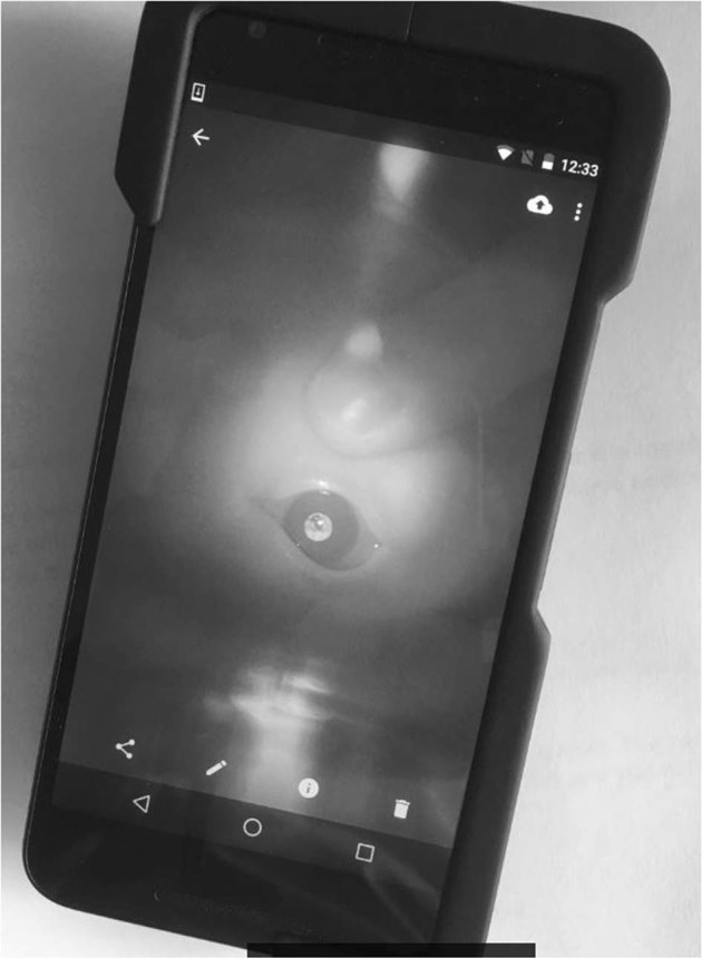
The IR-reflex imaging prototype device used was a Nexus (Google LLC, CA, US) smartphone, modified with a co-axial IR-emitting diode (wavelength 950 nm) and IR camera (Fig. 1). Used one-handed at a distance of 15–25 cm from the baby’s eye, it does not require visual fixation and enables the examiner to use the fingers on their other hand to part the baby’s eyelids. A standard Keeler Ltd. (Windsor, UK) DO was used for red-reflex assessment, with “0” lens dialled in and the baby’s eye viewed from ~50 cm, according to the American Academy of Pediatrics recommendations [15].
Fig. 1.

The modified smartphone IR-reflex imaging camera showing an image of a childhood cataract
Results
Newborn screening cohort
Ninety newborn babies were recruited, 65 (72%) of whom were Caucasian. IR and red-reflex testing were possible in all 180 eyes tested and no cataracts were identified with either screening tool by the student or midwife. Subtle ocular media opacities were detected only on IR-reflex imaging in nine (5%) eyes. These characteristic and visually insignificant anomalies were subsequently identified as bilateral persistent pupillary fibres in three babies (Fig. 2A) and unilateral Mittendorf dots in three babies (Fig. 2b) after specialist review with slit-lamp biomicroscopy (the anomalies were undetectable on retinoscopy or indirect ophthalmoscopy).
Fig. 2.
The IR-reflex is white on imaging and media opacities are silhouetted on the reflex. These images, taken during the newborn screening study illustrate: a pupillary fibres and b Mittendorf dot
Enriched clinic cohort
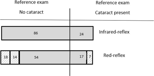
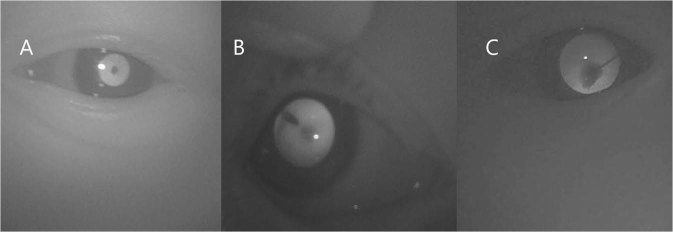
One hundred and ten children, median age 2 years (range 1 week to 13 years), were enrolled in the study: 89 (81%) were Caucasian, 21 (19%) were of Asian or African-Caribbean ethnicity. Specialist reference examination detected cataract in 24 eyes of the 110 eyes (22%) of which 2 were in non-Caucasian children. The screening results are shown in Table 1 and the number of false-negatives and positives is illustrated in Fig. 3. Red-reflex screening failed to detect 7 cataracts in Caucasian eyes: 5 of these were mild anterior polar or peripheral wedge-shaped peripheral cataracts requiring monitoring only, 2 were visually significant posterior plaque opacities both of which required subsequent surgery (Fig. 4c).
Table 1.
Results of matched diagnostic tests. A total of 95% confidence intervals are quoted in brackets after the relevant parameter
| IR-reflex (all ethnicities) | Red-reflex (all ethnicities) | IR-reflex in Caucasian eyes | Red-reflex in Caucasian eyes | IR-reflex in non-Caucasian eye | Red-reflex in non-Caucasian eyes | |
|---|---|---|---|---|---|---|
| Specificity | 100% (95–100%) | 63% (53–73%) | 100% | 72% | 100% | 32% |
| Sensitivity | 100% (83–100%) | 71% (53–89%) | 100% | 68% | 100% | 100% |
Fig. 3.

Comparison of IR and red-reflex findings to reference assessment in the enriched population trial. Grey shading represents true positives and negatives. A total of 32 eyes had false-positive red-reflex tests: 14 (cross-hatched) were non-Caucasian eyes, 18 (hatched) were Caucasian eyes. Seven eyes had false negative red-reflex tests, all were Caucasian eyes
Fig. 4.
Examples of false-negatives from red-reflex screening, imaged with IR-reflex camera. a Anterior polar lens opacity, b wedge shaped and mild posterior lens opacities, c posterior plaque due to persistent foetal vasculature
The diagnostic accuracy of IR-reflex assessment was significantly higher than red-reflex assessment (p < 0.05 using McNemar’s test). The specificity (32%) of red-reflex testing was significantly lower in the 21 non-Caucasian eyes than in Caucasian eyes (Fisher’s exact test, p < 0.05), whereas IR-reflex assessment was consistent across ethnic subgroups.
Discussion
The aim of this study was to determine the potential of using IR-reflex imaging for neonatal and childhood cataract screening. The UK newborn red-reflex assessment is performed by experienced midwives who develop considerable skill in the technique. This may not be the case internationally or for the community healthcare staff performing subsequent childhood examinations, who are unlikely to examine a baby with cataracts during their professional lives. To represent the relative inexperience of this group, medical student examiners were used in this study.
IR-reflex screening was possible in all newborns tested; none of the 180 eyes had congenital cataracts. Embryological remnants causing subtle, visually insignificant media opacities were detected in 5% of newborn eyes with IR retro-illumination. Although the characteristic appearance of pupillary fibres makes them unlikely to be mistaken for cataract, 1.6% of neonatal eyes examined had Mittendorf dots which may be difficult to distinguish from small posterior polar lens opacities on IR-reflex imaging alone. Screening newborns with the high-resolution IR camera used in this study therefore has the potential of causing unnecessary referral in these cases.
The enriched population study demonstrated the superior sensitivity and specificity of inexperienced screeners in the detection of childhood cataract when using IR-reflex imaging compared to red-reflex examination. The sensitivity and specificity of the clinic-based red-reflex assessment for cataract diagnosis were 71% and 63%, respectively. All IR-reflex assessments matched the reference examination. The poor diagnostic accuracy of red-reflex assessment in this study was surprising given previous reports of very high levels of accuracy, albeit with experienced paediatricians or ophthalmologists performing the assessment [6]. The lower specificity of red-reflex assessment in non-Caucasian children compared to Caucasians reached significance despite the small number of subjects studied, with 14 of the 16 abnormal red-reflex assessments in non-Caucasian children being false positives. The high proportion of non-Caucasian children amongst false positive referrals from primary care has previously been reported [11].
This study population had a cataract prevalence of 22% whereas the prevalence of cataract in the UK newborn population is ~0.03%. If the sensitivity of red-reflex examination in this study reflects that of the NIPE eye check, it is possible that up to a third of congenital or infantile cataracts might not be detected at the newborn and infant screen, a finding which may be supported by the surveillance data of Rahi et al. [10]. Conversely, the specificity of red-reflex examination in this enriched population would suggest a potentially extremely low positive predictive value in the low-prevalence screening population and thousands of false positive referrals, particularly in non-Caucasian babies, would be expected. Although there is no published data quantifying the number of false positive red-reflex referrals to specialist clinics per year, a high false-positive rate of primary care referrals for abnormal red-reflex, particularly in non-Caucasian infants and children, has been reported and there is concern that some equivocal assessments do not result in referral [11, 12].
This study demonstrates that digital IR-reflex imaging has potential advantages over the current method of red-reflex screening for childhood cataract. However, an important limitation of using IR illumination in isolation is the risk that the IR reflection from a white media opacity such as retinoblastoma is misinterpreted as a normal bright IR-reflex. The incidence of retinoblastoma in children under 5 years is 1 per 20,000; it is a curable cancer and the ability to detect the characteristic leukocoria on testing is an important consideration [16]. Green light is relatively poorly reflected by the normal fundus resulting in absence of a green-reflex (seen as a dark pupil). In comparison, a white media opacity, such as retinoblastoma, will reflect green light, giving an abnormal bright reflex [13]. Dual-wavelength sequential illumination that utilizes the different reflective properties of the normal fundus and different types of ocular media opacity, may be the solution.
The modified smartphone prototype used in this study has the advantage of being easy to use with one hand, with minimal training required. Its disadvantages are the potential of false positive referrals for insignificant opacities such as Mittendorf dots due to the smartphone camera’s very high resolution and, most importantly, the potential issue with retinoblastoma detection. To resolve these problems, a purpose-built new prototype has been developed with modified resolution dual-wavelength imaging: synchronized IR (890 nm) and green flash illumination (568 nm) reflex images are captured sequentially before pupil constriction can occur.
This proof-of-concept study, although small, has illustrated that the diagnostic accuracy of IR-reflex assessment for cataract is significantly better than standard red-reflex assessment in the hands of inexperienced screeners, particularly in non-Caucasian children. IR-reflex screening may have a global role to play in the prevention of child blindness due to cataract. Additional advantages of digital imaging include documentation, telemedicine and the potential development of diagnostic computer algorithms. The dual-wavelength prototype is currently being evaluated in a large African cluster trial supported by the International Agency for the Prevention of Blindness. Results of this and future studies will determine if the dual-wavelength device will be a cost-effective solution for population screening for cataract and retinoblastoma.
Summary
What was known before
Childhood cataract causes 20% of world child blindness. Congenital cataract red-reflex screening during the newborn and infant eye examination may miss up to 50% of cases. Severe congenital cataracts require surgery before 10 weeks of age to prevent permanent visual impairment due to amblyopia.
What this study adds
The diagnostic accuracy of red-reflex assessment by inexperienced health-care professionals is relatively poor. The specificity is especially poor in non-Caucasian eyes. Infrared-reflex testing may be a more accurate screening test for cataract, particularly in non-Caucasian populations.
Acknowledgements
We thank the donors to Addenbrooke's Charitable Trust who supported the development of the smartphone prototype.
Funding
LEA reports funding from Addenbrooke's Charitable Trust (Innovation Grant) during the conduct of the study. The Addenbrooke’s Charitable Trust had no input in the study design, in the collection, analysis and interpretation of the data, in the writing of the report, or in the decision to submit the paper for publication.
Compliance with ethical standards
Conflict of interest
LEA reports grants from NeoCam Ltd, outside the submitted work. In addition, LEA has a patent for the imaging device and method of imaging ocular media opacities pending.
Footnotes
Publisher’s note: Springer Nature remains neutral with regard to jurisdictional claims in published maps and institutional affiliations.
References
- 1.Holmes JM, Leske DA, Burke JP, Hodge DO. Birth prevalence of visually significant infantile cataract in a defined U.S. population. Ophthalmic Epidemiol. 2003;10:67–74. doi: 10.1076/opep.10.2.67.13894. [DOI] [PubMed] [Google Scholar]
- 2.Rahi JS, Dezateux C, British Congenital Cataract Interest Group. Measuring and interpreting the incidence of congenital ocular anomalies: lessons from a national study of congenital cataract in the UK. Investig Ophthalmol Vis Sci. 2001;42:1444–8. [PubMed] [Google Scholar]
- 3.Sheeladevi S, Lawrenson JG, Fielder AR, Suttle CM. Global prevalence of childhood cataract: a systematic review. Eye. 2016;30:1160–9. doi: 10.1038/eye.2016.156. [DOI] [PMC free article] [PubMed] [Google Scholar]
- 4.Gilbert C, Foster A. Childhood blindness in the context of Vision 2020: The Right to Sight. Bull World Health Organ. 2001;79:227–32. [PMC free article] [PubMed] [Google Scholar]
- 5.Birch EE, Cheng C, Stager DR, Weakley DR, Stager DR. The critical period for surgical treatment of dense congenital bilateral cataracts. J AAPOS. 2009;13:67–71. doi: 10.1016/j.jaapos.2008.07.010. [DOI] [PMC free article] [PubMed] [Google Scholar]
- 6.Sun M, Mis A, Li F, Cheng K, Zhang M, Yang H, et al. Sensitivity and specificity of red reflex test in newborn eye screening. J Pediatrics. 2016;179:192–6. doi: 10.1016/j.jpeds.2016.08.048. [DOI] [PubMed] [Google Scholar]
- 7.Mussavi M, Asadollahi K, Janbaz F, Mansoori E, Abbasi N. The Evaluation of Red Reflex Sensitivity and Specificity among Neonates in Different Conditions. Iran J Pediatrics. 2014;24:697–702. [PMC free article] [PubMed] [Google Scholar]
- 8.Rahi JS, Lynn R. A survey of paediatricians’ practice and training in routine infant eye examination. Arch Dis Child. 1998;78:364–6. doi: 10.1136/adc.78.4.364. [DOI] [PMC free article] [PubMed] [Google Scholar]
- 9.Green K, Oddie S. The value of the postnatal examination in improving child health. Arch Dis Child Fetal Neonatal Ed. 2008;93:F389–93. doi: 10.1136/adc.2007.122465. [DOI] [PubMed] [Google Scholar]
- 10.Rahi JS, Dezateux C, British Congenital Cataract Interest Group. National cross sectional study of detection of congenital and infantile cataract in the UK: role of childhood screening and surveillance. Br Med J. 1990;318:362–5. doi: 10.1136/bmj.318.7180.362. [DOI] [PMC free article] [PubMed] [Google Scholar]
- 11.Muen WJ, Hindocha M, Reddy MA. The role of education in the promotion of red reflex assessments. J R Soc Med Short Rep. 2010;1:46. doi: 10.1258/shorts.2010.010036. [DOI] [PMC free article] [PubMed] [Google Scholar]
- 12.Morgan S. In screening for congenital cataract, many false positive referrals will occur. Br Med J. 1999;319:122. doi: 10.1136/bmj.319.7202.122. [DOI] [PMC free article] [PubMed] [Google Scholar]
- 13.International Agency for the Prevention of Blindness. Global Action Plan. https://www.iapb.org/advocacy/global-action-plan-2014-9/.
- 14.Delori FC, Pflibsen KP. Spectral reflectance of the human ocular fundus. Appl Opt. 1989;28:1061–77. doi: 10.1364/AO.28.001061. [DOI] [PubMed] [Google Scholar]
- 15.American Academy of Pediatrics Policy Statement. Red reflex examination in neonates, infants and children. Pediatrics. 2008;122:1401–4. doi: 10.1542/peds.2008-2624. [DOI] [PubMed] [Google Scholar]
- 16.Broaddus E, Topham A, Singh AD. Incidence of retinoblastoma in the USA: 1975–2004. Br J Ophthalmol. 2009;93:21–3. doi: 10.1136/bjo.2008.138750. [DOI] [PubMed] [Google Scholar]


