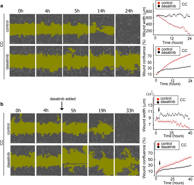
Fig. 6. Scratch assay confirms that Src inhibition prevents recolonization of scratch-wound area by EGFRvIII/EGFRwt CC.
a Left panel; U87EGFRwt cells were co-cultured with U87EGFRvIII cells (CC), in a ratio of 9:1 for 24 h. 100 nM dasatinib was added and movement of the cells into the scratch area was imaged every hour. Representative images for 0, 4, 5, 14, and 24 h are shown for CC and CC + dasatinib. The green shadow indicates the area with very sparse density due to the initial scratch boundary lines. a Right panel; wound width and confluence were calculated using the IncuCyte® S3 Software (see also video 3(A and B)). b Left panel; U87EGFRwt cells were co-cultured with U87EGFRvIII cells (CC), in a ratio of 9:1 for 40 h. Dasatinib (100 nM) was added between the time points 4 h and 5 h (see black arrow). Movement of the cells into the scratch area was imaged every hour. Representative images for 0, 4, 5, 19, and 33 h are shown for CC and CC + dasatinib. b Right panel: quantification of wound width and confluence. P value = 0.7 (between the control and treated groups at 4 h), P value = 0.005 (between the control and treated groups at 5 h), P value = 0.01 (between the treated cells at 4 and 5 h).

