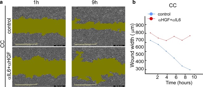
Fig. 9. Scratch assay confirms that αHGF and αIL6 inhibit recolonization of the scratch area by EGFRwt/EGFRVIII CC.
a 0.5 μg/ml αHGF and 0.5 μg/ml αIL6 were added to CC, and movement of the cells into the scratch was imaged for CC or CC + αHGF + αIL6 cells every hour. Representative images for 1 h and 9 h are shown. Green area represents an area unoccupied by the cells (IncuCyte S3 System). b The plot describes a change in the width of the “wound” over time. It represents a mean distance between the edges of the wound (in each vertical line resolution) as a function of time. The graph shows one representative experiment out of eight duplicates.

