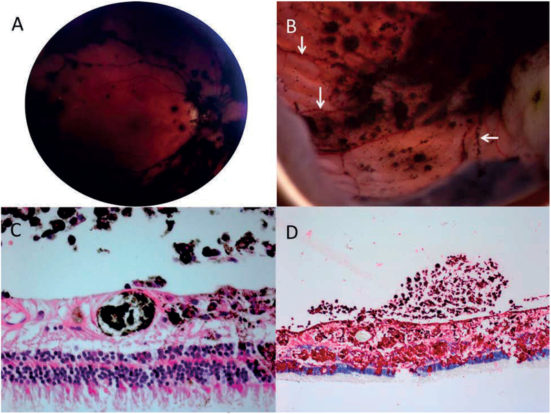
Figure 3:
Representative eye 14. A: The post-vitrectomy fundus image shows diffuse pigment on the surface of the retina, particularly in the distribution of the blood vessels. B: Gross pathologic examination of enucleated specimen revealed the retina to be carpeted with budding threads of fine, pigmented clumps, including in linear arrangements along the distribution of retinal blood vessels (white arrows). C: Melanoma cells and pigmented macrophages are present in a retinal vessels, the retina and vitreous (haematoxylin eosin stain, 100X) D: Immunohistochemical stains show melanoma cells in the retina and vitreous (peroxidase anti-peroxidase, HMB45 red chromagen, 100X)

