Abstract
We have known the accommodation phenomenon since 400 BC. Hypotheses about its mechanisms varied widely for two millennia. Early in the 17th century, when people became more aware of ophthalmic optics, Scheiner and Descartes were close to solving that accommodation worked by changes in the lens. Others rejected their idea, and people even denied the existence of accommodation because there was no clear proof. In the early 19th century, evidence accumulated for accommodation mechanisms studying bird, fish, insect, mammal and human eyes. On the discovery of muscle fibres in the ciliary body, attention shifted to its role in accommodation. Around 1850, came the proof that accommodation occurs by a change in the anterior lens curvature. Still for another 50 years, controversies remained about the exact changes in the lens and the precise accommodation mechanism. On looking back, this is not surprising because only late in the 20th century did it become clear that one cannot extrapolate from the multitude of accommodation mechanisms in the animal kingdom to human eyes.
Keywords: animal ocular accommodation, ciliary body, ciliary ligament, ciliary muscle, human ocular accommodation, ophthalmic history
Introduction
Huygens seems to have coined the word ‘accommodation’ in ophthalmic optics by writing AD 1703 that the eye: ‘ita nunc ad has nunc ad illas res se accommodet’ (adapts itself now to this, now to yonder matter) (Huygens 1703). That is in essence what ocular accommodation means; the potential of an eye to change its refractive power to maintain a clear focus both on distant and nearby objects. For many years, people knew from daily experience that accommodation existed but could not establish its mechanism. The aim of this historical review is to show the difficulties they encountered while finding definite proof how accommodation functions.
Accommodation from 500 BC until AD 1650
The Greeks and the Romans had no idea about the refraction of light in the eye. The Greeks thought that we see by a ‘pneuma’ escaping from the eye in the shape of a cone or by ether that moved from objects to the eye. The symptoms of accommodation were known around 500–300 BC but people explained accommodation by efforts of a ‘soul’ in the eye, in analogy to a brain thinking about difficult questions (Magnus 1877). They considered accommodation paresis with pupillary dilatation to be due to fluid accumulation in the iris. In Galen's era, AD 200, senile accommodation loss was known, and also that accommodation is hampered by abuse of opium, mandrake or hyoscyamine (Magnus 1901). Galen assumed that there were seven external eye muscles inserted around the optic nerve. He explained accommodation by muscle activity of the seventh (non‐existent) external choanoides or retractor muscle. This muscle survived another 1300 years in medical texts and thus Vesalius still drew it in the middle of the 16th century (Fig. 1) (Vesalius 1555; Magnus 1901). Over the years, the ciliary body became associated with accommodation, so we will first have a look at this body.
Figure 1.
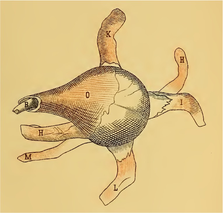
The (non‐existent) choanoides or retractor external eye muscle (O) responsible for accommodation according to Galen and Vesalius. Al six external eye muscles (H, I, K, L, M) had their insertion around the optic nerve B (Vesalius 1555; Magnus 1901).
Nomenclature, structure and some functions of the ciliary body, ciliary ligament and ciliary muscle
Duke‐Elder wrote that the name of this body stems from cilia, hair (Duke‐Elder 1961). Cilium is the Latin word for eyelid and Zinn mentioned that even before Galen, anatomists compared the ciliary body with an eyelid having lashes (Zinn 1755). He complained about the inconsequent nomenclature of many anatomists and used the term ciliary body that Falloppius had introduced (Falloppius 1562). Lucretius, just before the beginning of our era, considered this body a belt that connected different tissues and strengthened the eye wall. Vesalius named the ciliary body a tunic derived from the uvea, resembling eyelashes attached to the lens equator (Vesalius 1555). Kepler hypothesized that the ciliary processes contract during accommodation and become shorter, pulling the lateral parts of the eye inwards and thus elongating the eye (Kepler 1611). According to Duke‐Elder, Boerhaave mentioned in 1708 muscular fibres in the ciliary muscle and English scientists described these fibres before Brücke (Duke‐Elder 1961). Camper, however, did so already earlier (Camper 1746). Boerhaave described in the various editions of his published lectures muscle fibres in the iris and it remains unclear whether he saw these or assumed them to be there because of the pupillary reactions (Boerhaave 1708; Glauder 1751; Haller 1758). Porterfield refered to the muscularity of the ciliary ligament mentioned by many anatomists and did not find muscle fibres himself (Porterfield 1759). A century later, many animals, from fish to lynx and rhinoceros, were shown to have a ciliary ligament (Wallace 1836). Wallace could not obtain human eyes and surmised that contraction of these (hypothetical) muscle fibres compressed the ciliary veins, thus erecting and expanding the ciliary processes (Wallace 1835). He referred to Knox who wrote extensively on the ciliary muscle (the white ring as he called it) but also Knox could not find muscle fibres, even with a microscope (Knox 1826). Therefore, it seems that Brücke indeed described for the first time in the human eye the choroidal tensor muscle running in an axial direction in the ciliary body. This muscle was partly attached by an elastic mesh to the inner wall of Schlemm's canal and to the corneal basement membrane (Fig. 2a) (Brücke 1847). His teacher Müller added circular smooth muscle fibres parallel to the corneal limbus (Fig. 2b) (Müller 1857). The ciliary muscle seems to have three sections: a. on the scleral side a longitudinal layer running from the tendon attached to Schlemm's canal to the choroid; b. oblique fibres from the same tendon dividing in b1, towards the tails of the ciliary processes and b2 forming the smooth ring in the heads of these processes near Schlemm's canal (see g in Fig. 2b); c, meridional fibres running forward with subsections c1 going into the heads of the processes and c2 running into the iris (Duke‐Elder 1961). According to Donders, the ciliary muscle functions also as origin of the dilating muscle of the iris (Donders 1864).
Figure 2.
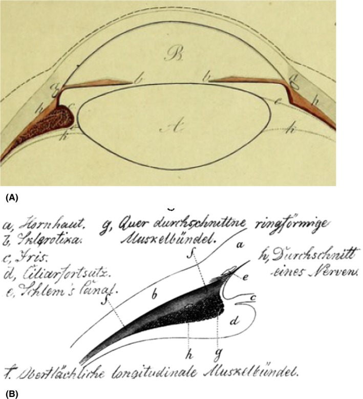
(A) The human choroidal tensor muscle (h) described by Brücke. On the left, the section through the eye runs through a ciliary process, on the right in between two of these. A Lens; B anterior chamber; a Schlemm's canal; b iris; c Zinn's zonules; k hyaloid tunic (Brücke 1847). (B) Superficial, longitudinal (f) and circular (g) human ciliary muscle fibres. The oblique fibres b1 and longitudinal ones c1 and c2 are not there (Müller 1857).
Confusion about accommodation mechanisms from 1619 until 1850
In the early 17th century, the ciliary processes were accredited to move the vitreous and the lens forwards or backwards by contraction and relaxation and thus to flatten or bulge the lens, according to whether one is looking at objects far away or close by (Fig. 3). Descartes considered accommodation a voluntary process, even when a person is unaware of the fact that he accommodates, because he intends to see close objects well. Van Leeuwenhoek, 100 years later, mistook fibres in the lens (and even fibres in the vitreous) for muscular fibre tendons (Van Leeuwenhoek 1704). This may have put subsequent researchers like Young & Brocklesby (1793) on the wrong track. Jurin hypothesised, spurred by a thesis of Pemberton, (Pemberton 1719) that ‘For many reasons the most advantageous and convenient method for the eye to be accommodated to near objects seems by rendring the anterior surface of the crystalline more convex, while the hinder surface grows flatter. But this surely is too great a change for a substance of such a consistence as the crystalline humour to admit of’ (Jurin 1738). Thus, he found ‘No satisfaction in any of the hypotheses above related’ and next focused on the cornea and uvea as the sites where accommodation took place. The Table 1 gives an overview of the wide variation in hypotheses and results of animal and human research on accommodation. Home tore instead of cut the rectus muscles from a human eye after death. Thus, he found that the rectus tendons became broader on approaching the cornea, forming a circle of which the cornea seemed to be the central continuation. This explained in his view the change in corneal radius of curvature during accommodation (Fig. 4) (Home 1795). Young & Brocklesby (1793) assumed that accommodation is a voluntary process leading to nerve impulses that ran via the lenticular ganglion and the ciliary processes to the crystalline muscle, making the lens more convex. In a later paper, Young described improvements on Porterfield's optometer, demonstrated that accommodation does not exist in aphakia, and excluded corneal, axial length or electrical changes as its mechanism. With data from his optometer, he predicted on theoretical grounds that during accommodation the axial length of the lens increases, leading to a greater relative convexity of the posterior lens surface than that of the anterior one. He thought that this expansion occurred by swelling of the muscular fibres in the lens (Young 1801).
Figure 3.
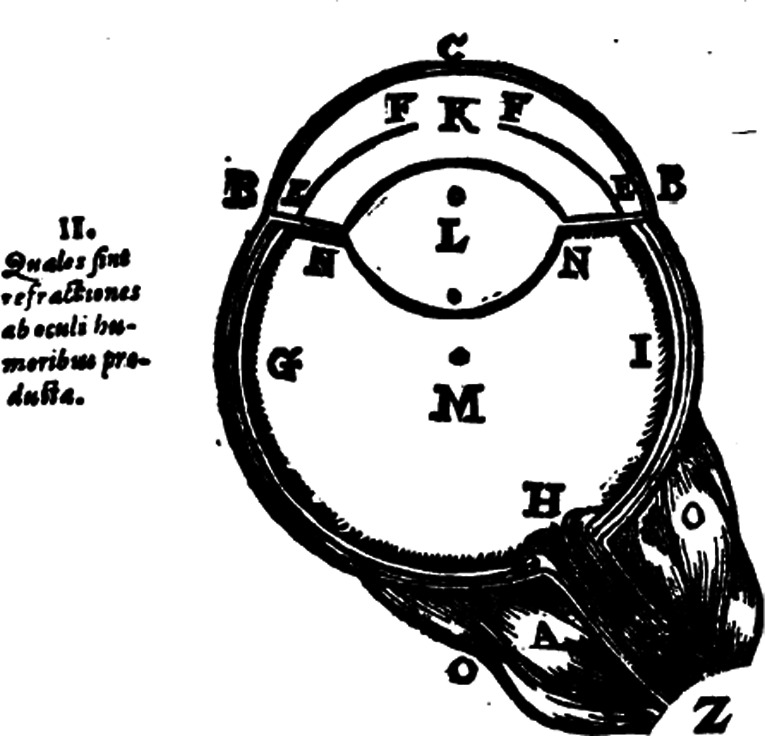
EN—EN are several small black strings that enfold the fluid marked with L (the lens), resembling many small tendons, by which L sometimes becomes more vaulted, sometimes flatter, according to whether one is looking at objects close by or far away. OO are six or seven muscles attached to the eye around the optic nerve Z (Descartes 1642).
Table 1.
Hypotheses or research results re accommodation mechanisms.
| Year | Mechanism | First authora | Later references |
|---|---|---|---|
| 1611 | Axial eye elongation on inward pull of ciliary processes | Kepler (1611) | |
| 1611 | Retinal movement by contraction of ciliary ligament leading to narrowing of eye equator | ||
| 1619 | Lens dislocation or bulging by ciliary processes | Scheiner (1619) | |
| 1637 | Lens bulging | Descartes (1642) | Pemberton (1719); Home (1794); Young (1801); Purkinje (1825); Hueck (1839); Camper (1913) |
| 1685 | No change in lens form or position. No muscles in ciliary ligament | de la Hire (1685) | |
| 1703 | Lens dislocation or bulging by pressure from external eye muscles | Huygens (1703) | Camper (1913) |
| 1738 | Changes in cornea and uvea | Jurin (1738) | Pemberton (1719) |
| 1743 | Contraction of oblique muscles | Le Camus (1743) | |
| 1745 | Air inflation in eyeball | Poupart (1745) | |
| 1746 | Contraction of muscular fibres in ciliary fibres. | Camper (1746) | Smith (1833); Hueck (1839) |
| 1755 | Constriction of eye ball by rectus muscles | Boerhaave (Glauder 1751) | Arnold (Cramer 1853) |
| 1759 | Lens becomes convex by muscular fibres in lens. Ciliary ligament contraction pulls lens forward, compresses vitreous and makes cornea more convex | Porterfield (1759) | Young (1801) |
| 1780 | Change in refractive index of ocular fluids | Grimm (Cramer 1853) | |
| 1795 | Diminishing corneal radius. Better accommodation in aphakic eye | Home (1795) | |
| 1801 | Lens swelling; relatively more at posterior than anterior surface | Young (1801) | |
| 1801 | Orbicular muscle flattening the cornea or shortening the visual axis | Monro (Brewster 1824) | Hueck (1839) |
| 1802 | Movement of macula or central plica | Albers (Cramer 1853) | |
| 1809 | Ciliary body force on lens rim or aqueous forwarding lens capsule | Grafe (1809) | |
| 1813 | Contraction of tissue between scleral bony ring and tendinous corneal ring | Crampton (1813) | |
| 1821 | No existent accommodative mechanism but mental brain process | Weller (Cramer 1853) | |
| 1824 | Both voluntary and involuntary processes | Brewster (1824) | |
| 1826 | Pupillary dilatation and narrowing. Denial of accommodation existence | Mile (1826) | Magendie (Hueck 1839) (de la Hire 1685) (Treviranus 1835) |
| Refractive index of vitreous on lens side different from that on fundus side | |||
| 1826 | Contraction of ciliary muscle and of pupil | Knox (1826) | |
| 1832 | Fluid congestion in the iris | Treviranus (1835) | Arnold (Cramer 1853) |
| 1835 | Shortening of the visual axis | Serre (Cramer 1853) | |
| 1835 | No lens changes. Accommodation possible due to laminated lens structure | Treviranus (1835) | |
| 1839 | Forward movement and greater convexity of lens | Hueck (1839) | |
| 1841 | Elongation of visual axis | Bonnet (Cramer 1853) | Henle (Cramer 1853) |
| 1842 | Corneal bulging and pupillary narrowing | Pappenheim (Cramer 1853) | |
| 1849 | Recti muscles, by pulling eye against orbital fat padding, pushing vitreous and lens forwards, increase corneal convexity | Szokalsky (1848) | |
| 1849 | Lens compressing muscle. Lens movement and bulging anterior capsule | Langenbeck (1849) | |
| 1850 | Movement of lens nucleus within capsule | Hannover (Cramer 1853) | |
| 1853 | Bulging of anterior lens capsule and iris pressure by simultaneous sphincter and dilatator contraction. Ciliary muscle contraction hinders posterior lens movement. No accommodation in hyperopic eyes | Cramer (1853) | |
| 1855 | Ciliary contraction and thickening of lens centre plus anterior bulging | Helmholtz (1855) | |
| 1855 | Positive accommodation in myopic eyes, negative in hyperopic ones | Weber (1855) | |
| 1857 | Increasing thickness of longitudinal ciliary muscle, slackening zonules, circular muscle pressing on lens rim, and pressure of peripheral iris | Müller (1857) | |
| 1904 | Backwards and downwards lens movement; central vitreous liquefaction and dilatation of the canal of Cloquet | Tscherning (1904) |
First time a mechanism was encountered in a manuscript.
Figure 4.
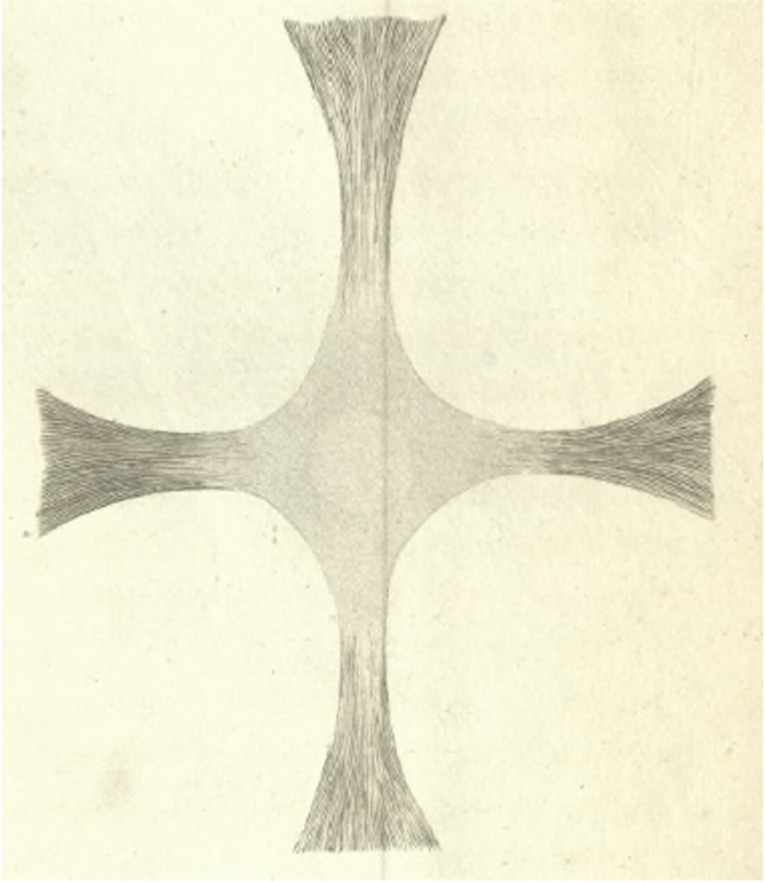
Tendons of the external recti muscles, encircling the cornea (vague circle in the centre of the cross) and pulling at the cornea during accommodation (Home 1795).
The final spurt to solving the accommodation puzzle
In 1848, a contest in the Netherlands was organized to solve the accommodation mechanism. In order to see how researchers overcame the deadlock in contradictions (Table 1), we have to go back 200 years. Scheiner described the reflection of a candle on the cornea (Scheiner 1619). Purkinje, a great (myopic) observer, discovered with bare eyes that there were, apart from this corneal image, more ocular candle reflections. These originated from the corneal endothelium and from the anterior lens surface, both acting as a convex mirror, as well as from the posterior lens or anterior vitreous surface (acting as a concave mirror) (Purkinje 1823). The endothelial image and more secondary images were hard to see. For practical purposes, authors restricted themselves to an upright image 1 from the corneal epithelium, upright image 2 from the anterior lens surface and an inverted image 3 from the posterior lens surface (Fig. 5). Sanson independently re‐discovered images 2 and 3 and described how one could use these images to differentiate between vision loss due to cataract or to other causes deeper in the eye (Sanson 1838). Bear in mind that this was before the invention of the slitlamp or the ophthalmoscope. The German surgeon Langenbeck stressed a year later the diagnostic value of the size, colour and relative distance of the Purkinje‐Sanson images from each other. He examined, also bare‐eyed, these images with a candle in front of an eye instead of to its side, thus hampering their observation because the images were nearly superimposed. Langenbeck wrote about the (in humans non‐existent) ‘musculus compressor lentis accommodatorius,’ and mentioned that accommodation was due to a change in lens position but also that the anterior lens surface became more convex during accommodation (Langenbeck 1849).
Figure 5.

(A) Reflections from a candlelight lateral to an accommodating eye, as seen from the contralateral side. a Upright corneal epithelial image 1 (brightest); b upright image 2 (weakest) from the anterior lens surface; c inverted image 3 (medium bright) from the posterior lens surface. While looking in an axial direction in the eye, a is in front, b is seen deepest and c is halfway between a and b. On moving the candle, a and b move in the same direction and c in the opposite one. (B) Pupil of a non‐accommodating eye. a upright corneal image; b upright image from the anterior lens surface; c inverted image from the posterior lens surface. (C) During accommodation: a and c remain in place, proof that the lens position does not change; b changed its position and became slightly smaller (Cramer 1853).
Donders calculated that displacement of the lens could not account for the normal range of accommodation and published his hypothesis that by carefully measuring the Purkinje images under telescopic magnification, one could solve the accommodation mystery. He predicted that during accommodation, the first and second Purkinje images would remain in place and that the third (middle one) would move, pointing to a change in curvature of the anterior lens surface (Fig. 5) (Donders 1849). He wrote, ‘The mechanism of the accommodation capacity is still unclear. I believe I have sufficient reasons to position its origin inside the eye, without completely thus clarifying its mechanism. The hypothesis that the root of the accommodation capacity lies in the oblique eye muscles is unjustified’ (Donders 1851). Cramer published his preliminary results in the accommodation contest describing the increasing curvature of the anterior lens plane (Cramer 1851). In 1852, he received the first prize including a gold medal, and his prize winning manuscript was published in 1853, in which he wrote that hyperopic eyes cannot accommodate (Cramer 1853). Cramer, who acknowledged Donders's predictions in both publications, built an ‘ophthalmoscope’ (Fig. 6) and performed many experiments to prove that the weak parts of the lens create the change in its anterior curvature during accommodation. Only then did it become clear that the 200‐year‐old hypotheses of Scheiner and Descartes and the one rejected by Jurin were correct. In addition, Langenbeck's observation now became better known to the public. Cramer found that in accommodation, the middle image becomes smaller indicating a smaller radius of curvature of the anterior lens surface.
Figure 6.
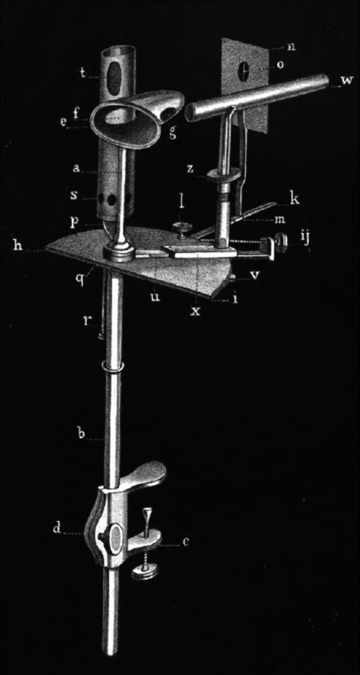
Ophthalmoscope of Anthonie Cramer. Cone‐shaped 8‐cm‐long tube fg with holes at its base and apex and on the sides. On the left side, candle light from tube s t enters the cone, on the right side the eye pressed to the wide end of the cone, can be observed via telescope w. Plate n can be moved along horizontal rod k and its opening o is in line with the axis through the openings the cone fg. In front of o is a taught perpendicular thread and behind o is a slider on a string that can be lowered, covering gap o. In the tube st a candle can be moved up and down by r. Microscope w can be adjusted in three directions with x, ij and z. Black bronzed apparatus in order to prevent reflections. Cramer wrote that a hyperopic person cannot accommodate, so one should select an emmetropic person (Cramer 1853).
Donders had three modifications made of the ‘ophthalmoscope’ of Cramer, who died in 1855, (Swaagman 1855) and named these a ‘phacoidoscope.’ By using his phacoidoscope, Donders could see tiny changes in the distance of the posterior Purkinje image, sometimes approaching the corneal image, sometimes increasing its distance. The lens equator remained more or less in the same position during accommodation. Helmholtz started his article on accommodation by claiming priority over the discovery of Cramer and Donders because he discovered late in 1852 changes in the reflections of the anterior lens surface during accommodation and sent this discovery to the Academy of Sciences in Berlin (Helmholtz 1855). He went on, however, writing that he overlooked the earlier publications of Donders and Cramer as well as Langenbeck's one on this matter. ‘After obtaining Cramer's work by the kindness of Mr. Donders, I convinced myself that the enigma of accommodation, in which so many researchers have in vain practiced their ingenuity, mainly was solved, and the intended investigation left me little more to do’(Helmholtz 1855). Helmholtz measured more accurately the Purkinje images in the eyes of three humans aged 30–35 years with an ophthalmometer. Its construction was based on the heliometer of astronomers, by which he obtained an accuracy of 0.01 mm on a moving eye. He measured all he could; for example, the distance from the corneal apex to the pupillary plane was 3.7–4.0 mm and to the posterior lens surface, 6.9–7.1 mm. After death, lenses become thicker. During accommodation, the pupillary plane came 0.36–0.44 mm forwards. Helmholtz confirmed Cramer's reduction of the middle image and wrote that the posterior lens radius of curvature became a little smaller (Helmholtz 1855).
Having reached consensus about the change in lenticular curvature during accommodation, there remained controversy about how exactly this took place. Helmholtz agreed with Young, Cramer and Donders that the corneal curvature does not change during accommodation. He thought together with Brücke that ciliary muscle contraction pulls the choroid and the zonules forward towards Descemet's membrane, receding the iris, slackening the zonules and thus the anterior lens surface bulges through its capsular elasticity. Helmholtz assumed that in the relaxed state of the eye while looking in the distance, the zonules are tightened and thus flatten the lens. He was uncertain whether the circular fibres in the ciliary muscle were the main active fibres and the radial fibres only auxiliary ones. His conclusion was: ‘So we hardly can deny the ciliary body some function in the accommodation process’ (Brücke 1847; Helmholtz 1855). The posterior surface of the lens remains in place, and the lens volume does not change, so the centre of the lens becomes thicker (Helmholtz 1855; Henke 1860). Donders was the first to show that even before puberty the accommodation range starts to diminish both in myopia, emmetropia and hyperopia (Donders 1860).
After examining various bird eyes, Müller thought that the ciliary muscle increased in thickness by contraction of the longitudinal fibres, thus slackening the anterior part of the zonules together with pressure of the circular ciliary muscle and the iris on the peripheral lens part (Müller 1857; Duke‐Elder 1961). Donders did not believe in this pressure of the circular fibres and the iris on the lens rim and considered it essential to measure first the circumference of the lens during accommodation (Donders 1864). Cramer used electrical currents in the ciliary region of enucleated seal and bird eyes to show that changes during accommodation occurred as long as the iris was intact, but nothing happened when he removed the iris or made radial cuts in it. Weber and Von Graefe assumed that there was a separate positive accommodation mechanism in myopic eyes and a negative one in hyperopic ones (Weber 1855). Knapp found a high concordance between his measurements and the visual determination of accommodation (the push‐up method of Donders). Accommodation in aphakia was questionable, not only by his measurements but also by the various experiments of Donders in Utrecht in which Knapp could participate (Knapp 1860). Tscherning challenged Helmholtz's suspicion that the lens is flatter seeing in the distance through the pull of the zonules. He considered the function of the iris for accommodation not proven and mentioned that Von Graefe demonstrated intact accommodation in complete aniridia (Tscherning 1883). According to Tscherning, Helmholtz and Donders took insufficient account of the peculiar structure of the ciliary muscle. Henle stressed that the circular and meridional muscle fibres of the ciliary body had a separate function and Iwanoff, Arlt and Sattler, who found hypertrophy of the circular fibres in hyperopia and of the meridional fibres in myopia, confirmed this (Tscherning 1883). Tscherning also mentioned the lack of knowledge about innervation of the ciliary muscle. He thought that the oculomotor nerve and perhaps also the sympathetic nerve were the accommodation nerves (Tscherning 1883). Cramer thought that the trigeminal and sympathetic nerve were involved (Cramer 1853). At present, the parasympathetic nerve is considered to the main one for accommodation (Drexler et al. 1997). Tscherning postulated a downwards and backwards lens movement during accommodation as well as central vitreous liquefaction with dilation of Cloquet's canal (Tscherning 1904).
At present, one finds in PubMed over 200 publications on the mechanism of the human ocular accommodation mechanism and these are beyond the scope of this review. Preliminary data, however, obtained with anterior segment optical coherent tomography showed the complexity of the human ciliary muscle action. During a 4 diopter accommodation stimulus, the maximum ciliary muscle thickness increased by 69.2 μm (18.1 μm per diopter) at about 1 mm posterior to the scleral spur but the muscle thickness decreased by 45.9 μm (−12.0 μm per diopter) at 3 mm from this spur. So indeed the portion of the ciliary body closest to the cornea bulges most. Unfortunately the zonules were absent on the images provided (Lossing 2012). The most sophisticated measuring instrument, a scanning partial coherence interferometer, found that in an emmetropic 30‐year‐old human eye, the anterior pole of the lens moved 228 μm forwards and the posterior lens pole 75 μm backwards when changing from distant vision to focusing on the near point. This ratio of three to one held for all 10 eyes tested (Drexler et al. 1997). Most remarkably, it seems that the absolute values found 160 years ago differed only by 0.1–0.2 mm from the present ones.
Only recently a fascinating review on accommodation mechanisms in various animals appeared (Ott 2006). Nearly all options mentioned over the centuries for these mechanisms in humans (Table 1) occur in the animal kingdom. They range from independent monocular accommodation between paired chameleon eyes, combined as well as independent accommodation between the two eyes of hawks and vultures, influence of retinal thickness on accommodation in small eyes, corneal changes and anterior lenticonus to shifting lens positions in cats. A sea otter is emmetropic above water and can see well under water because of a 60 diopter accommodation range. Humans and fish have a less perfect stimulus response function for accommodation than lizards and turtles (Ott 2006). It is no wonder that our predecessors were for so long groping in the dark, comparing animal eyes with human ones.
I am greatly indebted to P. Stoutenbeek MD, ophthalmologist, for his major help in translating Neo‐Latin from the Latin books in the reference list.
References
- Boerhaave H (1708): Institutiones medicae, in usus annuae exercitationis domesticos. Leiden: J. van der Linden; 101–109. [Google Scholar]
- Brewster D (1824): Ueber die Fähigkeit des Auges, sich den verschiedenen Entfernungen der Gegenstände anzupassen. Ann Phys 78: 271–281. [Google Scholar]
- Brücke E (1847): Anatomische Beschreibung des menschlichen Augapfels. Berlin: Reimer; 18. [Google Scholar]
- Camper P (1746): Dissertatio physiologica de quibusdam oculi partibus. Medicinae facultas. Leiden: 28.
- Camper P (1913): De oculorum fabrica et morbis. Opuscula selecta Neerlandicorum de arte medica. Amsterdam: Van Rossum: 408. [Google Scholar]
- Cramer A (1851): Mededeelingen uit het gebied der Ophthalmologie. Tijdschr Ned Maatsch bevord: Geneeskunst; 99–119. [Google Scholar]
- Cramer A (1853): Het accommodatievermogen der oogen, physiologisch toegelicht. Natuurkundige verhandelingen van de Hollandsche maatschappij der wetenschappen te Haarlem. Haarlem: de Erven Loosjes; 1–139. [Google Scholar]
- Crampton P (1813): The description of an organ by which the eyes of birds are accommodated to the different distances of objects. Ann Philos 1: 170–174. [Google Scholar]
- Descartes R (1642): Specimina philosophiae seu dissertatio de methodo. Dioptrice et meteora. Frankfurt am Main: Knochii; 53–55. [Google Scholar]
- Donders FC (1849): Reflexie‐proef van Purkinje en Sanson en accommodatie van het oog naar Max Langenbeck. Nederlands Lancet 5: 132–147. [Google Scholar]
- Donders FC (1851): Ophthalmologische aantekeningen. Accommodatie‐vermogen. Nederlandsch Lancet 2: 600–612. [Google Scholar]
- Donders FC (1860): Ametropia en hare gevolgen; Utrecht: Van der Post; 133 [Google Scholar]
- Donders FC (1864): On the anomalies of accommodation and refraction of the eye. London: New Sydenham Society; 635. [Google Scholar]
- Drexler W, Baumgartner A, Findl O, Hitzenberger CK & Fercher AF (1997): Biometric investigation of changes in the anterior eye segment during accommodation. Vision Res 37: 2789–2800. [DOI] [PubMed] [Google Scholar]
- Duke‐Elder S (1961): The anatomy of the visual system. The ciliary body In: Duke Elder S. (ed.) System of ophthalmology. Vol II London: Kimpton; 146–156. [Google Scholar]
- Falloppius G (1562): Observationes anatomicae. Cologne: Birckman; 329. [Google Scholar]
- Glauder GF (1751): Hermann Boerhavens Abhandlung von Augenkrankheiten und derselben Kur. Nürnberg: Endterischen Consorten; 252–276. [Google Scholar]
- Grafe D (1809): Ueber die Bestimmung der Morgagnischen Feuchtigkeit, der Linsenkapsel und des Faltenkranzes, als ein Beytrag zur Physiologie des Auges In: Reil DJ. (ed.) Archiv für die Physiologie. Halle: Curtsch; 225–236. [Google Scholar]
- Haller A (1758): Hermanni Boerhaave praelectiones academicae. Leiden: Sumptibus Societatis; 64–205. [Google Scholar]
- Helmholtz H (1855): Ueber die Accommodation des Auges. Arch Ophthalmol 1: 1–74. [Google Scholar]
- Henke W (1860): Der Mechanismus der Accommodation für Nähe und Ferne. Arch Ophthalmol 6: 53–72. [Google Scholar]
- de la Hire P (1685): Dissertation sur la conformite de l'oeil. J Scavans 23: 279–290. [Google Scholar]
- Home E (1794): III. Some facts relative to the late Mr. John Hunter's preparation for the Croonian lecture. Phil Trans R Soc Lond 84: 21–27. [Google Scholar]
- Home E (1795): I. The Croonian Lecture on muscular motion. Phil Trans R Soc Lond 85: 1–23. [Google Scholar]
- Hueck A (1839): Die Bewegung der Krystallinse. Dorpat: Kluge; 108. [Google Scholar]
- Huygens C (1703): Opuscula postuma quae continent dioptricam. Leiden: Boutesteyn; 112–118. [Google Scholar]
- Jurin J (1738): An essay upon distinct and indistinct vision In: Smith R. (ed.) A complete system of opticks in four books. Cambridge: Crownfield: 115–171. [Google Scholar]
- Kepler J (1611): Dioptrice seu demonstratio eorum quae visui & visibilibus propter conspicilla non ita pridem inventa accidunt. Augsburg: Francus; 83. [Google Scholar]
- Knapp JH (1860): Ueber die Lage und Krümmung der Oberflächen der menschlichen Kristallinse und den Einfluss ihrere Veränderungen bei der Accomodation auf die Dioptrik des Auges. Arch Ophthalmol 6: 1–52. [Google Scholar]
- Knox R (1826): Observations on the comparative anatomy of the eye. Trans Roy Soc Edinb 10: 43–78. [Google Scholar]
- Langenbeck M (1849): Klinische Beiträge aus dem Gebiete der Chirurgie und Ophthalmologie. Göttingen: Dieterich; 57–58. [Google Scholar]
- Le Camus A (1743): An obliqui oculorum musculi retinam a crystallino removeant. Paris: 133–140. [Google Scholar]
- Lossing LA, Sinnott LT, Kao CY, Richdale K & Bailey MD (2012): Measuring changes in ciliary muscle thickness with accommodation in young adults. Optom Vis Sci. 89: 719–726. [DOI] [PMC free article] [PubMed] [Google Scholar]
- Magnus H (1877): Die Kenntniss der Sehstörungen bei den Griechen und Römern. Graefes Arch Clin Exp Ophthalmol 23: 24–48. [Google Scholar]
- Magnus H (1901): Die Augenheilkunde der Alten. Breslau: Kern; 115–162. [Google Scholar]
- Mile J (1826): De la cause qui dispose l'oeil pour voir distinctement les objets placés a différentes distances. In: Magendie F. (ed.) Journal de physiologie expérimentale et patholoqique. Paris: Méquignon‐Marvis; 166–177. [Google Scholar]
- Müller H (1857): Anatomische Beiträge zur Ophthalmologie. Ueber einen ringförmigen Muskel am Ciliar‐Körper des Menschen und über den Mechanismus der Accommodation. Arch Ophthalmol 3: 1–98. [Google Scholar]
- Ott M (2006): Visual accommodation in vertebrates: mechanisms, physiological response and stimuli. J Comp Physiol A 192: 97–111. [DOI] [PubMed] [Google Scholar]
- Pemberton H (1719): Dissertatio physico‐medica inauguralis de facultate oculi, qua ad diversas rerum conspectarum distantias se accomodat. Facultas medicae. Lugduni Batavorum: 37.
- Porterfield W (1759): A treatise of the eye, the manner and phaenomena of vision in two volumes. Edinburgh: Hamilton and Balfour; 4–8. [Google Scholar]
- Poupart M (1745): The libella. Memoirs Royal Soc 3: 489–491. [Google Scholar]
- Purkinje JE (1823): Commentatio de examine physiologico organi visus et systematis cutanei.Bratislava: Typis Universitatis; 58. [Google Scholar]
- Purkinje JE (1825): Neue Beiträge zur Kenntniss des Sehens in subjectiver Hinsicht. Berlin: Reimer; 192. [Google Scholar]
- Sanson MLJ (1838): Leçons sur les maladies des yeux, faites a l'Hopital de la Pitié. Paris: Ebrard; 135. [Google Scholar]
- Scheiner C (1619): Oculus hoc est: fundamentum opticum. Insbruck: Agricola; 254. [Google Scholar]
- Smith T (1833): On the muscular structure and functions of the capsule of the crystalline lens and ciliary zone. Lond Edinb Phil Mag J of Sci 3: 5–15. [Google Scholar]
- Swaagman AH (1855): Herinnering aan Antonie Cramer. Tijdschr Ned Maatsch Bevord. Geneeskunst 6: 54–64. [Google Scholar]
- Szokalsky WF (1848): Das Anpassungsvermögen des Auges, vom pathologischen Gesichtspunkte aus betrachtet. Arch Physiol Hlk 7: 695–709. [Google Scholar]
- Treviranus GR (1835): Ueber die blättrige Textur der Crystallinse des Auges als Grund des Vermögens einerlei Gegenstand in verschiedener Entfernung deutlich zu sehen. Bremen: Heyse; 55–56. [Google Scholar]
- Tscherning M (1883): Studien über die Aetiologie der Myopie. Graefes Arch Clin Exp Ophthalmol 29: 201–271. [Google Scholar]
- Tscherning M (1904): The mechanism of accommodation. Ophthal Rev 23: 95–104. [Google Scholar]
- Van Leeuwenhoek A (1704): A letter from Mr Anthony van Leeuwenhoek F.R.S. concerning the flesh of whales, crystaline humour of the eye of whales, and other creatures, and of the use of eyelids. Phil Trans R Soc Lond 24: 1723–1730. [Google Scholar]
- Vesalius A (1555): De corporis humani fabrica. Basel: Oporinus; 285. [Google Scholar]
- Wallace WC (1835): Dissection of the eye of the streaked bass, with observations on the accommodation of the eye to distances. Am J Sci Arts 27: 216–222. [Google Scholar]
- Wallace WC (1836): The structure of the eye, with reference to natural theology. New York: Wiley & Long; 27. [Google Scholar]
- Weber T (1855): Unterscheidung zweier wesentlich verschiedener Arten der Accommodation des Auges und deren Einfluss beim Gebrauche des Augenspiegels. Arch Physiol Heilk 14: 479–490. [Google Scholar]
- Young T (1801): On the mechanism of the eye. Phil Trans R Soc Lond 91: 23–88. [Google Scholar]
- Young T & Brocklesby R (1793): Observations on vision. Phil Trans R Soc Lond 83: 169–181. [Google Scholar]
- Zinn JG (1755): Descriptio anatomica oculi humani. Göttingen: Vandenhoeck; 60–61. [Google Scholar]


