Abstract
Aim:
The study was conducted to evaluate the effect of increased vertical dimension on the lip positions at smile in dentulous subjects.
Settings and Design:
Invivo - comparative study.
Materials and Methods:
Thirty individuals aged between 22 and 30 years were selected for the study. Poly-vinyl siloxane (Jet Bite, Coltene, Switzerland) interocclusal bite records of varying thickness of +1, +2, +3, and +4 mm were made using articulated stone casts for all the participants, respectively. Posed smile photographs at different increased vertical dimensions of +1, +2, +3, and +4 mm were captured with D-SLR camera (Nikon D3200 of 18 megapixels with macro lens, Japan) mounted on tripod stand keeping a uniform distance of five feet from the face. Head positioning device (Genoray CBCT Machine Papaya 3D Plus, Unicorn DenMart, India) was used to stabilize the head position of the participants. Interlabial gap height, intercommissural width, smile index (width/height ratio), incisal edge-to-upper lip distance, incisal edge-to-lower lip distance, and display zone area measurements were made in AutoCAD software (Autodesk, Inc., California, USA).
Statistical Analysis Used:
One-way repeated measures ANOVA tests (α = 0.05) and Bonferroni's post hoc tests were performed for statistical analysis.
Results:
With increasing occlusal vertical dimension, the interlabial gap height, incisal edge-to-lower lip distance, and display zone area increased significantly (P < 0.001). The smile index decreased significantly as the occlusal vertical dimension increased (P < 0.001). No significant difference was found in intercommissural width and incisal edge-to-upper lip distance.
Conclusion:
It was found that an increase in occlusal vertical dimension led to an increase in interlabial gap height, incisal edge-to-lower lip distance, and display zone area measurements, whereas the width of smile and incisal edge-to-upper lip distance did not change with increasing occlusal vertical dimension.
Keywords: Display zone area, full-mouth rehabilitation, lip positions, smile index, vertical dimension
INTRODUCTION
Dental and facial attractiveness plays an important role in boosting one's self-confidence. Smile is the essence of facial attractiveness. Various orofacial musculatures are responsible for producing smile, amongst which lips are significant structures as they form the curtain of oral cavity affecting the facial esthetics in the lower third of the face.[1]
Vertical dimension at occlusion (VDO) can be described as a comfort zone which is formed by the musculoskeletal balance during growth. The facial morphology is influenced by the muscle organization surrounding the face and the underlying skeletal morphology.[2] In a dentate patient, as age advances, there are changes in the orofacial musculature, teeth, and periodontium eventually affecting the smile.[3] With increasing age, muscles lose tonicity and elasticity, causing smile to become narrower vertically and wider transversely.[4] Diminished facial contour, thin lips with narrow vermillion borders, and drooping commissures are associated with reduction in VDO.[5] Hard tissue changes due to reduced VDO include resorption of alveolar bone and loss of teeth.[4] Increasing VDO may provide greater interocclusal restorative space and may reduce the need for clinical crown lengthening or endodontic procedures.[6] In addition to that, increasing the VDO may change the overjet and overbite of the anterior teeth and may alter facial esthetics in a positive manner.
A moderate amount of increase in VDO can be easily adapted by the masticatory system.[7] A recent systematic review reported that increasing the VDO, particularly with fixed restorations, is a predictable and stable procedure.[8] Gross and Ormianer found that increasing the VDO significantly increases the measurement of the lower face height.[9] A similar study on edentate individuals by Ushijima et al. reported that a deficient VDO with extensive lip support can curve the oral fissure form upward, whereas a deficient VDO with deficient lip support can reduce the vermilion height of lips.[10]
Predicting an esthetic outcome while planning full-mouth rehabilitation becomes a difficult task for the clinician. Hence, this study was undertaken to clinically evaluate the effect of different occlusal vertical dimensions on the lip positions at smile.
MATERIALS AND METHODS
Thirty dentulous adults were selected for the study after obtaining ethical clearance from the ethical review board of the institute. The inclusion criteria of the participants were that they should be between 22 and 30 years of age, be volunteering to participate, have all anterior teeth and at least three teeth in occlusion in both posterior segments, have normal occlusion of maxillary and mandibular teeth, and those who had never undergone any prosthetic or restorative treatment in the area of interest. The exclusion criteria included history of trauma and injury to facial structures, history of neurological disorders, a centric occlusion – maximum intercuspation (MI) discrepancy more than 1 mm, an inability or unwillingness to smile, and an allergy to silicone, nitrile, or irreversible hydrocolloid material.
Determination of sample size for conducting the study with repeated measures ANOVA was done using power analysis software. The total sample size of 30 was achieved with a power of 90%, to detect differences among means with a significance level of 0.05. Thirty dentate individuals, 13 males and 17 females (mean age: 25.8 years; range, 22–30 years), participated in the study. All the participants belonged to Asian Indian ethnicity. The study was conducted in two phases.
In the first phase, maxillary and mandibular dentulous impressions were made with irreversible hydrocolloid impression material (DPI Imprint, India) using perforated stock metal rim-lock trays [Figure 1]. Arbitrary facebow transfer using Hanau™ springbow earpiece facebow was obtained [Figure 2]. Silicone bite registration (Jet Bite, Coltene, Switzerland) in MI was done for articulating the cast on semi-adjustable articulator (Hanau™ wide-vue arcon articulator, USA) [Figure 3]. The impressions were disinfected and poured into with Type III dental stone (Kaldent, Kalabhai Pvt. Ltd., India).
Figure 1.

Irreversible hydrocolloid maxillary and mandibular impressions
Figure 2.

Arbitrary facebow transfer done using Hanau™ springbow earpiece facebow
Figure 3.

Fabrication of bite record in maximum intercuspation
Occlusal bite record fabrication
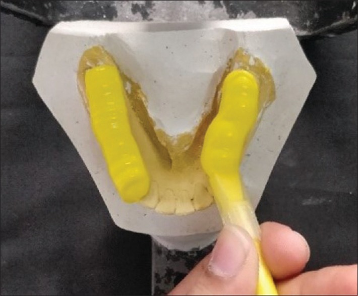
Fabrication of occlusal records was done extraorally in order to prevent any subjective discrepancies induced during the procedure. To fabricate occlusal records, silicone occlusal registration material was injected onto the occlusal surfaces from the first mandibular premolar to the second mandibular molar on the casts mounted on articulator [Figure 4]. The vertical dimension was increased by +1, +2, +3, and +4 mm on the articulator using incisal pin, and the corresponding occlusal bite records were fabricated using the technique stated above. These occlusal records were utilized to open the bite to the desired amount of increase in VDO in the participant's mouth. Since the records were fabricated on the articulator, the desired amount of increase in vertical dimension was achievable and maintained during the preparation of the occlusal record. The occlusal records were minimally scraped along the gingival margins to ensure proper seating of the bite record intraorally. All occlusal records were disinfected (Surfasept S.A., Septodont, India) and were stored in labeled plastic bags.
Figure 4.

Fabrication of bite records at different vertical dimensions at occlusion
In the second phase, head positioning device (Genoray CBCT Machine – Papaya 3D Plus, Unicorn DenMart, India) was utilized for orienting the head position of the participant for capturing the photographs. A millimeter ruler was attached to the head positioning device for calibration of photographs by referring to the magnification of ruler [Figure 5]. The height of camera was adjusted on tripod stand according to the height of the participant, and the horizontal distance between the face and camera lens was kept at five feet.
Figure 5.

Head positioning device of Genoray CBCT Machine – Papaya 3D Plus utilized for uniform orientation of the head
A digital single reflex camera (Nikon D3200, Japan) with a macro lens (18 megapixels APC-C size sensor) was used to acquire photographic data. The camera was mounted on a tripod (Simpex, VCT-691RM). For accurate positioning of the tripod between sessions, the tripod was secured to the floor with an adhesive (GG5; IE Dev Adhesive, India).
The participant was asked to close gently on the posterior teeth, say, “M, M, M,” relax, and smile. Two posed smile photographs were taken at VDO (MI) [Figure 6]. This procedure was repeated with bite registration records placed intraorally. The order of the placement of the occlusal registration records was +1, +2, +3, and +4 mm.
Figure 6.

Posed smile photograph at vertical dimension at occlusion (maximum intercuspation)
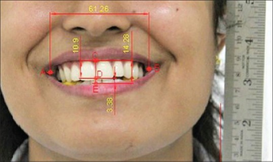
The digital images were imported into AutoCAD Software(Autodesk, Inc., California, USA), and the following measurements were made: interlabial gap height (the vertical distance between the upper and lower lips, which intersects the midpoint of incisal embrasure between the maxillary central incisors), intercommissural width (the distance between left and right commissures), incisal edge-to-upper lip distance (the vertical distance between the midpoint of the incisal embrasure and the upper lip), and incisal edge-to-lower lip distance (the vertical distance between the midpoint of the incisal embrasure and the lower lip). The measurements were made in millimeters (mm). The intercommissural width measurement was divided by interlabial gap height measurement to obtain smile index [Figure 7]. The outline of the borders of display zone was traced, and its area was recorded in AutoCAD software in mm2 [Figure 8].
Figure 7.

Parameters measured in AutoCAD software: (1) Interlabial gap height (C-E), (2) intercommissural width (A-B), (3) incisal edge-to-upper lip distance (C-D), and (4) incisal edge-to-lower lip distance (D-E)
Figure 8.

Tracing of display zone area in AutoCAD software
This procedure was repeated for all images by a single investigator. The data collected were tabulated in a spreadsheet (Excel 2010; Microsoft Corp.), and statistical analysis was conducted using SPSS software (IBM Corp, ver. 23, New York, USA). One-way repeated measures ANOVA was used and level of significance was kept at 0.05. Groups with a statistically significant difference were further analyzed for intergroup comparison with Bonferroni's corrected paired t-test.
RESULTS
The results of this study are shown in Tables 1 and 2.
Table 1.
Results of smile measurements
| Measurement | Occlusal vertical dimension | Mean±SD | P |
|---|---|---|---|
| Interlabial gap height (mm) | MI | 10.27±2.20 | <0.001 (significant) |
| +1 | 11.13±2.12 | ||
| +2 | 13.05±2.52 | ||
| +3 | 13.55±2.62 | ||
| +4 | 16.13±2.26 | ||
| Intercommissural width (mm) | MI | 61.69±4.82 | 0.839 (not significant) |
| +1 | 62.56±4.20 | ||
| +2 | 62.85±4.50 | ||
| +3 | 62.61±4.37 | ||
| +4 | 63.04±4.10 | ||
| Smile index | MI | 5.60±1.66 | <0.001 (significant) |
| +1 | 5.86±1.45 | ||
| +2 | 4.98±1.19 | ||
| +3 | 4.90±1.49 | ||
| +4 | 3.99±0.68 | ||
| Incisal edge-to-upper lip distance (mm) | MI | 7.28±2.16 | 0.530 (not significant) |
| +1 | 7.63±2.38 | ||
| +2 | 7.68±2.17 | ||
| +3 | 8.06±1.97 | ||
| +4 | 7.76±1.86 | ||
| Incisal edge-to-lower lip distance (mm) | MI | 2.90±2.07 | <0.001 (significant) |
| +1 | 4.08±2.02 | ||
| +2 | 5.73±3.12 | ||
| +3 | 6.41±2.74 | ||
| +4 | 7.51±2.18 | ||
| Display zone area (mm2) | MI | 509±133 | <0.001 (significant) |
| +1 | 630±166 | ||
| +2 | 694±165 | ||
| +3 | 731±190 | ||
| +4 | 803±159 |
MI: Maximum intercuspation, SD: Standard deviation
Table 2.
Results of one-way repeated measures ANOVA
| Source | Type III sum of squares | Df | Mean square | F | P |
|---|---|---|---|---|---|
| Interlabial gap height (mm) | 417.647 | 4 | 104.412 | 93.734 | <0.001 |
| Intercommissural width (mm) | 21.442 | 4 | 5.361 | 0.356 | 0.839 |
| Smile index | 42.322 | 4 | 10.581 | 11.300 | <0.0001 |
| Incisal edge-to-upper lip distance (mm) | 6.12 | 4 | 1.53 | 0.79735 | 0.530 |
| Incisal edge-to-lower lip distance (mm) | 271.945 | 4 | 67.986 | 20.901 | <0.001 |
| Display zone area (mm2) | 989,294.860 | 4 | 247,323.715 | 16.250 | <0.001 |
For interlabial gap height, the mean measurement in MI was 10.27 ± 2.20 mm [Table 1]. A statistically significant difference (P < 0.001) was found in interlabial gap height with increasing VDO [Table 2]. For intergroup comparison, post hoc Bonferroni's corrected paired t-tests revealed all the groups to be significantly different from each other [Table 3].
Table 3.
Post hoc Bonferroni’s corrected paired t-tests for intergroup comparison of change in interlabial gap height at different vertical dimensions at occlusion
| Group | Mean difference (mm) | P | Significance |
|---|---|---|---|
| MI versus +1 mm | 0.87 | <0.001 | Highly significant |
| MI versus +2 mm | 2.78 | <0.001 | Highly significant |
| MI versus +3 mm | 3.28 | <0.001 | Highly significant |
| MI versus +4 mm | 5.86 | <0.001 | Highly significant |
| +1 mm versus +2 mm | 1.91 | 0.001 | Significant |
| +1 mm versus +3 mm | 2.41 | <0.001 | Highly significant |
| +1 mm versus +4 mm | 4.99 | <0.001 | Highly significant |
| +2 mm versus +3 mm | 1.81 | 0.001 | Significant |
| +2 mm versus +4 mm | 3.07 | <0.001 | Highly significant |
| +3 mm versus +4 mm | 2.58 | <0.001 | Highly significant |
MI: Maximum intercuspation
The mean smile index at MI was 5.60 ± 1.66 [Table 1]. A statistically significant difference (P < 0.001) was found with increasing VDO [Table 2]. Pairwise comparisons revealed all groups to be significantly different from each other except for groups comparing MI and +1, +2, and +3 mm and +2 and +3 mm [Table 4].
Table 4.
Post hoc Bonferroni’s corrected paired t-tests for intergroup comparison of change in smile index at different vertical dimensions at occlusion
| Group | Mean difference | P | Significance |
|---|---|---|---|
| MI versus +1 mm | 0.260 | 1.0 | Not significant |
| MI versus +2 mm | −0.620 | 1.0 | Not significant |
| MI versus +3 mm | −0.697 | 1.0 | Not significant |
| MI versus +4 mm | −1.614 | 0.003 | Significant |
| +1 mm versus +2 mm | −0.880 | 0.002 | Significant |
| +1 mm versus +3 mm | −0.958 | 0.001 | Significant |
| +1 mm versus +4 mm | −1.874 | <0.001 | Highly significant |
| +2 mm versus +3 mm | −0.078 | 1.0 | Not significant |
| +2 mm versus +4 mm | −0.994 | <0.001 | Highly significant |
| +3 mm versus +4 mm | −0.917 | 0.008 | Significant |
MI: Maximum intercuspation
The incisal edge-to-lower lip distance at MI was 2.90 ± 2.07 mm [Table 1]. A statistically significant difference was found with increasing VDO (P < 0.001) [Table 2]. Pairwise comparisons revealed all groups to be significantly different from each other between MI and +1 mm, +1 and +2 mm, +2 and +3 mm, and between +3 and +4 mm [Table 5].
Table 5.
Post hoc Bonferroni’s corrected paired t-tests for intergroup comparison of change in incisal edge-to-lower lip distance at different vertical dimensions at occlusion
| Group | Mean difference (mm) | P | Significance |
|---|---|---|---|
| MI versus +1 mm | 1.18 | 0.272 | Not significant |
| MI versus +2 mm | 2.84 | 0.01 | Significant |
| MI versus +3 mm | 3.51 | <0.001 | Highly significant |
| MI versus +4 mm | 4.61 | <0.001 | Highly significant |
| +1 mm versus +2 mm | 1.65 | 0.066 | Not significant |
| +1 mm versus +3 mm | 2.34 | 0.003 | Significant |
| +1 mm versus +4 mm | 3.44 | <0.001 | Highly significant |
| +2 mm versus +3 mm | 0.68 | 1.0 | Not significant |
| +2 mm versus +4 mm | 1.92 | 0.04 | Significant |
| +3 mm versus +4 mm | 1.10 | 0.991 | Not significant |
MI: Maximum intercuspation
The mean measurement of the display zone area was 509 ± 133 mm2 [Table 1]. A statistically significant difference in the values was found with increasing VDO (P < 0.001) [Table 2]. Pairwise comparisons revealed all groups to be significantly different from each other between MI and +1 mm, +1 and +2 mm, +2 and +3 mm, and between +3 and +4 mm [Table 6].
Table 6.
Post hoc Bonferroni’s corrected paired t-tests for intergroup comparison of change in display zone area at different vertical dimensions at occlusion
| Group | Mean difference (mm) | P | Significance |
|---|---|---|---|
| MI versus +1 mm | 121.60 | 0.23 | Not significant |
| MI versus +2 mm | 185.55 | <0.001 | Highly significant |
| MI versus +3 mm | 222.05 | 0.001 | Highly significant |
| MI versus +4 mm | 294.15 | <0.001 | Highly significant |
| +1 mm versus +2 mm | 63.95 | 0.723 | Not significant |
| +1 mm versus +3 mm | 100.45 | 0.603 | Not significant |
| +1 mm versus +4 mm | 172.55 | 0.001 | Highly significant |
| +2 mm versus +3 mm | 36.50 | 1.0 | Not significant |
| +2 mm versus +4 mm | 108.60 | <0.001 | Highly significant |
| +3 mm versus +4 mm | 72.10 | 1.0 | Not significant |
MI: Maximum intercuspation
DISCUSSION
An esthetically pleasing smile depends significantly on the surrounding lips. As age advances, lips undergo several predictable changes such as decreased lip volume, loss of lip architecture, and lip lengthening.[11] These changes lead to less amount of the maxillary tooth display on smile.[12] Minimal and gradual attrition of the occlusal surfaces is generally compensated by passive eruption of teeth.[13] However, excessive occlusal attrition may result in pulpal pathology, occlusal disharmony, impaired function, and esthetic disfigurement of the face due to loss of VDO.[10] This clinical situation often requires extensive restorative treatment for increasing the VDO in order to restore facial esthetics in a positive manner. Bloom and Padayachy had stated that facial esthetics can be considered as a rationale for altering VDO.[14] Changes in soft tissue and hard tissue with varying VDO should be evaluated and quantified before performing the treatment for obtaining a predictable esthetic outcome.
Hence, this study was conducted to determine the effect of an increase in VDO on the dimensions of smile and changes in the relationship of lips to the teeth. Dimensions of the face vary according to the race or ethnicity of an individual.[15,16] In order to avoid any discrepancy in the values of results, the participants belonging to Asian Indian ethnicity were only selected for the study.
A significant increase in interlabial gap height was noted for all the groups when compared to that found at VDO (MI) [Table 2], whereas no significant change was noted in the values of intercommissural width with increasing VDO [Table 4]. An increase in the interlabial gap height denotes more amount of tooth display with increasing VDO, whereas an increase in intercommissural width denotes wider buccal corridor on smile.[17] Assessing the results obtained from this study, it can be concluded that with increasing VDO, the exposure of teeth increases on smile, whereas the width of buccal corridor remains unchanged. These findings were found similar to that reported by Chou et al.[18] This can be justified by the action of buccinator muscles and not the circumoral muscles responsible for pulling the corners of the mouth laterally.[19] McNamara et al. performed video analysis of posed smile at MI and found the average interlabial gap height and intercommissural width to be 10.4 ± 3.7 mm and 61.1 ± 5.4 mm, respectively.[20] Gross et al. reported that an increase in VDO in the range of 2–6 mm did not show any significant change in the lower face height measurement when viewed in anterior direction at clinical rest position.[21]
The average value of smile index at VDO at MI was observed to be 5.60 ± 1.66 for the studied age group (22–30 years). This value is less when compared to the results reported by Desai et al. and Chou et al. who found the smile index to be 6.73 ± 2.09 and 6.58 ± 1.92, respectively.[18,22] It was observed that increasing VDO resulted in a significant decrease in the smile index. These results can help us in assessment of smile index in patients requiring full-mouth rehabilitation at increased vertical dimension before starting the treatment.
A significant increase in the values of incisal edge-to-lower lip distance was noted for all the groups. However, a difference in these values when compared with those recorded by Chou et al. was noted [Table 7].[18]
Table 7.
Comparison of incisal edge-to-lower lip distance recorded by Chou et al.[18] and the present study
| Increase in VDO (mm) | Chou et al.[18] | Present study |
|---|---|---|
| 0 | 2.28±1.99 | 2.90±2.07 |
| 2 | 4.14±2.52 | 5.73±3.12 |
| 4 | 5.29±2.95 | 7.51±2.18 |
VDO: Vertical dimension at occlusion
No significant difference was found in the values of incisal edge-to-upper lip distance at increased VDO. This implies that as the VDO increases, the exposure of mandibular teeth increases whereas that of maxillary anterior teeth remains constant.
The display zone area can be described as a framework comprising upper and lower lips forming the boundaries, whereas the teeth and surrounding gingivae forming the components of the display zone.[23] The display zone area was quantified in the present study by determining the area between upper and lower lips at smile using the AutoCAD software.[24]
AutoCAD is a computer-aided design software that has been utilized by architects and engineers for precise, accurate, and reliable measurements. The use of AutoCAD software in research related to dentistry was first advocated by Brunetto et al.[24] Therefore, in order to obtain precise measurements, this software was utilized to record the display zone area in this study.
The mean display zone area was found to be 509 ± 133 mm2 for VDO at MI. This result was found similar to the results reported by Chou et al. (509.08 ± 190.08 mm2).[18] A significant increase in the values of display zone area was observed with increasing the VDO by +2 mm and above suggesting a greater amount of tooth display on smile for patients restored at increased VDO [Table 6].
The difference in the values reported by Chou et al. and the present study in smile parameters such as smile index and incisal edge-to-lower lip distance can be attributed to the difference in ethnicity of the population being studied.[18] Chou et al. conducted a study on predominantly white population, whereas in the present study, all the participants belonged to Asian Indian ethnicity.[18]
CONCLUSION
In clinical conditions requiring full-mouth rehabilitation and/or complex prosthodontic treatment, an increase in VDO does not reduce maxillary gingival display or buccal corridor display but shifts the lower lip downward on smile. Hence, care should be taken to match the width-to-height ratio (smile index) and display zone area to the patient's age when an increase in the occlusal vertical dimension is contemplated.
Scope for further studies
A study can be conducted for evaluating the long-term adaptation of the facial muscles to increased VDO
A study on an older age group can be performed to compare the effect of aging of muscles on the smile parameters.
Declaration of patient consent
The authors certify that they have obtained all appropriate patient consent forms. In the form, the patients have given their consent for their images and other clinical information to be reported in the journal. The patients understand that their names and initials will not be published and due efforts will be made to conceal their identity, but anonymity cannot be guaranteed.
Financial support and sponsorship
Nil.
Conflicts of interest
There are no conflicts of interest.
REFERENCES
- 1.Dion K, Berscheid E, Walster E. What is beautiful is good. J Pers Soc Psychol. 1972;24:285–90. doi: 10.1037/h0033731. [DOI] [PubMed] [Google Scholar]
- 2.Vinnakota DN, Kanneganti KC, Pulagam M, Keerthi GK. Determination of vertical dimension of occlusion using lateral profile photographs: A pilot study. J Indian Prosthodont Soc. 2016;16:323–7. doi: 10.4103/0972-4052.176531. [DOI] [PMC free article] [PubMed] [Google Scholar]
- 3.Johnson FB, Sinclair DA, Guarente L. Molecular biology of aging. Cell. 1999;96:291–302. doi: 10.1016/s0092-8674(00)80567-x. [DOI] [PubMed] [Google Scholar]
- 4.Spear F. Approaches to vertical dimension. Adv Esthet Interdiscip Dent. 2006;2:2–12. [Google Scholar]
- 5.Heartwell CN, Rahn AO. 3rd ed. Philadelphia: Lea & Febiger; 1980. Syllabus of Complete Dentures; p. 214. [Google Scholar]
- 6.Keough B. Occlusion-based treatment planning for complex dental restorations: Part 1. Int J Periodontics Restorative Dent. 2003;23:237–47. [PubMed] [Google Scholar]
- 7.Krishna MG, Rao KS, Goyal K. Prosthodontic management of severely worn dentition: Including review of literature related to physiology and pathology of increased vertical dimension of occlusion. J Indian Prosthodont Soc. 2005;5:89–93. [Google Scholar]
- 8.Abduo J. Safety of increasing vertical dimension of occlusion: A systematic review. Quintessence Int. 2012;43:369–80. [PubMed] [Google Scholar]
- 9.Gross MD, Ormianer Z. A preliminary study on the effect of occlusal vertical dimension increase on mandibular postural rest position. Int J Prosthodont. 1994;7:216–26. [PubMed] [Google Scholar]
- 10.Ushijima M, Kamashita Y, Nishi Y, Nagaoka E. Changes in lip forms on three-dimensional images with alteration of lip support and/or occlusal vertical dimension in complete denture wearers. J Prosthodont Res. 2013;57:113–21. doi: 10.1016/j.jpor.2012.11.003. [DOI] [PubMed] [Google Scholar]
- 11.Krajicek D. Guides for natural facial appearance as related to complete denture construction. J Prosthet Dent. 1969;21:654–62. doi: 10.1016/0022-3913(69)90014-6. [DOI] [PubMed] [Google Scholar]
- 12.Mohindra NK, Bulman JS. The effect of increasing vertical dimension of occlusion on facial aesthetics. Br Dent J. 2002;192:164–8. doi: 10.1038/sj.bdj.4801324. [DOI] [PubMed] [Google Scholar]
- 13.Turner KA, Missirlian DM. Restoration of the extremely worn dentition. J Prosthet Dent. 1984;52:467–74. doi: 10.1016/0022-3913(84)90326-3. [DOI] [PubMed] [Google Scholar]
- 14.Bloom DR, Padayachy JN. Increasing occlusal vertical dimension-why, when and how. Br Dent J. 2006;200:251–6. doi: 10.1038/sj.bdj.4813305. [DOI] [PubMed] [Google Scholar]
- 15.van Sickels JE, Rugh JD, Chu GW, Lemke RR. Electromyographic relaxed mandibular position in long-faced subjects. J Prosthet Dent. 1985;54:578–81. doi: 10.1016/0022-3913(85)90439-1. [DOI] [PubMed] [Google Scholar]
- 16.Mack MR. Vertical dimension: A dynamic concept based on facial form and oropharyngeal function. J Prosthet Dent. 1991;66:478–85. doi: 10.1016/0022-3913(91)90508-t. [DOI] [PubMed] [Google Scholar]
- 17.Frush JP, Fisher RD. The dynesthetic interpretation of the dentogenic concept. J Prosthet Dent. 1958;8:558–81. [Google Scholar]
- 18.Chou JC, Thompson GA, Aggarwal HA, Bosio JA, Irelan JP. Effect of occlusal vertical dimension on lip positions at smile. J Prosthet Dent. 2014;112:533–9. doi: 10.1016/j.prosdent.2014.04.009. [DOI] [PubMed] [Google Scholar]
- 19.Terry DA, Pirtle PL. Learning to smile: The neuroanatomic basis for smile training. J Esthet Restor Dent. 2001;13:20–7. doi: 10.1111/j.1708-8240.2001.tb00248.x. [DOI] [PubMed] [Google Scholar]
- 20.McNamara L, McNamara JA, Jr, Ackerman MB, Baccetti T. Hard-and soft-tissue contributions to the esthetics of the posed smile in growing patients seeking orthodontic treatment. Am J Orthod Dentofacial Orthop. 2008;133:491–9. doi: 10.1016/j.ajodo.2006.05.042. [DOI] [PubMed] [Google Scholar]
- 21.Gross MD, Nissan J, Ormianer Z, Dvori S, Shifman A. The effect of increasing occlusal vertical dimension on face height. Int J Prosthodont. 2002;15:353–7. [PubMed] [Google Scholar]
- 22.Desai S, Upadhyay M, Nanda R. Dynamic smile analysis: Changes with age. Am J Orthod Dentofacial Orthop. 2009;136:310e1–10. doi: 10.1016/j.ajodo.2009.01.021. [DOI] [PubMed] [Google Scholar]
- 23.Ackerman MB, Ackerman JL. Smile analysis and design in the digital era. J Clin Orthod. 2002;36:221–36. [PubMed] [Google Scholar]
- 24.Brunetto J, Becker MM, Volpato CA. Gender differences in the form of maxillary central incisors analyzed using AutoCAD software. J Prosthet Dent. 2011;106:95–101. doi: 10.1016/S0022-3913(11)60102-9. [DOI] [PubMed] [Google Scholar]


