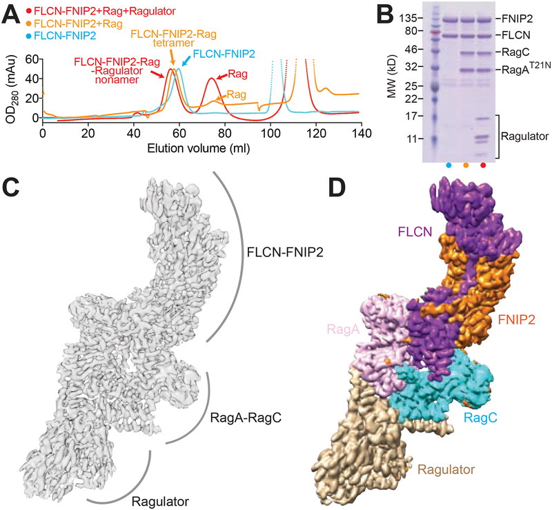
Figure 1. Structural determination of the FLCN-FNIP2-Rag-Ragulator nonamer.
A. Gel filtration profiles for the assembled FLCN-FNIP2-Rag-Ragulator supercomplex on a Hiload 16/60 Superdex 200 column. Cyan, FLCN-FNIP2 heterodimer only. Orange, FLCN-FNIP2 in complex with the Rag GTPases. Red, FLCN-FNIP2 in complex with the Rag GTPases and Ragulator.
B. Coomassie blue stained gel analyses to identify the protein subunits within the peaks of the gel filtration profiles in A. Lanes are labeled based on the coloring pattern in A.
C & D. Cryo-EM density map (C) and segmented map (D) for the FLCN-FNIP2-Rag-Ragulator nonamer. Subunits within the FLCN-FNIP2-Rag-Ragulator nonamer are differentiated by color as indicated: FLCN, purple; FNIP2, orange; RagA, pink; RagC, cyan; Ragulator, light brown.

