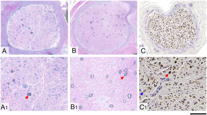
Figure 3.
Nerve biopsy findings in RFC1 biallelic repeat expansions. Severe loss of large and small myelinated fibres with no evidence of ongoing axonal degeneration, no signs of regeneration and no features of demyelination is seen in all CANVAS patients with sensory neuropathy due to RFC1 biallelic expansion. (A, A1, B and B1) Resin-embedded, semi-thin sections stained with methylene blue azure-basic fuchsin are shown from two different patients. Occasional large remaining fibres in both cases are highlighted with a red arrow in A1 and B1. The unmyelinated fibres in the peripheral nerves from CANVAS patients are comparably much better preserved. (C) Axons in formalin-fixed paraffin-embedded tissue are shown on immunostaining with SMI31 antibody. (C1) A large residual axon is highlighted with red arrow and one of multiple clusters of small unmyelinated fibres is shown with a blue arrow. Scale bars = 100 µm in A–C; 40 µm in A1–C1.

