PLOS ONE logoLink to PLOS ONE
. 2020 Feb 10;15(2):e0220756. doi: 10.1371/journal.pone.0220756

TSG-6 in extracellular vesicles from canine mesenchymal stem/stromal is a major factor in relieving DSS-induced colitis

Ju-Hyun An 1, Qiang Li 1,¤, Min-Ok Ryu 1, A-Ryung Nam 1, Dong-Ha Bhang 2, Yun-Chan Jung 3, Woo-Jin Song 1,*,#, Hwa-Young Youn 1,*,#
Editor: Hossam MM Arafa4
PMCID: PMC7010233  PMID: 32040478

Abstract

Adipose tissue derived mesenchymal stem/stromal cell (ASC)-derived extracellular vesicles (EV) have been reported to be beneficial against dextran sulfate sodium (DSS)-induced colitis in mice. However, the underlying mechanisms have not been fully elucidated. We hypothesize that the tumor necrosis factor-α-stimulated gene/protein 6 (TSG-6) in EVs is a key factor influencing the alleviation of colitis symptoms. DSS-induced colitis mice (C57BL/6, male, Naïve = 6, Sham = 8, PBS = 8 EV = 8, CTL-EV = 8, TSG-6 depleted EV = 8) were intraperitoneally administered EVs (100 ug/mice) on day 1, 3, and 5; colon tissues were collected on day 10 for histopathological, RT-qPCR, western blot and immunofluorescence analyses. In mice injected with EV, inflammation was alleviated. Indeed, EVs regulated the levels of pro- and anti-inflammatory cytokines, such as TNF-α, IL-1β, IFN-γ, IL-6, and IL-10 in inflamed colons. However, when injected with TSG-6 depleted EV, the degree of inflammatory relief was reduced. Furthermore, TSG-6 in EVs plays a key role in increasing regulatory T cells (Tregs) and polarizing macrophage from M1 to M2 in the colon. In conclusion, this study shows that TSG-6 in EVs is a major factor in the relief of DSS-induced colitis, by increasing the number of Tregs and macrophage polarization from M1 to M2 in the colon.

Introduction

Inflammatory bowel disease (IBD) is a chronic debilitating disease that affects both humans and dogs, characterized by abdominal pain and diarrhoea. It may result in significant morbidity and mortality, with compromised quality of life and life expectancy. Clinical signs may be controlled by single or combination therapy, including dietary modifications, antibiotics and immune-suppressants. However, since there is no clear treatment method, clinical recurrence frequently occurs even after treatment, and thus, new therapeutic agents need to be sought [1].

Adipose tissue derived mesenchymal stem/stromal cells (ASCs) are of great interest as novel therapeutics for IBD patients because of their unique ability to regulate immune cells and heal damaged colonic tissue [2]. Particularly, it has been found that extracellular vesicles (EVs, 40–1000 nm sized circular membrane fragments shed from the cell surface) secreted from cells can mediate the delivery of secreted molecules in cell-to-cell communication. Thus, studies on the use of EVs as an alternative to stem cells have been actively conducted [3, 4]. Recently, various studies have been carried out on the application of EVs as therapeutic agents in various pre-clinical models such as acute kidney injury, hepatitis, cystitis and uveitis[58]. In addition, injecting EVs into DSS-induced colitis mouse models has shown that not only does it improve activity and appetite, but it also alleviates inflammation in the colon [9]. Although these studies reported that damaged tissues were improved following treatment with EVs, the factors responsible for the protective effects have yet to be elucidated. If stem cell-derived EVs are to be used as therapeutic agents in the future, in-depth mechanistic studies to determine which factors are most highly associated with the ability of EVs to alleviate inflammation must be conducted.

Tumor necrosis factor (TNF)-α stimulated gene/ protein 6 (TSG-6) secreted from stem cells is a major factor responsible for regulation of inflammatory responses [1012]. Moreover, several studies have shown that TSG-6 plays important roles in attenuating DSS-induced colitis in mice by altering the composition of immune cells in the colon [11, 13]. However, studies on TSG-6 in EVs have not yet been conducted, and, thus, further studies are required.

In particular, IBD is related to an immunological imbalance in the intestinal mucosa, which is primarily associated with cells of the adaptive immune system that respond to self-antigens produced under inflammatory conditions in such patients [14, 15]. Among the intestinal immune cells, regulatory T cells (Tregs) control the balance of immune cell functions and play critical roles in self-tolerance and homeostasis in the colon [16]. Additionally, macrophages of the colon are essential for local homeostasis and play an important role in inflammation and protective immunity[17]. And they are classically divided into two major types: A very basic dichotomous view classified M1 as having an inflammatory phenotype, while M2 was considered an anti-inflammatory macrophage[18]. For this reason, various methods of increasing Tregs and macrophage M2 types in the colon in colitis experimental models have been proposed as treatment options, with stem cell derived EV being one of them[9, 19]. However, there is still a lack of research on mechanisms, and there is a need for further study.

Therefore, this study focused on elucidating the role of TSG-6 in EVs in mitigating colitis and as well as to describe potential mechanisms responsible for any protective effects observed in DSS-induced mouse models of colitis. We also investigated the effect that EVs have on Treg and M2 within the colon and how TSG-6 in EVs affects the Treg and M2 population in inflamed colons.

Materials and methods

All animal experimental procedures were approved by the Institutional Animal Care and Use Committee of Seoul National University (SNU), Republic of Korea, and all protocols were in accordance with approved guidelines (SNU; protocol no. SNU-180829-2-1).

Isolation, culture, and characterization of cASCs

The adipose tissue was obtained from a healthy adult female dog during ovariohysterectomy at the SNU Veterinary Medicine Teaching Hospital, with the owner’s consent. ASCs were isolated from tissues and cultures as previous described [11, 20]. The cells were characterized for the expression of several stem cell markers by flow cytometry before they were used in the experiments. Additionally, the differentiation ability of cells was confirmed for cASCs at passages 3 and 4, and these cells were used in subsequent experiments. The methods used for isolating, culturing, and characterizing stem cells are described in detail in S1 File, and corresponding results were described in detail in S1 Fig. This experiment was repeated a minimum of three times to confirm reproducibility.

Small interfering RNA (siRNA) transfection of cASCs

To obtained TSG-6 depleted EV, when cASCs reached approximately 70% confluence, they were transfected for 48 h with TSG-6 siRNA or control siRNA (sc-39819 and sc-27007, respectively, Santa Cruz Biotechnology, Dallas, TX, USA) using Lipofectamine RNAiMAX (Invitrogen, Carlsbad, CA, USA) according to the manufacturer’s instructions [11, 20, 21]. TSG-6 knockdown was confirmed by gel PCR and RT-qPCR. The cells were washed twice with PBS, and the media was exchanged with DMEM containing exosome-depleted FBS for an additional 48 h before collecting TSG-6-depleted EVs. EVs were obtained using an ultracentrifuge as above described, and the relative TSG-6 protein levels in EVs were measured by western blot analysis. Protein concentrations were determined by performing BCA assays. The total protein content (20 μg) of each sample was subjected to sodium dodecyl sulfate-polyacrylamide gel electrophoresis (SDS-PAGE) and immunoblotting with antibodies against CD63 (NBP2-42225; Novus Inc., Littleton, CO, USA)) and TSG-6 (sc-30140; Santa Cruz Biotechnology). These results were described in S2 Fig. This experiment was repeated a minimum of three times to confirm reproducibility.

Isolation and characterization of EVs

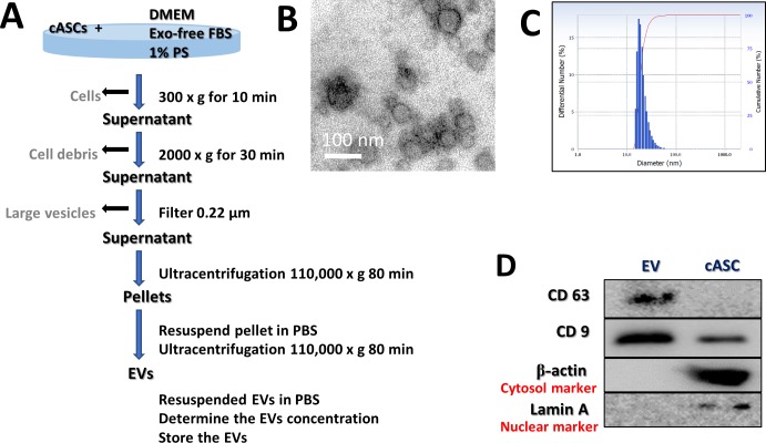
cASCs were cultured for 48 h in Dulbecco's Modified Eagle's Medium (DMEM; PAN-Biotech, Aidenbach, Germany) supplemented with 10% Exosome-depleted Fetal bovine serum (FBS; Systembio, CA, USA) and 1% penicillin-streptomycin (PS; PAN-Biotech). The supernatant from each cultured cASC sample was collected on ice and centrifuged at 300 × g for 10 min to remove the cells. Each supernatant was transferred to a fresh tube, centrifuged at 2000 × g for 30 min to remove cellular debris, and then passed through a 0.22-μm filter (Millipore, Billerica, MA, USA) to remove the large vesicles. Each supernatant was transferred to a fresh tube and centrifuged at 110,000 × g (Beckman Avanti Centrifuge J-26XP with 70Ti rotor, Brea, CA, USA) for 80 min, washed with Dulbecco’s phosphate-buffered saline (DPBS), and purified by centrifugation at 110,000 × g for 80 min. All centrifugation steps were performed at 4°C. Each pellet was resuspended in DPBS and sterilized by filtration through a 0.22-μm filter (Fig 1C). The total protein concentration in each EV preparation was quantified by performing bicinchoninic acid (BCA) assays, and the samples were stored at −80°C until use. Protein markers of purified EVs were determined by western blotting with antibodies against CD63 (Novus Inc.) and CD9 (GTX76185; GeneTex, Irivine, CA, USA). The EV morphology was characterized by transmission electron microscopy. Briefly, 10 μL of an EV suspension was placed on clean parafilm. A 300-mesh formvar/carbon-coated electron microscopy grid was floated on the drop, with the coated side facing the suspension, and allowed to adsorb for 20 min at 23 ± 2ଌ. The grid was transferred to a 100 μL drop of distilled water and left to stand for 2 min. The grid was then transferred to a 50 μL drop of 2% uranyl acetate for negative staining for 10 min, followed by observation under a TEM (LIBRA 120, Carl Zeiss, Germany) at 120 kV. The size distribution of the particles was measured using a Zeta-potential & Particle size Analyzer (ELSZ-1000ZS, Otsuka Electronics, Osaka, Japan). This experiment was repeated a minimum of three times to confirm reproducibility.

Fig 1. Characterization of cASC-EVs.

Fig 1

(A) Schematic overview of the UC-based purification protocol (B) Scanning electron microscopy micrographs of cASC-EV showed spheroid shaped vesicles with diameters of approximately 20–100 nm. Scale bar, 50 nm. (C) Size-distribution analysis of purified cASC-EVs showed that the vesicle diameters were approximately 20–100 nm. (D) Immunoblotting analysis of common EV markers, where 10 μg of total protein was loaded in each lane. cASCs-EVs expressed CD63 and CD9, while beta actin and lamin A showed lower expression. The displayed data represent at least three repeated experiments with consistent results.

Isolation of peripheral blood mononuclear cells (PBMCs) and non-adherent cells

Using citrate phosphate dextrose adenine-containing tubes, we collected blood samples (30 mL) from three healthy dogs. The blood samples were diluted with an equal volume of phosphate-buffered saline (PBS) and then layered over Ficoll-Plaque PLUS (GE Healthcare Life Sciences, Little Chalfont, UK) in a conical tube. After centrifugation at 400 × g for 30 min, the buffy coat layer was carefully collected. The collected samples were incubated with red blood cell-lysis buffer at room temperature for 10 min. After adding PBS, each sample was centrifuged at 400 × g for 10 min, and the washing and centrifugation steps were repeated. Canine PBMCs (cPBMCs) were resuspended in Roswell Park Memorial Institute (RPMI) medium (Pan-Biotech, Dorset, Germany) supplemented with 10% EV-free FBS and 1% PS. Non-adherent cells were obtained after 24 hours.

Co-culture experiments

DH82 cells, a canine macrophage-like cell line, were purchased from the Korean Cell Line Bank (Seoul, Korea). DH82 cells were seeded in 6-well plates (1×10⁶ cells/well), then incubated for 24 h. After adherence to the plates was confirmed, the DH82 cells were treated with LPS (200 ng/mL; Sigma-Aldrich) or control for 24 h. Similarly, canine lymphocytes were seeded in 6-well plates (1×10⁶ cells/well) and exposed to Con A (5 μg/mL) or control for 24 h. Next, the medium was removed and replaced with media containing EV (100 μg/well) derived from naïve, si RNA and si TSG-6 cASCs. Next, the cells were incubated for 48 h and then harvested for RNA extraction and flow cytometry analysis.

RNA extraction, cDNA synthesis, and reverse transcription-quantitative polymerase chain reaction

RNA was extracted using the Easy-BLUE Total RNA Extraction Kit (Intron Biotechnology). Next, cDNA was synthesized using LaboPass M-MuLV Reverse Transcriptase (Cosmogenetech, Seoul, Korea), according to the manufacturer’s instructions. cDNA samples were assayed using AMPIGENE® qPCR Green Mix Hi-ROX with SYBR Green Dye (Enzo Life Sciences, Farmingdale, NY, USA), according to the manufacturer’s instructions. Expression levels were normalized to those of glyceraldehyde 3-phosphate dehydrogenase (GAPDH). The sequences of the primers used in this experiment are shown in Table 1.

Table 1. Sequences of PCR primers used in this study.

Gene Forward (5’-3’) Reverse (5’-3’) Reference
cGAPDH TTAACTCTGGCAAAGTGGATATTGT GAATCATACTGGAACATGTACACCA [2]
cTNF-α TCATCTTCTCGAACCCCAAG ACCCATCTGACGGCACTATC [2]
cIFN-γ TTCAGCTTTGCGTGATTTG CTGCAGATCGTTCACAGGAA [22]
cIL-1β AGTTGCAAGTCTCCCACCAG TATCCGCATCTGTTTTGCAG [23]
cIL-6 ATGATCCACTTCAAATAGTCTACC AGATGTAGGTTATTTTCTGCCAGTG [2]
cIL-10 ATTTCTGCCCTGTGAGAATAAGAG TGTAGTTGATGAAGATGTCAAGCTA [2]
cTSG-6 TCCGTCTTAATAGGAGTGAAAGATG AGATTTAAAAATTCGCTTTGGATCT [2]
cCD4 TGCTCCCAGCGGTCACTCCT GCCCTTGCAGCAGGCGGATA [24]
cCD25 GGCAGCTTATCCCACGTGCCAG ATGGGCGGCGTTTGGCTCTG [24]
cFOXP3 AAACAGCACATTCCCAGAGTTC AGGATGGCCCAGCGGATCAG [25]
ciNOS GAGATCAATGTCGCTGTACTCC TGATGGTCACATTTTGCTTCTG [23]
cCD206 GGAAATATGTAAACAGGAATGATGC TCCATCCAAATAAACTTTTTATCCA [23]
mGAPDH AGTATGTCGTGGAGTCTACTGGTGT AGTGAGTTGTCATATTTCTCGTGGT [14]
mTNF-α CCAGGAGAAAGTCAGCCTCCT TCATACCAGGGCTTGAGCTCA [14]
mIFN-γ GATGCATTCATGAGTATTGCCAAGT GTGGACCACTCGGATGAGCTC [14]
mIL-1β CACCTCTCAAGCAGAGCACAG GGGTTCCATGGTGAAGTCAA [14]
mIL-6 TCCAGTTGCCTTCTTGGGAC GTACTCCAGAAGACCAGAGG [14]
mIL-10 TGGCCCAGAAATCAAGGAGC CAGCAGACTCAATACACACT [14]
mCD4 GAGAGTCAGCGGAGTTCTC CTCACAGGTCAAAGTATTGTTG [26]
mCD25 CTCCCATGACAAATCGAGAAAGC ACTCTGTCCTTCCACGAAATGAT [26]
mFOXP3 TTGGCCAGCGCCATCTT TGCCTCCTCCAGAGAGAAGTG [26]
miNOS AAAGGAAATAGAAACAACAGGAACC GCATAAAGTATGTGTCTGCAGATGT [27]
mCD206 AACGGAATGATTGTGTAGTTCTAGC TACAGGATCAATAATTTTTGGCATT [28]
mArg CAGAAGAATGGAAGAGTCAG CAGATATGCAGGGAGTCACC [29]

Flow cytometry analysis

To evaluate Treg polarization, PBMC-derived lymphocytes cocultured with cASC-EVs were harvested. Obtained cells (1 × 10⁶) were suspended in 100μL DPBS and 1 μL of each primary antibody against the following proteins: FOXP3-PE (eBioscience, San Diego, CA, USA; 1:100), CD3-FITC (MCA1774F; Bio-Rad, San Diego, CA, USA; 1:100), CD206-FITC (eBioscience, San Diego, CA, USA; 1:100) and CD11c-PE (eBioscience, San Diego, CA, USA; 1:100). After incubation for 1 h at 23 ± 2°C, the cells were washed with DPBS. Unstained cells were used as controls for autofluorescence. Cell fluorescence was analyzed with a flow cytometer (FACS Aria Ⅱ; BD bioscience). The results were analyzed using FlowJo 7.6.5 software (Tree Star, Inc., Ashland, OR, USA).

ELISA analysis

The protein levels of interleukin 10 (IL-10) in each cell culture supernatant was detected using an IL-10 Enzyme-Linked Immunosorbent Assay Kit (ELISA; eBioscience), according to the manufacturer’s instructions.

Mice

To determine the therapeutic effect of ASC-derived EVs on colitis, we induced mouse colitis with DSS (36–50 kDa; MP Biomedical, Solon, OH, USA). Male C57BL/6 mice (6 to 8-week-old and weighing 18 to 20 g) were purchased from Nara Bio (Gyeonggi, Korea) and acclimatized for 7 days with a 12 h light/dark cycle at 22°C and 60% humidity before performing the experiments. For environmental enrichment, 3 to 4 mice were raised in polycarbonate cages (324 × 221.5 × 130 mm) containing clean bedding (shavings; Nara Biotech), cardboard boxes, and tunnels. Mice were fed a standard laboratory rodent diet (Central Lab Animal Inc., Seoul, Korea) and water ad libitum. At the start of the experiments, the health status of the mice was evaluated by measuring their weight, vitality, and defecation; the experiments were conducted on mice with no abnormal symptoms. The mice were administered 3% DSS in their drinking water from days 0 to 7. The studies were conducted using 46 animals, and the mice were randomly divided into 6 groups with 6–8 mice per group (naïve = 6, Sham = 8, PBS = 8, EV = 8, CTL-EV = 8, TSG-6 depleted EV = 8). The mice were treated with 100 μL DPBS, with or without cASC-EV (100 μg/mouse), by intraperitoneal (IP) injection on days 1, 3 and 5. The disease-activity index (DAI) represents the combined score of weight loss relative to the initial body weight (grades 0–4; 0, no weight loss; 1, < 10% loss; 2, 10–20% loss; 3, 20–30% loss; and 4, 30–40% loss), stool consistency (grades 0–2; 0, normal; 1, soft; and 2, liquid), the presence of blood in the feces and anus (grades 0–2; 0, negative fecal occult bleeding; 1; positive fecal occult bleeding; and 3, visible fecal occult bleeding), and general activity (grades 0–2; 0, normal; 1, mildly to moderate depressed and 2, severely depressed). The DAI score of colitis was calculated independently by two blinded investigators. The score for each parameter was summed from day 0 to the day of sacrifice, and the summed score was averaged to yield the final score. Since this model has been verified in several studies, it is unlikely to be accompanied by unexpected pain. However, should three or more of the following abnormal behaviors be observed: behaviorally excessive waist bending, self-cutting, aggressive behavior, stabbing of other mice, injury due to fall, failure to build a nest, abnormally rough hair, abnormal posture, or convulsions, the animal was euthanized within one day and the experiment terminated. On day 10 of the study, the veterinarian euthanized all mice with xylazine infusion and CO2 inhalation according to the approved institutional animal ethics protocol.

Histological evaluation

Colon tissues were fixed in 10% formaldehyde for 48 h, embedded in paraffin, cut into 4-μm sections, and stained with hematoxylin and eosin (H&E). Histological scores are provided in Table 2. Because DSS-related injury varies, two slides from each colon section were assessed per mouse, and at least three areas on each slide were examined.

Table 2. Histological assessment.

Score Mucosal thickness & hyperplasia Inflammatory cell extent Damaged to the crypt
0 Normal Normal An intact crypt
1 Minimal 11–25% of mucosa Loss of the basal 1/3 of crypt
2 Mild 26–50% of mucosa Loss of basal 2/3 of crypt
3 Moderate Mucosa and submucosa Entire loss of crypt

Immunofluorescence analysis

Colon sections were deparaffinized and rehydrated, and antigen retrieval was carried out in 10 mM citrate buffer. Sections were then washed and blocked with blocking buffer containing 1% bovine serum albumin and 0.1% tween 20 for 30 min. The sections were then incubated overnight at 4ଌ with mouse monoclonal anti-Forkhead box (Fox) P3 (1:100; Santa Cruz Biotechnology) and mouse monoclonal anti CD206 (1:100; Santa Cruz Biotechnology). The colon sections were washed three times with DPBS. Then, the sections were incubated FITC conjugated anti mouse (1:500; Santa Cruz) for 1 hr. After that, they were washed three times with DPBS. All samples were mounted using Vectashield mounting medium containing 4’,6-diamidino-2-phenylindole (DAPI; Vector Laboratories, Burlingame, CA, USA). The samples were observed using an EVOS FL microscope (Life Technologies, Darmstadt, Germany). Immunoreactive cells were counted in 20 random fields per group, and the percentage of CD206+ positive cells and FOXP3+ positive cells was calculated in colon sections from the same mice.

Statistical analysis

GraphPad prism (version 6.01) software (GraphPad, Inc., La Jolla, CA, USA) was used for statistical analysis. the differences between two groups were analyzed using Student’s t-tests and differences between more than two groups were analyzed using one-way analysis of variance (ANOVA) followed by Bonferroni multiple comparison test. The results are presented as the mean value ± standard deviation (SD). Differences with a value of P < 0.05 were considered as statistically significant.

Results

Characterization of cASC-EV

The characterization of stem cells used for these studies are described in detail in S1 Fig. The EVs were separated from stem cell culture media by ultracentrifugation. Schematic overview describing this protocol is presented in Fig 1A. Approximately 100 μg of EVs was produced in the media in which 1 × 10⁶ cells were seeded and grown for 2 days. Electron microscopic analysis demonstrated that the EVs were round-shaped and 50–100 nm in diameter (Fig 1B). Using a particle-size analyzer, the EVs were confirmed to be less than 100 nm in diameter (Fig 1C). In addition, positive markers of EVs such as CD63 and CD9 were identified by western blotting, whereas negative markers of EVs such as Lamin A (a nuclear marker) and beta actin (a cytosolic marker) were present in lower abundance (Fig 1D). Whole-cell lysates were used as a positive control. Our findings suggest that the EVs contained little or no cellular matrix and nuclei, which are intracellular components.

Production of EVs containing less TSG-6 from cASCs

To reduce TSG-6 in EVs, cASCs were transfected with si-TSG-6. No differences were observed in the cell viability and cell differentiation potentials between transfected and untreated stem cells. Furthermore, the size and shape of the EVs secreted from the transfected stem cells did not differ from those of the untreated stem cells, and no difference occurred in the amount of EVs produced (S1 Fig). Further, the TSG-6 mRNA levels in the transfected cASCs were reduced by over 50% (S2 Fig) while the TSG-6 protein levels in the EVs were reduced to less than half of those in the naïve and control siRNA-treated groups (S2 Fig)).

IP administration of cASC-EVs containing TSG-6 played a crucial role in alleviating IBD

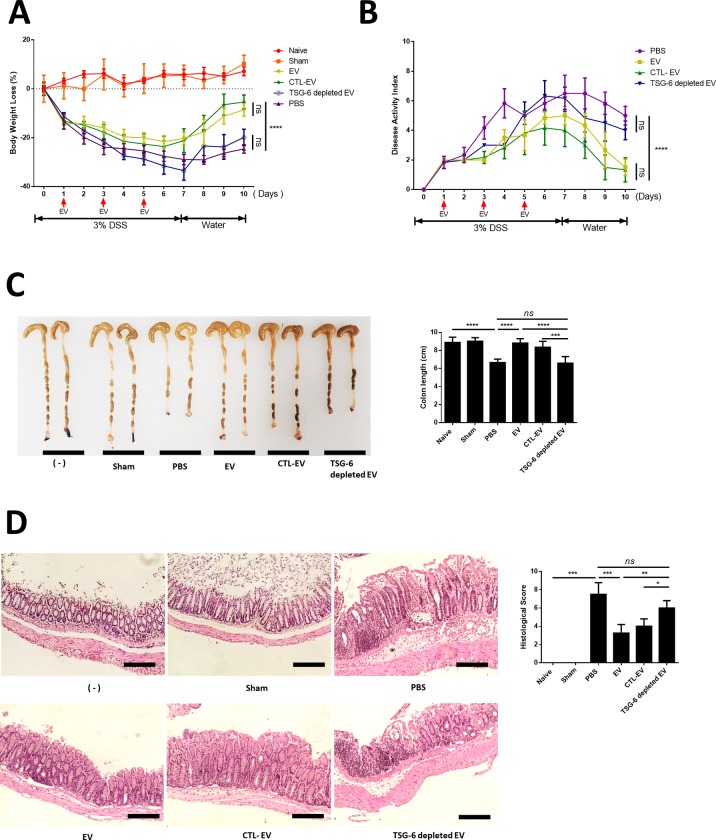
In this study, all animals met the euthanasia criteria before being sacrificed. Injecting EV into mice that did not induce colitis (sham group) showed no difference in vitality, weight, and stool consistency from naive mice. In addition, the sham group was not different from the naive group in the colon length and histological examination. In the DSS-administered group, significant weight loss and clinical indices including DAI which is based on body weight (Fig 2A), stool consistency, bloody diarrhea and general activity were found to worsen compared to the healthy group; while the EV group exhibited improved weight and DAI compared to the DSS group (Fig 2B). Moreover, shortening of the colon length significantly improved in the EV group compared with that in the PBS-treated group. However, in TSG-6 depleted EV group was found to have a shorter colon length than the EV and CTL-EV groups (Fig 2C). The EV group showed greatly decreased histological colitis scores for mucosal thickness, mucosal hyperplasia, extent of inflammation, and crypt damage. In addition, the anti-inflammatory effect of CTL-EV was similar to that of naive-EV, whereas the effect of TSG-6-depleted EV was insignificantly decreased. (Fig 2D).

Fig 2. cASC-EV injection ameliorated DSS-induced colitis in mice.

Fig 2

EVs (100 μg), TSG-6 depleted EVs (100 μg), control EVs (100 μg), or vehicle control were injected IP one day after mice were administered 3% DSS. On days 3 and 5, the mice in each group were re-injected with EVs (100 μg), TSG-6 depleted EVs (100 μg), control EVs (100 μg), or vehicle control (PBS). Mice were monitored for changes in (A) body weight, (B) DAIs, and (C) colon lengths. (D) H&E staining of colon sections and histological scores are shown. Scale bars, 100 μm. The results are shown as mean ± standard deviation (n = 6–8 in each group, *P < 0.05, **P < 0.01, ***P < 0.001, ****P < 0.0001, as determined by one-way ANOVA)

TSG-6 in cASC-EV modulate pro- and anti- inflammatory cytokine inflamed colon

The colon of EV-treated mice showed reduced levels of inflammatory cytokines (TNF-α, IFN-γ, IL-1β, IL-6 and iNOS) and elevated levels of an anti-inflammatory/regulatory cytokine (IL-10), compared to that of untreated mice with colitis and TSG-6-depleted EV-treated mice (Fig 3).

Fig 3. EVs from cASCs inhibited inflammatory responses in the colon.

Fig 3

mRNA-expression levels of pro- and anti-inflammatory cytokines in the colon were determined by qRT-PCR. These data show that TSG-6 in EVs played a major role in regulating inflammatory cytokine levels in the colon. The results are shown as the mean ± standard deviation (n = 6–8 in each group, *P < 0.05, **P < 0.01, ***P < 0.001, as determined by one-way ANOVA).

TSG-6 was a major factor in increasing Tregs in Con A stimulated lymphocytes in vitro

We found that CD4, CD25 mRNA expression levels increased in the EV group, but decreased in the TSG-6 depleted EV group (Fig 4A). Additionally, the protein levels of IL-10 were measured in the lymphocyte cultured medium. The EV group showed increased IL-10 expression, whereas the TSG-6-depleted EV group showed decreased IL-10 expression (Fig 4B). In addition, to determine whether the increased number of Tregs among total T cells was associated with TSG-6 in the EVs, the degree of Treg activation was confirmed by fluorescence-activated cell sorting. Accordingly, the number of FOXP+ cells among CD3+ cells increased in the EV group. However, in the TSG-6-depleted EV group, the proportion of FOXP+ cells decreased (Figs 4C and S5). These results demonstrate that the immunomodulatory effects of EV were related to TSG-6.

Fig 4. cASC-EV TSG-6 increased Treg proliferation in vitro.

Fig 4

Con A-stimulated canine lymphocytes were cocultured for 48 h with cASC-EVs transfected with TSG-6 siRNA (si-TSG6) or scrambled siRNA (siCTL), or naïve EVs. (A) CD4 and CD25 mRNA-expression levels were measured, confirming that TSG-6 was associated with increased Treg production. (B) IL-10, which is known to be secreted from Tregs, was also measured in the supernatant medium, and the results confirmed that IL-10 production in lymphocytes was associated with TSG-6 (n = 6 in each group). (C) The Treg population was determined by measuring FOXP3 and CD3 double-positive cells by flow cytometry (n = 6 in each group). The results are presented as the mean ± standard deviation (*P < 0.05, **P < 0.01, ***P < 0.001, ****P < 0.0001, as determined by one-way ANOVA).

TSG-6 in EV is a major factor in macrophage polarization from M1 to M2 type in vitro

LPS-stimulated macrophages cocultured with EVs showed reduced levels of TNF-α and increased IL-10 levels, compared to untreated and TSG-6-depleted EV-treated. Moreover, we found that CD206 and Arg mRNA expression levels increased in the EV group but decreased in the TSG-6 depleted EV group (Fig 5A). To determine the effect TSG-6 contained in EV on macrophage polarization, the extent of M1 and M2 were confirmed. Accordingly, the number of CD206+cells increased in the EV group. However, in the TSG-6-depleted EV group, the proportion of CD206+cells decreased (Figs 5B and S5). Contrary, the number of CD11c+cells decreased in the EV group compared to PBS group. However, in the TSG-6-depleted EV group, the proportion of CD11c+cells increased compared to EV group.

Fig 5. cASC-EV TSG-6 induced macrophage polarization from M1 to M2 type in vitro.

Fig 5

LPS-stimulated canine macrophage (DH82) were cocultured for 48 h with cASC-EVs transfected with TSG-6 siRNA (si-TSG6) or scrambled siRNA (siCTL), or naïve EVs. (A) TNF-α, IL-10, CD206 and Arg mRNA-expression levels were measured (C) The M1 and M2 population were determined by measuring CD206 and CD11c positive cells by flow cytometry (n = 6 in each group). The results are presented as the mean ± standard deviation (*P < 0.05, **P < 0.01, ***P < 0.001, ****P < 0.0001, as determined by one-way ANOVA).

TSG-6 in EV induced phenotypic enhancement of Tregs and M2 macrophage in inflamed colon

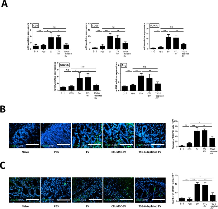
The FOXP3, CD4, CD25, CD206 and Arg mRNA levels were evaluated to examine whether stem cell EVs affected the activation of Tregs and polarization of macrophage in the inflamed colon. FOXP3 (6.8 fold), CD4 (3.0 fold), CD25 (4.0 fold), CD206 (3.0 fold) and Arg (2.7 fold) mRNA levels increased in the EV group compared to the PBS group. However, their levels significantly decreased in the TSG-6-depleted EV group compared to the EV and CTL-EV group (Fig 6A). To determine whether the increase in the number of Tregs and M2 was associated with TSG-6 in the EVs, quantitative analysis of FOXP3+ cells and CD206+ cells detected in colon tissue sections by immunofluorescence showed that the percentage of FOXP+ cells (4.76 fold) and (7.61 fold) increased significantly in the cASC-EV group compared to that in the PBS group. However, the enhancement of the number of FOXP3+ cells (0.54 fold) and CD206+ cells (0.35 fold) in the EVs of the colon tissue decreased when TSG-6 was inhibited (Fig 6B).

Fig 6. TSG-6 increases regulatory T cells in the inflamed colon.

Fig 6

TSG-6 in EVs increased the proportion of Tregs in the inflamed colon. (A) Relative gene-expression levels of CD4, CD25, FOXP3, CD206 and Arg in the inflamed colon. (B) FOXP3+ (green) cells (C) CD206+ (gren)cells were detected in colon tissue sections by immunofluorescence. The data shown demonstrated that TSG-6 in EVs played a major role in increasing the number of Tregs and M2 in the colon. Scale bar, 50 μm. The results are shown as the mean ± standard deviation (n = 6–8 in each group, *P < 0.05, ****P < 0.0001, as determined by one-way ANOVA).

Discussion

Our data suggests the following important points: (1) TSG-6 in the stem cell derived EVs is a key factor in immune regulation and relieving inflammation in the DSS-induced mouse model of colitis. (2) TSG-6 in EVs alleviates inflammation by enhancing colonic Tregs and polarizing colonic macrophage from M1 and M2 in an IBD mouse model.

Previous studies have shown that stem cells affect recipient cells in a paracrine manner, considering that EVs largely account for the paracrine effect of stem cells [3, 4, 30]. In our, preliminary study, the immune regulatory capacity of stem cells was significantly decreased when GW4869 (a noncompetitive neutral sphingomyelinase (N-SMase) inhibitor; exosome inhibitor) was applied to cASCs (S3 Fig). Moreover, EVs and stem cells have similar immunomodulatory effects (S4 Fig).

In addition, with this tendency, various studies have been conducted since the introduction of EV as a therapeutic agent [3, 31]. However, the exact mechanism by which they relieve colitis has not been revealed. Therefore, it is noteworthy that this study demonstrated that TSG-6 in EVs is a major factor in relieving colitis symptoms.

Previous studies have shown that immune cells play a role in controlling inflammation in the colon [32]. Among these immune cells, Tregs have been described as having important roles in regulating the pathogenesis of IBD [33, 34] and the balance between Tregs and other T cells in the intestinal tract is known to influence IBD pathogenesis [35]. To a large extent, FOXP3 amplifies and stabilizes the molecular features of Treg precursor cells, which is beneficial for their function and maintenance, and attenuates features that are deleterious to Treg functions [36]. FOXP3-expressing Tregs, which belong to a suppressive subset of CD4+ T cells, can regulate infection, tumor development, allergy, and autoimmunity [37]. It was reported that FOXP3+ Tregs are lower in patients with IBD progression than in healthy controls. It was also reported that an increase in the number of Tregs after treating IBD patients correlated with relief of IBD symptoms [35]. In other words, increasing the number of Tregs may serve as a method for treating IBD.

Colon macrophages are essential for maintaining mucosal homeostasis for the ongoing need for epithelial regeneration but are also an important component of protective immunity and are involved in the pathology of IBD [38]. M1 preferentially metabolize arginine to nitric oxide via inducible nitric oxide synthase (iNOS; NOS2), while M2 preferentially metabolize arginine to ornithine via arginase -1. Therefore, it is known that M1 are involved in pre-inflammatory and M2 are involved in anti-inflammatory [39]. The importance of macrophages in maintaining immune homeostasis has shown that IL-10 secreted from M2 acts on Treg to maintain Foxp3 expression [40]. These Foxp3-expressed Tregs relieved inflammation by suppressing the activity of Th1 and Th17 cells [41]. Therefore, increasing M2 in the colon is noted as a way to alleviate colitis [21]. Accumulated evidence shows that infusion of EVs into colitis models relieves inflammation [42], but the relationship between colonic immune cells and EVs is not clear. Therefore, our study is valuable in that it clarifies the relationship between TSG-6 in EV and colonic immune cells such as Tregs and macrophages.

Although proteins other than TSG-6, such as TGF-beta, IDO and PGE2 may also be contributing to the protective effect of EVs in relieving inflammation [43]. Previous studies have reported that TGF-β plays a role in inhibiting activated immunity by inducing FoxP3+ regulatory t cells in an inflammatory environment and has been shown to play an important role in relieving inflammation in colitis models [44]. Zhang et al. reported that TGF-beta in bone marrow derived stem cells plays a major role in polarizing macrophage from M1 to M2 [45]. IDO expression and activity is an important mediator of intestinal homeostasis both in health and disease [46]. In addition, IDO appears to be the most promising candidate, which plays an important role in the immunomodulatory effects of stem cells by inhibiting T cell activation and enhancing Tregs [47]. Also, IDO has been shown to play a major role in suppressing immunity by polarizing macrophage from M1 to M2.[48]. Furthermore, IDO as an anti-inflammatory agent has been reported to reduce inflammation in colitis models [49]. In addition, in our previous study, we confirmed the efficacy of adipose-derived stem cells in murine-derived macrophage cell lines in inflammatory environments and demonstrated that PGE2 secreted from stem cells is a key factor in polarizing macrophage [50, 51]. Moreover, we previous showed that PGE2 secreted feline ASC is a key factor for enhancing regulatory T cell in inflamed colon [52]

Although further research on the correlation between these immunomodulatory factors of EV and immune cell regulation is needed, results of the current study confirm that TSG-6-depleted EVs significantly reduce the immunoregulatory ability, which clearly indicates that TSG-6 is a major factor in immune regulation and anti-inflammatory action. Furthermore, the finding that TSG-6 in EVs plays an important role in immune regulation will serve as evidence to support increasing the level of TSG-6 in EVs as a strategy to develop EVs with enhanced immunomodulating properties.

Although EV-specific studies have not been conducted, other studies have shown that pretreatment of stem cells with TNF-alpha (TNF-α) resulted in an increase in mRNA levels of TSG-6 in stem cells as well as increased levels of TSG-6 protein in the culture medium [21, 53]. Further studies on pre-treated stem cell-derived EVs are needed.

Nuclear transcription factor kappaB (NF-κB) is a central mediator of pro-inflammatory gene induction and function in immune cells and has a significant effect on mucosal inflammatory process [54]. Moreover, in IBD patients, its activation is markedly induced. Therefore, the NF-κB pathway is considered to be an attractive target of therapeutic intervention in IBD [55]. In our previous study, TSG-6 from stem cells significantly suppressed nuclear factor kappa B (NF-κB) activity and alleviated inflammation and reduced apoptosis in acute pancreatitis model [20]. However, the relationship between EV and NF-kB has not been studied in this study, and it is necessary to confirm whether EV's TSG-6 lowers NF-kB in inflammatory colon.

This study is also an important basis for future transitional studies. Like human IBD, canine idiopathic IBD is a commonly observed chronic IBD that occurs spontaneously with similar multifocal etiology due to the interactions between abnormal host immune responses, and genetic and environmental factors. Histological evaluation of intestinal biopsies reveals extensive or multifocal inflammatory cell infiltration (most commonly lymphoid evolutive, eosinophilic, and neutrophilic), with simultaneous changes in mucosal structures (e.g. villous atrophy and fusion). In severe cases, intestinal protein loss, similar to in human disease, can be observed [1, 56, 57]. This study has been carried out with EVs derived from canine cells and is of great value in facilitating subsequent experiments in dogs. Therefore, evaluating the efficacy of TSG-6 in EVs conducted in this study is valuable for applications in veterinary medicine, particularly for intractable immune-mediated diseases such as IBD, however, much of these finds may also be applicable to human IBD in the future.

Conclusion

We demonstrated that TSG-6 in EVs secreted from cASCs ameliorated DSS-induced colitis in mice by enhancing the Treg population and polarizing macrophage from M1 to M2 in the inflamed colon. Our findings provide an insight to improve the current understanding of the role that EVs have in immunoregulation and serve as a foundation for applying EVs as a therapeutic agent in IBD. Also, this study is the basis of a strategy for developing EVs with improved immunomodulatory properties by increasing TSG-6 levels in EVs.

Supporting information

S1 Fig. Identification of cASCs and si TSG-6 cASC.

(A) Immunophenotypes of the cultured cASCs were examined by flow cytometry. The vast majority of cells were positive for CD90, CD44, CD29, and CD73, but a few cells expressed CD34 and CD45. (B) The naïve and si TSG-6 cASCs were maintained in specific differentiation media for 3 weeks, and the differentiated cells were stained by Oil red O to identify adipocytes, Alizarin Red S for osteocytes and Alcian Blue for chondrocytes. Scale bars, 200 μm. (C) Cell-viability assays of naïve and siTSG6-cASCs. si TSG-6 transfection was not cytotoxic when applied to stem cells. (n = 6 in each group) (D) Morphology of EVs from siTSG6-cASCs, as studied by transmission electron microscopy. EV was identified as a circular particle with a diameter of less than 100 nm. (E) EV production by naïve and siTSG6-cASCs. The production of exosome does not differ between naive and siTSG-6 groups. (n = 6 in each group) The results are shown as the mean ± standard deviation (ns, not significant, were analyzed using Student’s t-tests)

(TIF)

S2 Fig. Production of TSG-6 depleted EV.

(A) TSG-6 mRNA-expression levels in naïve cASCs, cASCs transfected with a scrambled siRNA (CTL-cASC), or cASCs transfected with TSG-6 (siTSG-6-cASC) was determined by agarose gel electrophoresis and RT-qPCR. (Lane 1 and 2: Naïve, Lane 3 and 4: CTL-cASC, Lane 5 and 6: si TSG-6 cASC in gel PCR) (B) TSG-6 protein-expression levels in naïve cASC-EVs, EVs from cASCs transfected with a scrambled siRNA (CTL-EV), or EVs from cASCs transfected with TSG-6 (TSG-6 depleted-EV) were determined by western blot analysis. The results are presented as the mean ± standard deviation. (n = 6 in each group) (ns = Not Statistically Significant *P < 0.05, **P < 0.01, ***P < 0.001 by one-way ANOVA analysis).

(TIF)

S3 Fig. Immunological biomarkers observed upon co-culturing total lymphocytes with cASCs.

(A) Treatment with 0.005% DMSO, 10 μM, 20 μM GW4869, or 1% DMSO showed no cytotoxic effects on cASCs, as shown by similar viability rates following all treatments, compared to the non-treated group (n = 6 in each group) (B) Pre-treatment with GW4869(10 μM, for 12h) significantly reduced production of EV proteins by cASCs. EV production was reduced by more than 70% in the GW4869-treated group (n = 6 in each group ) (* p< 0.05, were analyzed using Student’s t-tests)(C) The mRNA levels of TNA-α, IL-1β, IL-6, IFN-γ, and IL-10 were detected by qRT-PCR. Con A-treated lymphocytes showed significantly increased levels of pro-inflammatory cytokines, such as TNF-α, IFN-γ, IL-1β, and IL-6, compared to the untreated group. cASCs depressed activated lymphocyte. however, pre-treatment with GW4869 significantly reduced the modulatory effects of cASCs. (n = 6 in each group)The results are presented as the mean ± standard deviation (**P < 0.01, ***P < 0.001, ****P < 0.0001 as determined by one-way ANOVA).

(TIF)

S4 Fig. Immunomodulatory effects of cASC-EVs.

(A) Changes in the expression levels of mRNAs encoding several canine lymphocyte-derived cytokines including TNF-α, IL-1β, IFN-γ, IL-6, and IL-10 in the presence of cASC-Evs (100ug/well). After Con A-stimulated lymphocytes were cocultured with EV (100 ug), the levels of activated pro-inflammatory cytokines (TNF-α, IFN-γ, IL-1β, and IL-6) decreased significantly. Production of the anti-inflammatory cytokine IL-10 significantly increased, compared to that in the untreated group. (B) Changes in the expression levels of mRNAs encoding several canine macrohage-derived cytokines including TNF-α, IL-6, iNOS and IL-10 in the presence of cASC-Evs (100ug/well). After LPS-stimulated DH82 were cocultured with EV (100 ug), the levels of activated pro-inflammatory cytokines (TNF-α, IL-6 and iNOS) decreased significantly. Production of the anti-inflammatory cytokine IL-10 significantly increased, compared to that in the untreated group. The data show that EVs exerted immunosuppressive effects as much as stem cells. The results are presented as the mean ± standard deviation (n = 6 in each group), **P < 0.01, ***P < 0.001, ****P < 0.0001, as determined by one-way ANOVA).

(TIF)

S5 Fig. TSG-6 in EV enhance regulatory T cells and regulate the M1/M2 balance in vitro.

TSG-6 in EV plays an important role in the increase of regulatory t cells and macrophage polarization. (A) Tregs (FOXP3+CD3+ cells) level in canine lymphocytes (B) M1 (CD11+cells) and M2 macrophages (CD206+ cells) level in canine macrophage cell line (DH82). FACS plots (right panel) show representative examples and bar graphs (left panel) represent mean values +SD (ns = Not Statistically Significant *P < 0.05, **P < 0.01, ***P < 0.001 by one-way ANOVA analysis)

(TIF)

S1 Raw Images. protein marker of cASC derived EV were analysis by western blot.

The original underlying images of CD63, CD9, Lamin A and β-actin in Fig 1D.

(TIF)

S1 File. Isolation, Culture and Characterization of cASCs.

(DOCX)

Abbreviations

ANOVA

Analysis of variance

BCA

Bicinchoninic acid

cASC

Canine adipose tissue-derived mesenchymal stem/stromal cell

CD

Cluster of differentiation

Con A

Concanavalin A

CTL

Control

cPBMC

Canine peripheral blood mononuclear cell

DAI

Disease-activity index

DMEM

Dulbecco's modified Eagle's medium

DMSO

Dimethyl sulfoxide

DPBS

Dulbecco’s phosphate-buffered saline

DSS

Dextran sulfate sodium

ELISA

Enzyme-linked immunosorbent assay

EV

Extracellular vesicle

FBS

Fetal bovine serum

FITC

Fluorescein isothiocyanate

FOXP3

Forkhead box P3

GAPDH

Glyceraldehyde 3-phosphate dehydrogenase

H&E

Hematoxylin and eosin

IBD

Inflammatory bowel disease

IFN

Interferon

IL

Interleukin

IP

Intraperitoneal

LPS

Lipopolysaccharides

M1

Macrophage M1 type

M2

Macrophage M2 type

NO

Nitric oxide

PBMC

Peripheral blood mononuclear cell

PBS

Phosphate-buffered saline

PE

Phycoerythrin

RBC

Red blood cell

RT-qPCR

Reverse transcription quantitative polymerase chain reaction

siRNA

Small-interfering RNA

siTSG-6

Small-interfering TSG-6

TEM

Transmission Electron Microscope

TGF-β

Transforming growth factor-β

TNF-α

Tumor necrosis factor-α

Treg

Regulatory T cell

TSG-6

Tumor necrosis factor-α-stimulated gene/protein-6

Data Availability

All relevant data are within the manuscript and its Supporting Information files.

Funding Statement

This study received support from the Research Institute for Veterinary Science, Seoul National University and Basic Science Research Program of the National Research Foundation of Korea. Author YCJ receives support in the form of salary from the commercial company the Chaon Corporation. The funders did not have any additional role in the study design, data collection and analysis, decision to publish, or preparation of the manuscript. The specific roles of YCJ are articulated in the ‘author contributions’ section.

References

  • 1.Cerquetella M, Spaterna A, Laus F, Tesei B, Rossi G, Antonelli E, et al. Inflammatory bowel disease in the dog: differences and similarities with humans. World journal of gastroenterology: WJG. 2010;16(9):1050 10.3748/wjg.v16.i9.1050 [DOI] [PMC free article] [PubMed] [Google Scholar]
  • 2.Yi Z, Stunz LL, Bishop GA. CD40-mediated maintenance of immune homeostasis in the adipose tissue microenvironment. Diabetes. 2014:DB_131657. [DOI] [PMC free article] [PubMed] [Google Scholar]
  • 3.Katsuda T, Kosaka N, Takeshita F, Ochiya T. The therapeutic potential of mesenchymal stem cell‐derived extracellular vesicles. Proteomics. 2013;13(10–11):1637–53. 10.1002/pmic.201200373 [DOI] [PubMed] [Google Scholar]
  • 4.Burrello J, Monticone S, Gai C, Gomez Y, Kholia S, Camussi G. Stem cell-derived extracellular vesicles and immune-modulation. Frontiers in cell and developmental biology. 2016;4:83 10.3389/fcell.2016.00083 [DOI] [PMC free article] [PubMed] [Google Scholar]
  • 5.Hartjes TA, Mytnyk S, Jenster GW, van Steijn V, van Royen ME. Extracellular Vesicle Quantification and Characterization: Common Methods and Emerging Approaches. Bioengineering. 2019;6(1):7. [DOI] [PMC free article] [PubMed] [Google Scholar]
  • 6.Qian X, Xu C, Fang S, Zhao P, Wang Y, Liu H, et al. Exosomal microRNAs derived from umbilical mesenchymal stem cells inhibit hepatitis C virus infection. Stem Cells Translational Medicine. 2016;5(9):1190–203. 10.5966/sctm.2015-0348 [DOI] [PMC free article] [PubMed] [Google Scholar]
  • 7.Bai L, Shao H, Wang H, Zhang Z, Su C, Dong L, et al. Effects of mesenchymal stem cell-derived exosomes on experimental autoimmune uveitis. Scientific reports. 2017;7(1):4323 10.1038/s41598-017-04559-y [DOI] [PMC free article] [PubMed] [Google Scholar]
  • 8.Bruno S, Grange C, Collino F, Deregibus MC, Cantaluppi V, Biancone L, et al. Microvesicles derived from mesenchymal stem cells enhance survival in a lethal model of acute kidney injury. PloS one. 2012;7(3):e33115 10.1371/journal.pone.0033115 [DOI] [PMC free article] [PubMed] [Google Scholar]
  • 9.Mao F, Wu Y, Tang X, Kang J, Zhang B, Yan Y, et al. Exosomes derived from human umbilical cord mesenchymal stem cells relieve inflammatory bowel disease in mice. BioMed research international. 2017;2017. [DOI] [PMC free article] [PubMed] [Google Scholar]
  • 10.Lee RH, Yu JM, Foskett AM, Peltier G, Reneau JC, Bazhanov N, et al. TSG-6 as a biomarker to predict efficacy of human mesenchymal stem/progenitor cells (hMSCs) in modulating sterile inflammation in vivo. Proceedings of the National Academy of Sciences. 2014;111(47):16766–71. [DOI] [PMC free article] [PubMed] [Google Scholar]
  • 11.Song W-J, Li Q, Ryu M-O, Ahn J-O, Bhang DH, Jung YC, et al. TSG-6 secreted by human adipose tissue-derived mesenchymal stem cells ameliorates DSS-induced colitis by inducing M2 macrophage polarization in mice. Scientific Reports. 2017;7(1):5187 10.1038/s41598-017-04766-7 [DOI] [PMC free article] [PubMed] [Google Scholar]
  • 12.Wang N, Li Q, Zhang L, Lin H, Hu J, Li D, et al. Mesenchymal stem cells attenuate peritoneal injury through secretion of TSG-6. PloS one. 2012;7(8):e43768 10.1371/journal.pone.0043768 [DOI] [PMC free article] [PubMed] [Google Scholar]
  • 13.Milner CM, Day AJ. TSG-6: a multifunctional protein associated with inflammation. Journal of cell science. 2003;116(10):1863–73. [DOI] [PubMed] [Google Scholar]
  • 14.Arseneau KO, Tamagawa H, Pizarro TT, Cominelli F. Innate and adaptive immune responses related to IBD pathogenesis. Current gastroenterology reports. 2007;9(6):508–12. 10.1007/s11894-007-0067-3 [DOI] [PubMed] [Google Scholar]
  • 15.Sartor RB. Pathogenesis and immune mechanisms of chronic inflammatory bowel diseases. American Journal of Gastroenterology. 1997;92. [PubMed] [Google Scholar]
  • 16.Banz A, Peixoto A, Pontoux C, Cordier C, Rocha B, Papiernik M. A unique subpopulation of CD4+ regulatory T cells controls wasting disease, IL‐10 secretion and T cell homeostasis. European journal of immunology. 2003;33(9):2419–28. 10.1002/eji.200324205 [DOI] [PubMed] [Google Scholar]
  • 17.Mowat AM, Bain CC. Mucosal macrophages in intestinal homeostasis and inflammation. Journal of innate immunity. 2011;3(6):550–64. 10.1159/000329099 [DOI] [PMC free article] [PubMed] [Google Scholar]
  • 18.Atri C, Guerfali FZ, Laouini D. Role of human macrophage polarization in inflammation during infectious diseases. International journal of molecular sciences. 2018;19(6):1801. [DOI] [PMC free article] [PubMed] [Google Scholar]
  • 19.Liu H, Liang Z, Wang F, Zhou C, Zheng X, Hu T, et al. Exosomes from Mesenchymal Stromal Cells Reduce Murine Colonic Inflammation via a Macrophage-Dependent Mechanism. Available at SSRN 3408050. 2019. [DOI] [PMC free article] [PubMed] [Google Scholar]
  • 20.Li Q, Song W-J, Ryu M-O, Nam A, An J-H, Ahn J-O, et al. TSG-6 secreted by human adipose tissue-derived mesenchymal stem cells ameliorates severe acute pancreatitis via ER stress downregulation in mice. Stem cell research & therapy. 2018;9(1):255. [DOI] [PMC free article] [PubMed] [Google Scholar]
  • 21.Song W-J, Li Q, Ryu M-O, Ahn J-O, Bhang DH, Jung YC, et al. TSG-6 released from intraperitoneally injected canine adipose tissue-derived mesenchymal stem cells ameliorate inflammatory bowel disease by inducing M2 macrophage switch in mice. Stem cell research & therapy. 2018;9(1):91. [DOI] [PMC free article] [PubMed] [Google Scholar]
  • 22.Schmitz S, Garden OA, Werling D, Allenspach K. Gene expression of selected signature cytokines of T cell subsets in duodenal tissues of dogs with and without inflammatory bowel disease. Veterinary immunology and immunopathology. 2012;146(1):87–91. 10.1016/j.vetimm.2012.01.013 [DOI] [PubMed] [Google Scholar]
  • 23.Manning K, Rachakonda PS, Rai MF, Schmidt MF. Co-expression of insulin-like growth factor-1 and interleukin-4 in an in vitro inflammatory model. Cytokine. 2010;50(3):297–305. 10.1016/j.cyto.2010.01.010 [DOI] [PubMed] [Google Scholar]
  • 24.Lee K, Cha S-H, Kang H, Song J-Y, Lee K, Ko K, et al. Effects of serial passage on the characteristics and chondrogenic differentiation of canine umbilical cord matrix derived mesenchymal stem cells. Asian-Australasian journal of animal sciences. 2013;26(4):588 10.5713/ajas.2012.12488 [DOI] [PMC free article] [PubMed] [Google Scholar]
  • 25.Biller B, Elmslie R, Burnett R, Avery A, Dow S. Use of FoxP3 expression to identify regulatory T cells in healthy dogs and dogs with cancer. Veterinary immunology and immunopathology. 2007;116(1–2):69–78. 10.1016/j.vetimm.2006.12.002 [DOI] [PubMed] [Google Scholar]
  • 26.Jo H, Eom YW, Kim H-S, Park HJ, Kim HM, Cho M-Y. Regulatory dendritic cells induced by mesenchymal stem cells ameliorate dextran sodium sulfate-induced chronic colitis in mice. Gut and liver. 2018;12(6):664 10.5009/gnl18072 [DOI] [PMC free article] [PubMed] [Google Scholar]
  • 27.Maloney J, Keselman A, Li E, Singer SM. Macrophages expressing arginase 1 and nitric oxide synthase 2 accumulate in the small intestine during Giardia lamblia infection. Microbes and infection. 2015;17(6):462–7. 10.1016/j.micinf.2015.03.006 [DOI] [PMC free article] [PubMed] [Google Scholar]
  • 28.Bruno S, Deregibus MC, Camussi G. The secretome of mesenchymal stromal cells: role of extracellular vesicles in immunomodulation. Immunology letters. 2015;168(2):154–8. 10.1016/j.imlet.2015.06.007 [DOI] [PubMed] [Google Scholar]
  • 29.Monk JM, Richard CL, Woodward B. A non-inflammatory form of immune competence prevails in acute pre-pubescent malnutrition: new evidence based on critical mRNA transcripts in the mouse. British Journal of Nutrition. 2012;107(9):1249–53. 10.1017/S0007114511004399 [DOI] [PubMed] [Google Scholar]
  • 30.Biancone L, Bruno S, Deregibus MC, Tetta C, Camussi G. Therapeutic potential of mesenchymal stem cell-derived microvesicles. Nephrology Dialysis Transplantation. 2012;27(8):3037–42. [DOI] [PubMed] [Google Scholar]
  • 31.Zhang B, Yin Y, Lai RC, Tan SS, Choo ABH, Lim SK. Mesenchymal stem cells secrete immunologically active exosomes. Stem cells and development. 2013;23(11):1233–44. [DOI] [PubMed] [Google Scholar]
  • 32.Cook MD, Allen JM, Pence BD, Wallig MA, Gaskins HR, White BA, et al. Exercise and gut immune function: evidence of alterations in colon immune cell homeostasis and microbiome characteristics with exercise training. Immunology and cell biology. 2016;94(2):158–63. 10.1038/icb.2015.108 [DOI] [PubMed] [Google Scholar]
  • 33.Asseman C, Mauze S, Leach MW, Coffman RL, Powrie F. An essential role for interleukin 10 in the function of regulatory T cells that inhibit intestinal inflammation. Journal of Experimental Medicine. 1999;190(7):995–1004. 10.1084/jem.190.7.995 [DOI] [PMC free article] [PubMed] [Google Scholar]
  • 34.Round JL, Mazmanian SK. Inducible Foxp3+ regulatory T-cell development by a commensal bacterium of the intestinal microbiota. Proceedings of the National Academy of Sciences. 2010;107(27):12204–9. [DOI] [PMC free article] [PubMed] [Google Scholar]
  • 35.Yamada A, Arakaki R, Saito M, Tsunematsu T, Kudo Y, Ishimaru N. Role of regulatory T cell in the pathogenesis of inflammatory bowel disease. World Journal of Gastroenterology. 2016;22(7):2195 10.3748/wjg.v22.i7.2195 [DOI] [PMC free article] [PubMed] [Google Scholar]
  • 36.Tang Q, Bluestone JA. The Foxp3+ regulatory T cell: a jack of all trades, master of regulation. Nature immunology. 2008;9(3):239 10.1038/ni1572 [DOI] [PMC free article] [PubMed] [Google Scholar]
  • 37.Sakaguchi S, Ono M, Setoguchi R, Yagi H, Hori S, Fehervari Z, et al. Foxp3+ CD25+ CD4+ natural regulatory T cells in dominant self‐tolerance and autoimmune disease. Immunological reviews. 2006;212(1):8–27. [DOI] [PubMed] [Google Scholar]
  • 38.Bain CC, Mowat AM. Macrophages in intestinal homeostasis and inflammation. Immunological reviews. 2014;260(1):102–17. 10.1111/imr.12192 [DOI] [PMC free article] [PubMed] [Google Scholar]
  • 39.Isidro RA, Appleyard CB. Colonic macrophage polarization in homeostasis, inflammation, and cancer. American Journal of Physiology-Gastrointestinal and Liver Physiology. 2016;311(1):G59–G73. 10.1152/ajpgi.00123.2016 [DOI] [PMC free article] [PubMed] [Google Scholar]
  • 40.Murai M, Turovskaya O, Kim G, Madan R, Karp CL, Cheroutre H, et al. Interleukin 10 acts on regulatory T cells to maintain expression of the transcription factor Foxp3 and suppressive function in mice with colitis. Nature immunology. 2009;10(11):1178 10.1038/ni.1791 [DOI] [PMC free article] [PubMed] [Google Scholar]
  • 41.Xavier R, Podolsky D. Unravelling the pathogenesis of inflammatory bowel disease. Nature. 2007;448(7152):427 10.1038/nature06005 [DOI] [PubMed] [Google Scholar]
  • 42.Wu Y, Qiu W, Xu X, Kang J, Wang J, Wen Y, et al. Exosomes derived from human umbilical cord mesenchymal stem cells alleviate inflammatory bowel disease in mice through ubiquitination. American journal of translational research. 2018;10(7):2026 [PMC free article] [PubMed] [Google Scholar]
  • 43.Soleymaninejadian E, Pramanik K, Samadian E. Immunomodulatory properties of mesenchymal stem cells: cytokines and factors. American journal of reproductive immunology. 2012;67(1):1–8. 10.1111/j.1600-0897.2011.01069.x [DOI] [PubMed] [Google Scholar]
  • 44.Becker C, Fantini MC, Neurath MF. TGF-beta as a T cell regulator in colitis and colon cancer. Cytokine & growth factor reviews. 2006;17(1–2):97–106. [DOI] [PubMed] [Google Scholar]
  • 45.Zhang F, Wang H, Wang X, Jiang G, Liu H, Zhang G, et al. TGF-β induces M2-like macrophage polarization via SNAIL-mediated suppression of a pro-inflammatory phenotype. Oncotarget. 2016;7(32):52294 10.18632/oncotarget.10561 [DOI] [PMC free article] [PubMed] [Google Scholar]
  • 46.Ciorba MA. Indoleamine 2, 3 dioxygenase (IDO) in intestinal disease. Current opinion in gastroenterology. 2013;29(2):146 10.1097/MOG.0b013e32835c9cb3 [DOI] [PMC free article] [PubMed] [Google Scholar]
  • 47.Yan Y, Zhang G-X, Gran B, Fallarino F, Yu S, Li H, et al. IDO upregulates regulatory T cells via tryptophan catabolite and suppresses encephalitogenic T cell responses in experimental autoimmune encephalomyelitis. The Journal of Immunology. 2010;185(10):5953–61. 10.4049/jimmunol.1001628 [DOI] [PMC free article] [PubMed] [Google Scholar]
  • 48.Wang X-F, Wang H-S, Wang H, Zhang F, Wang K-F, Guo Q, et al. The role of indoleamine 2, 3-dioxygenase (IDO) in immune tolerance: focus on macrophage polarization of THP-1 cells. Cellular immunology. 2014;289(1–2):42–8. 10.1016/j.cellimm.2014.02.005 [DOI] [PubMed] [Google Scholar]
  • 49.Coquerelle C, Oldenhove G, Acolty V, Denoeud J, Vansanten G, Verdebout J-M, et al. Anti-CTLA-4 treatment induces IL-10-producing ICOS+ regulatory T cells displaying IDO-dependent anti-inflammatory properties in a mouse model of colitis. Gut. 2009;58(10):1363–73. 10.1136/gut.2008.162842 [DOI] [PMC free article] [PubMed] [Google Scholar]
  • 50.Yang H-M, Song W-J, Li Q, Kim S-Y, Kim H-J, Ryu M-O, et al. Canine mesenchymal stem cells treated with TNF-α and IFN-γ enhance anti-inflammatory effects through the COX-2/PGE2 pathway. Research in veterinary science. 2018;119:19–26. 10.1016/j.rvsc.2018.05.011 [DOI] [PubMed] [Google Scholar]
  • 51.Chae H-K, Song W-J, Ahn J-O, Li Q, Lee B-Y, Kweon K, et al. Immunomodulatory effects of soluble factors secreted by feline adipose tissue-derived mesenchymal stem cells. Veterinary immunology and immunopathology. 2017;191:22–9. 10.1016/j.vetimm.2017.07.013 [DOI] [PubMed] [Google Scholar]
  • 52.AN J-H, Woo-Jin S, Qiang L, Sang-Min K, Ji-In Y, Min-Ok R, et al. Prostaglandin E 2 secreted from feline adipose tissue-derived mesenchymal stem cells alleviate DSS-induced colitis by increasing regulatory T cells in mice. BMC veterinary research. 2018;14(1):354 10.1186/s12917-018-1684-9 [DOI] [PMC free article] [PubMed] [Google Scholar]
  • 53.Xiao Q, Wang S-k, Tian H, Xin L, Zou Z-g, Hu Y-l, et al. TNF-α increases bone marrow mesenchymal stem cell migration to ischemic tissues. Cell biochemistry and biophysics. 2012;62(3):409–14. 10.1007/s12013-011-9317-y [DOI] [PubMed] [Google Scholar]
  • 54.Liu T, Zhang L, Joo D, Sun S-C. NF-κB signaling in inflammation. Signal transduction and targeted therapy. 2017;2:17023 10.1038/sigtrans.2017.23 [DOI] [PMC free article] [PubMed] [Google Scholar]
  • 55.Atreya I, Atreya R, Neurath M. NF‐κB in inflammatory bowel disease. Journal of internal medicine. 2008;263(6):591–6. 10.1111/j.1365-2796.2008.01953.x [DOI] [PubMed] [Google Scholar]
  • 56.Coelho LP, Kultima JR, Costea PI, Fournier C, Pan Y, Czarnecki-Maulden G, et al. Similarity of the dog and human gut microbiomes in gene content and response to diet. Microbiome. 2018;6(1):72 10.1186/s40168-018-0450-3 [DOI] [PMC free article] [PubMed] [Google Scholar]
  • 57.Vázquez-Baeza Y, Hyde ER, Suchodolski JS, Knight R. Dog and human inflammatory bowel disease rely on overlapping yet distinct dysbiosis networks. Nature microbiology. 2016;1(12):16177. [DOI] [PubMed] [Google Scholar]

Decision Letter 0

Hossam MM Arafa

4 Nov 2019

PONE-D-19-20533

TSG-6 in extracellular vesicles from canine mesenchymal stem/stromal is a major factor in relieving DSS-induced colitis

PLOS ONE

Dear Dr. Youn,

Thank you for submitting your manuscript to PLOS ONE. Your submission has been thoroughly peer-reviewed by two independent referees. After careful consideration, we feel that it has merit but does not fully meet PLOS ONE’s publication criteria as it currently stands. Therefore, we invite you to submit a revised version of the manuscript that addresses the points raised during the review process.

Please respond ASAP to the queries raised by reviewers and given hereunder. Besides, the following shortcomings should be addressed when revising the manuscript:

1- One should specify the animal sample size for each group enrolled in the study (Line 6; "the mice were randomly divided into 6 groups with 6-8 mice per group"- misleading and not accepted).

2- In the statistical analysis section; Line 14, the criterion for least significance (p < 0.05) should be narrated. The individual t-tests, in contrast to multiple comparison post-hoc tests, can estimate the variances only from 2 of the 6 groups, which is less precise and less reliable. Since the variance is estimated from the whole set of data (6 groups) as pooled estimate, such estimate is much more robust and precise than that estimated from just a part of the whole set of data. So, authors ought to use less conservative and more accurate post-hoc test such as Tukey or Bonferroni. As regards the histological scoring system, did the authors perform any non-parametric multiple comparison tests in similar analogy to ANOVA? if so- which test did they perform?

3- In results, one should not discuss his findings just narrate them. Hence, the following statements should be omitted and transferred to the discussion section:

- Lines 14-16/page 15; "Collectively, these results indicate that EV 14 administration alleviated DSS-induced colitis and indicate that TSG-6 in EV played a role in relieving colitis".

- Last paragraph in page 15; "These results indicate that EV markedly attenuated the inflammatory state in mice with induced colitis and suggest that TSG-6 in EV played an important role in relieving inflammatory conditions".

3- Lines 4-9/page 16; there is no room in results for such verbosity "TSG-6 was a major factor in increasing Tregs in Con A stimulated lymphocytes In vitro.The expression levels of CD4, and CD25 mRNA in lymphocytes derived canine PBMCs were assessed to determine whether TSG-6 in EVs affected Treg activation. Con A, a mannose/glucose-binding lectin, is a well-known T cell mitogen that can activate the immune system, recruit lymphocytes, and elicit cytokine production. Therefore, lymphocytes were stimulated with Con A to confirm the immune cell-control function of EVs". Authors either should omit it or incorporate in the introduction section.

4- Lines 1-2/page 17;  the sentence reads "FOXP3, CD4, and CD25 mRNA levels increased in the EV group. However, their levels decreased in the TSG-6-depleted EV group". Authors should specify the percentage changes whether steered up or down. 

5- Lines 5-7/page 17; the same holds true for the sentence  "the percentage of these cells increased significantly in the cAT-MSC-EV group compared to that in the PBS group. However, the enhancement of the number of FOXP3+ cells in the EVs of the colon tissue decreased when TSG-6 was inhibited". One should exactly clarify the percentage increase or decrease.

==============================

We would appreciate receiving your revised manuscript by 30 days. When you are ready to submit your revision, log on to https://www.editorialmanager.com/pone/ and select the 'Submissions Needing Revision' folder to locate your manuscript file.

If you would like to make changes to your financial disclosure, please include your updated statement in your cover letter.

To enhance the reproducibility of your results, we recommend that if applicable you deposit your laboratory protocols in protocols.io, where a protocol can be assigned its own identifier (DOI) such that it can be cited independently in the future. For instructions see: http://journals.plos.org/plosone/s/submission-guidelines#loc-laboratory-protocols

Please include the following items when submitting your revised manuscript:

  • A rebuttal letter that responds to each point raised by the academic editor and reviewer(s). This letter should be uploaded as separate file and labeled 'Response to Reviewers'.

  • A marked-up copy of your manuscript that highlights changes made to the original version. This file should be uploaded as separate file and labeled 'Revised Manuscript with Track Changes'.

  • An unmarked version of your revised paper without tracked changes. This file should be uploaded as separate file and labeled 'Manuscript'.

Please note while forming your response, if your article is accepted, you may have the opportunity to make the peer review history publicly available. The record will include editor decision letters (with reviews) and your responses to reviewer comments. If eligible, we will contact you to opt in or out.

We look forward to receiving your revised manuscript.

Kind regards,

Hossam MM Arafa

Academic Editor

PLOS ONE

Journal Requirements:

1. When submitting your revision, we need you to address these additional requirements. Please ensure that your manuscript meets PLOS ONE's style requirements, including those for file naming. The PLOS ONE style templates can be found at http://www.journals.plos.org/plosone/s/file?id=wjVg/PLOSOne_formatting_sample_main_body.pdf and http://www.journals.plos.org/plosone/s/file?id=ba62/PLOSOne_formatting_sample_title_authors_affiliations.pdf

2. PLOS ONE now requires that authors provide the original uncropped and unadjusted images underlying all blot or gel results reported in a submission’s figures or Supporting Information files. This policy and the journal’s other requirements for blot/gel reporting and figure preparation are described in detail at https://journals.plos.org/plosone/s/figures#loc-blot-and-gel-reporting-requirements and https://journals.plos.org/plosone/s/figures#loc-preparing-figures-from-image-files. When you submit your revised manuscript, please ensure that your figures adhere fully to these guidelines and provide the original underlying images for all blot or gel data reported in your submission. See the following link for instructions on providing the original image data: https://journals.plos.org/plosone/s/figures#loc-original-images-for-blots-and-gels.

In your cover letter, please note whether your blot/gel image data are in Supporting Information or posted at a public data repository, provide the repository URL if relevant, and provide specific details as to which raw blot/gel images, if any, are not available. Email us at plosone@plos.org if you have any questions.

3. In your Methods section, please provide additional details regarding the mice used in your study and ensure you have described the source. For more information regarding PLOS' policy on materials sharing and reporting, see https://journals.plos.org/plosone/s/materials-and-software-sharing#loc-sharing-materials.

4.  Please include captions for your Supporting Information files at the end of your manuscript, and update any in-text citations to match accordingly. Please see our Supporting Information guidelines for more information: http://journals.plos.org/plosone/s/supporting-information.

5. We note that you have included the phrase “data not shown” in your manuscript. Unfortunately, this does not meet our data sharing requirements. PLOS does not permit references to inaccessible data. We require that authors provide all relevant data within the paper, Supporting Information files, or in an acceptable, public repository. Please add a citation to support this phrase or upload the data that corresponds with these findings to a stable repository (such as Figshare or Dryad) and provide and URLs, DOIs, or accession numbers that may be used to access these data. Or, if the data are not a core part of the research being presented in your study, we ask that you remove the phrase that refers to these data.

6. Thank you for stating the following in the Financial Disclosure section:

H.Y. YOUN

The Research Institute for Veterinary Science, Seoul National University and Basic Science Research Program of the National Research Foundation of Korea

http://vetsci.snu.ac.kr/

These funds contributed to the collection, analysis, and interpretation of data generated in this study.

We note that one or more of the authors are employed by a commercial company: Chaon Corporation

1. Please provide an amended Funding Statement declaring this commercial affiliation, as well as a statement regarding the Role of Funders in your study. If the funding organization did not play a role in the study design, data collection and analysis, decision to publish, or preparation of the manuscript and only provided financial support in the form of authors' salaries and/or research materials, please review your statements relating to the author contributions, and ensure you have specifically and accurately indicated the role(s) that these authors had in your study. You can update author roles in the Author Contributions section of the online submission form.

Please also include the following statement within your amended Funding Statement.

“The funder provided support in the form of salaries for authors [insert relevant initials], but did not have any additional role in the study design, data collection and analysis, decision to publish, or preparation of the manuscript. The specific roles of these authors are articulated in the ‘author contributions’ section.”

If your commercial affiliation did play a role in your study, please state and explain this role within your updated Funding Statement.

2. Please also provide an updated Competing Interests Statement declaring this commercial affiliation along with any other relevant declarations relating to employment, consultancy, patents, products in development, or marketed products, etc.  

Within your Competing Interests Statement, please confirm that this commercial affiliation does not alter your adherence to all PLOS ONE policies on sharing data and materials by including the following statement: "This does not alter our adherence to  PLOS ONE policies on sharing data and materials.” (as detailed online in our guide for authors http://journals.plos.org/plosone/s/competing-interests). If this adherence statement is not accurate and  there are restrictions on sharing of data and/or materials, please state these. Please note that we cannot proceed with consideration of your article until this information has been declared.

Please include both an updated Funding Statement and Competing Interests Statement in your cover letter. We will change the online submission form on your behalf.

Please know it is PLOS ONE policy for corresponding authors to declare, on behalf of all authors, all potential competing interests for the purposes of transparency. PLOS defines a competing interest as anything that interferes with, or could reasonably be perceived as interfering with, the full and objective presentation, peer review, editorial decision-making, or publication of research or non-research articles submitted to one of the journals. Competing interests can be financial or non-financial, professional, or personal. Competing interests can arise in relationship to an organization or another person. Please follow this link to our website for more details on competing interests: http://journals.plos.org/plosone/s/competing-interests

7. Your ethics statement must appear in the Methods section of your manuscript. If your ethics statement is written in any section besides the Methods, please move it to the Methods section and delete it from any other section. Please also ensure that your ethics statement is included in your manuscript, as the ethics section of your online submission will not be published alongside your manuscript.

[Note: HTML markup is below. Please do not edit.]

Reviewers' comments:

Reviewer's Responses to Questions

Comments to the Author

1. Is the manuscript technically sound, and do the data support the conclusions?

The manuscript must describe a technically sound piece of scientific research with data that supports the conclusions. Experiments must have been conducted rigorously, with appropriate controls, replication, and sample sizes. The conclusions must be drawn appropriately based on the data presented.

Reviewer #1: Yes

Reviewer #2: Partly

**********

2. Has the statistical analysis been performed appropriately and rigorously?

Reviewer #1: Yes

Reviewer #2: Yes

**********

3. Have the authors made all data underlying the findings in their manuscript fully available?

The PLOS Data policy requires authors to make all data underlying the findings described in their manuscript fully available without restriction, with rare exception (please refer to the Data Availability Statement in the manuscript PDF file). The data should be provided as part of the manuscript or its supporting information, or deposited to a public repository. For example, in addition to summary statistics, the data points behind means, medians and variance measures should be available. If there are restrictions on publicly sharing data—e.g. participant privacy or use of data from a third party—those must be specified.

Reviewer #1: Yes

Reviewer #2: Yes

**********

4. Is the manuscript presented in an intelligible fashion and written in standard English?

PLOS ONE does not copyedit accepted manuscripts, so the language in submitted articles must be clear, correct, and unambiguous. Any typographical or grammatical errors should be corrected at revision, so please note any specific errors here.

Reviewer #1: Yes

Reviewer #2: Yes

**********

5. Review Comments to the Author

Please use the space provided to explain your answers to the questions above. You may also include additional comments for the author, including concerns about dual publication, research ethics, or publication ethics. (Please upload your review as an attachment if it exceeds 20,000 characters)

Reviewer #1: I would like to thank the authors for their great efforts in conducting the study in this well organised manner.

Overall, the manuscript emphasises on using EVs as a next generation therapeutic agent as an alternative to stem cells for immune-mediated diseases owing to their similar immunomodulatory effect which partly dependent on TSG6. This is beside the notable anti inflammatory effect exerted by EVs that was also attiributed to TSG6. The results were promising and provide an important basis for future transitional studies.

However, i have some minor comments:

1-What is the cause of investigating the effect of TSC6 in EVs on Tregs only without investigating the effect on other immune cells like M2 macrophages switch? A previous study in 2017 states that hAT-MSC-produced TSG-6 can ameliorate IBD by inducing M2 macrophage switch in mice.

2-What is the cause of selecting TSG6 although proteins other than TSG-6, such as TGF-beta, IDO, PGE2, and NO, may also be contributing to the protective effect of EVs in relieving inflammation? A detailed justification may be required.

3-Increasing the level of TSG-6 in EVs as a strategy to develop EVs with enhanced immunomodulating properties is thought to be a very promising idea that needs to be addressed as a major recommendation or even to be added in the conclusion section.

Some typographical errors:

The abbreviation TEM (page 14, line 17)is not included in the abbreviations list.

Page 13, line 9; as previously described not as previous.

Page 24, line 12; immune mediated diseases not disease.

Thank you

Best regards

Reviewer #2: you write in the experimental design that is 6 groups but in some figure 5 group only

CTl , si-tsg-6 are not present in the abbreviation list and not mentioned in experimental design

what is the difference between CTL-MSc-EV and TSG 6 depleted EV. what is the significance of using these two groups

experimental design needs to clarify (page no 12)

there is very high S.D in the results such as figure 4, 5 and 6.

why you didn't use DSS 5%

why you didn't analyze the stool EV, colon length

also why you didn't measure NF-KBp65, INOs, caspase 3

is a relation between TSG 6 and TSG 14 in colitis

in reference No 9 (the paper on renal not on colon ) you need to check the paper

https://www.ncbi.nlm.nih.gov/pmc/articles/PMC3303802/

**********

6. PLOS authors have the option to publish the peer review history of their article (what does this mean?). If published, this will include your full peer review and any attached files.

If you choose “no”, your identity will remain anonymous but your review may still be made public.

Do you want your identity to be public for this peer review? For information about this choice, including consent withdrawal, please see our Privacy Policy.

Reviewer #1: No

Reviewer #2: No

[NOTE: If reviewer comments were submitted as an attachment file, they will be attached to this email and accessible via the submission site. Please log into your account, locate the manuscript record, and check for the action link "View Attachments". If this link does not appear, there are no attachment files to be viewed.]

While revising your submission, please upload your figure files to the Preflight Analysis and Conversion Engine (PACE) digital diagnostic tool, https://pacev2.apexcovantage.com/. PACE helps ensure that figures meet PLOS requirements. To use PACE, you must first register as a user. Registration is free. Then, login and navigate to the UPLOAD tab, where you will find detailed instructions on how to use the tool. If you encounter any issues or have any questions when using PACE, please email us at figures@plos.org. Please note that Supporting Information files do not need this step.

PLoS One. 2020 Feb 10;15(2):e0220756. doi: 10.1371/journal.pone.0220756.r002

Author response to Decision Letter 0


25 Nov 2019

November 15, 2019

Hossam MM Arafa

Academic Editor

PLOSONE

Dear Prof. Joerg Heber

We are very pleased to have been given the opportunity to revise our manuscript entitled “TSG-6 in extracellular vesicles from canine mesenchymal stem/stromal is a major factor in relieving DSS-induced colitis” for PLOSONE. We want to extend our appreciation to you and the reviewers for taking the time and effort necessary to provide such insightful guidance. We have carefully considered comments offered by the reviewers. Herein, we explain how we revised the paper based on those comments and recommendations. The manuscript has certainly benefited from these revision suggestions. We look forward to working further with you and the reviewers to move this manuscript closer to publication.

COMMENT 1- One should specify the animal sample size for each group enrolled in the study (Line 6; "the mice were randomly divided into 6 groups with 6-8 mice per group"- misleading and not accepted).

RESPONSE: we revised this in abstract section.

“ DSS-induced colitis mice (C57BL/6, male, Naïve n= 6, Sham n =8, PBS n=8, EV n=8, CTL-EV n=8, TSG-6 depleted EV n=8) were intraperitoneally administered EVs (100 ug/mice) on day 1, 3, and 5; colon tissues were collected on day 10 for histopathological, RT-qPCR, western blot, and immunofluorescence analyses”

COMMENT 2- In the statistical analysis section; Line 14, the criterion for least significance (p < 0.05) should be narrated. The individual t-tests, in contrast to multiple comparison post-hoc tests, can estimate the variances only from 2 of the 6 groups, which is less precise and less reliable. Since the variance is estimated from the whole set of data (6 groups) as pooled estimate, such estimate is much more robust and precise than that estimated from just a part of the whole set of data. So, authors ought to use less conservative and more accurate post-hoc test such as Tukey or Bonferroni. As regards the histological scoring system, did the authors perform any non-parametric multiple comparison tests in similar analogy to ANOVA? if so- which test did they perform?

RESPONSE: we revised this in the statistical analysis section

“GraphPad prism (version 6.01) software (GraphPad, Inc., La Jolla, CA, USA) was used for statistical analysis. the differences between two groups were analyzed using Student’s t-tests and differences between more than two groups were analyzed using one-way analysis of variance (ANOVA) followed by Bonferroni multiple comparison test. The results are presented as the mean value ± standard deviation (SD). Differences with a value of P < 0.05 were considered as statistically significant.”

And the histological scoring section, we analyzed the group ANOVA except the naive and sham groups. The reason why the PBS group was compared with the naive group was to compare the well-induced colitis model with DSS and to see if EV has significantly improved DAI. I hope you agree.

COMMENT 3- In results, one should not discuss his findings just narrate them. Hence, the following statements should be omitted and transferred to the discussion section:

Lines 14-16/page 15; "Collectively, these results indicate that EV 14 administration alleviated DSS-induced colitis and indicate that TSG-6 in EV played a role in relieving colitis".

Last paragraph in page 15; "These results indicate that EV markedly attenuated the inflammatory state in mice with induced colitis and suggest that TSG-6 in EV played an important role in relieving inflammatory conditions".

RESPONSE: Thank you for the good comment. This paragraph has moved to the discussion section.

COMMENT 4- Lines 4-9/page 16; there is no room in results for such verbosity "TSG-6 was a major factor in increasing Tregs in Con A stimulated lymphocytes In vitro.The expression levels of CD4, and CD25 mRNA in lymphocytes derived canine PBMCs were assessed to determine whether TSG-6 in EVs affected Treg activation. Con A, a mannose/glucose-binding lectin, is a well-known T cell mitogen that can activate the immune system, recruit lymphocytes, and elicit cytokine production. Therefore, lymphocytes were stimulated with Con A to confirm the immune cell-control function of EVs". Authors either should omit it or incorporate in the introduction section.

RESPONSE: Thank you for the good comment. This paragraph has deleted in this section

COMMENT 5- Lines 1-2/page 17; the sentence reads "FOXP3, CD4, and CD25 mRNA levels increased in the EV group. However, their levels decreased in the TSG-6-depleted EV group". Authors should specify the percentage changes whether steered up or down.

RESPONSE: Thank you for the good comment. We revised this in result section. Revised paragraph is described below.

“The FOXP3, CD4, CD25, CD206 and Arg mRNA levels were evaluated to examine whether stem cell EVs affected the activation of Tregs in the inflamed colon. FOXP3 (6.8 fold), CD4(3.0 fold), CD25 (4.0 fold), CD206 (3.0 fold) and Arg (2.7 fold) mRNA levels increased in the EV group compared to the PBS group. However, their levels significantly decreased in the TSG-6-depleted EV group compared to the EV and CTL-EV group (Figure 6A).”

COMMENT 6- Lines 5-7/page 17; the same holds true for the sentence "the percentage of these cells increased significantly in the cAT-MSC-EV group compared to that in the PBS group. However, the enhancement of the number of FOXP3+ cells in the EVs of the colon tissue decreased when TSG-6 was inhibited". One should exactly clarify the percentage increase or decrease.

RESPONSE: Thank you for the good comment. We revised this in result section. Revised paragraph is described below.

“To determine whether the increase in the number of Tregs was associated with TSG-6 in the EVs, quantitative analysis of FOXP3+ cells detected in colon tissue sections by immunofluorescence showed that the percentage of these cells (4.76 fold) increased significantly in the cAT-MSC-EV group compared to that in the PBS group. However, the enhancement of the number of FOXP3+ cells in the EVs of the colon tissue (0.54 fold) decreased when TSG-6 was inhibited”

Ⅰ Journal Requirements:

COMMENT 1. When submitting your revision, we need you to address these additional requirements. Please ensure that your manuscript meets PLOS ONE's style requirements, including those for file naming. The PLOS ONE style templates can be found at

http://www.journals.plos.org/plosone/s/file?id=wjVg/PLOSOne_formatting_sample_main_body.pdf and http://www.journals.plos.org/plosone/s/file?id=ba62/PLOSOne_formatting_sample_title_authors_affiliations.pdf

RESPONSE: we revised this in revised manuscript.

COMMENT2. PLOS ONE now requires that authors provide the original uncropped and unadjusted images underlying all blot or gel results reported in a submission’s figures or Supporting Information files. This policy and the journal’s other requirements for blot/gel reporting and figure preparation are described in detail at https://journals.plos.org/plosone/s/figures#loc-blot-and-gel-reporting-requirements and https://journals.plos.org/plosone/s/figures#loc-preparing-figures-from-image-files. When you submit your revised manuscript, please ensure that your figures adhere fully to these guidelines and provide the original underlying images for all blot or gel data reported in your submission. See the following link for instructions on providing the original image data: https://journals.plos.org/plosone/s/figures#loc-original-images-for-blots-and-gels.

In your cover letter, please note whether your blot/gel image data are in Supporting Information or posted at a public data repository, provide the repository URL if relevant, and provide specific details as to which raw blot/gel images, if any, are not available. Email us at plosone@plos.org if you have any questions.

RESPONSE: we revised this in revised manuscript. These raw files were named S1_raw images (contained with figure 1 contents) and uploaded as a Supporting Information file.

COMMENT3. In your Methods section, please provide additional details regarding the mice used in your study and ensure you have described the source. For more information regarding PLOS' policy on materials sharing and reporting, see https://journals.plos.org/plosone/s/materials-and-software-sharing#loc-sharing-materials.

RESPONSE: we revised this in revised manuscript as follow in M&M section.

“To determine the therapeutic effect of ASC-derived EVs on colitis, we induced mouse colitis with DSS (36–50 kDa; MP Biomedical, Solon, OH, USA). Male C57BL/6 mice (6 to 8-week-old and weighing 18 to 20 g) were purchased from Nara Bio (Gyeonggi, Korea) and acclimatized for 7 days with a 12 h light/dark cycle at 22 ℃ and 60 % humidity before performing the experiments. For environmental enrichment, 3 to 4 mice were raised in polycarbonate cages (324 × 221.5 × 130 mm) containing clean bedding (shavings; Nara Biotech), cardboard boxes, and tunnels. Mice were fed a standard laboratory rodent diet (Central Lab Animal Inc., Seoul, Korea) and water ad libitum.”

COMMENT4. Please include captions for your Supporting Information files at the end of your manuscript, and update any in-text citations to match accordingly. Please see our Supporting Information guidelines for more information: http://journals.plos.org/plosone/s/supporting-information.

RESPONSE: we revised this in revised manuscript.

COMMENT5. We note that you have included the phrase “data not shown” in your manuscript. Unfortunately, this does not meet our data sharing requirements. PLOS does not permit references to inaccessible data. We require that authors provide all relevant data within the paper, Supporting Information files, or in an acceptable, public repository. Please add a citation to support this phrase or upload the data that corresponds with these findings to a stable repository (such as Figshare or Dryad) and provide and URLs, DOIs, or accession numbers that may be used to access these data. Or, if the data are not a core part of the research being presented in your study, we ask that you remove the phrase that refers to these data.

RESPONSE: Thank you for the good point. This phrase has been removed.

COMMENT 6. Thank you for stating the following in the Financial Disclosure section:

H.Y. YOUN

The Research Institute for Veterinary Science, Seoul National University and Basic Science Research Program of the National Research Foundation of Korea

http://vetsci.snu.ac.kr/

These funds contributed to the collection, analysis, and interpretation of data generated in this study.

We note that one or more of the authors are employed by a commercial company: Chaon Corporation

1. Please provide an amended Funding Statement declaring this commercial affiliation, as well as a statement regarding the Role of Funders in your study. If the funding organization did not play a role in the study design, data collection and analysis, decision to publish, or preparation of the manuscript and only provided financial support in the form of authors' salaries and/or research materials, please review your statements relating to the author contributions, and ensure you have specifically and accurately indicated the role(s) that these authors had in your study. You can update author roles in the Author Contributions section of the online submission form.

Please also include the following statement within your amended Funding Statement.

“The funder provided support in the form of salaries for authors [insert relevant initials], but did not have any additional role in the study design, data collection and analysis, decision to publish, or preparation of the manuscript. The specific roles of these authors are articulated in the ‘author contributions’ section.”

If your commercial affiliation did play a role in your study, please state and explain this role within your updated Funding Statement.

2. Please also provide an updated Competing Interests Statement declaring this commercial affiliation along with any other relevant declarations relating to employment, consultancy, patents, products in development, or marketed products, etc.

Within your Competing Interests Statement, please confirm that this commercial affiliation does not alter your adherence to all PLOS ONE policies on sharing data and materials by including the following statement: "This does not alter our adherence to PLOS ONE policies on sharing data and materials.” (as detailed online in our guide for authors http://journals.plos.org/plosone/s/competing-interests). If this adherence statement is not accurate and there are restrictions on sharing of data and/or materials, please state these. Please note that we cannot proceed with consideration of your article until this information has been declared.

Please include both an updated Funding Statement and Competing Interests Statement in your cover letter. We will change the online submission form on your behalf.

Please know it is PLOS ONE policy for corresponding authors to declare, on behalf of all authors, all potential competing interests for the purposes of transparency. PLOS defines a competing interest as anything that interferes with, or could reasonably be perceived as interfering with, the full and objective presentation, peer review, editorial decision-making, or publication of research or non-research articles submitted to one of the journals. Competing interests can be financial or non-financial, professional, or personal. Competing interests can arise in relationship to an organization or another person. Please follow this link to our website for more details on competing interests: http://journals.plos.org/plosone/s/competing-interests

RESPONSE: Thank you for your attention. In this study, funding was received only by HYY, and the rest is not applicable to funding. We described as follow

Conflicts of interest

The authors declare that no conflicts of interest exist regarding the publication of this article.

Availability of data and material

The datasets used and/or analyzed during the current study are available from the corresponding author on reasonable request.

Financial Disclosure

H.Y. YOUN

The Research Institute for Veterinary Science, Seoul National University and Basic Science Research Program of the National Research Foundation of Korea. These funds contributed to the collection, analysis, and interpretation of data generated in this study.

Acknowledgements

Not applicable

Authors’ contributions

JHA conceived and designed the study; collected, analyzed, and interpreted the data; and helped in writing the manuscript. QL participated in the conception and design of the study. MOR and ARN collected the data. DHB and YCJ provided administrative support, supported study materials and collected data. WJS and HYY contributed to the conception and design of the study, data analysis and interpretation, and granted final approval of the manuscript. All authors have read and approved the final manuscript.

And we described as follow in cover letter

“And the authors declare that no conflicts of interest exist regarding the publication of this article. This study is partially supported by the Research Institute for Veterinary Science, Seoul National University and Basic Science Research Program of the National Research Foundation of Korea.”

COMMENT 7. Your ethics statement must appear in the Methods section of your manuscript. If your ethics statement is written in any section besides the Methods, please move it to the Methods section and delete it from any other section. Please also ensure that your ethics statement is included in your manuscript, as the ethics section of your online submission will not be published alongside your manuscript.

RESPONSE: we revised this in revised manuscript.

Ⅱ. Review Comments to the Author

Reviewer #1:

COMMENT 1-What is the cause of investigating the effect of TSC6 in EVs on Tregs only without investigating the effect on other immune cells like M2 macrophages switch? A previous study in 2017 states that hAT-MSC-produced TSG-6 can ameliorate IBD by inducing M2 macrophage switch in mice.

RESPONSE: Thank you for your good feedback. During the under-review period, additional experiments were conducted, and it was confirmed that TSG-6 in EV not only increased regulatory T cells but also polarized macrophage to M2 type. In this regard, additional primers were added, additional in vitro experiments were performed using canine macrophage cell line, and macrophage related factors were additionally identified in colitis-induced mice. We described this as follow and in revised manuscript. We hope our approach acceptable.

1. in M&M section

Co-culture experiments

DH82 cells, a canine macrophage-like cell line, were purchased from the Korean Cell Line Bank (Seoul, Korea). DH82 cells were seeded in 6-well plates (1×10⁶ cells/well), then incubated for 24 h. After adherence to the plates was confirmed, the DH82 cells were treated with LPS (200 ng/mL; Sigma-Aldrich) or control for 24 h. Similarly, canine lymphocytes were seeded in 6-well plates (1×10⁶ cells/well) and exposed to Con A (5 μg/mL) or control for 24 h. Next, the medium was removed and replaced with media containing EV (100 μg/well) derived from naïve, si RNA and si TSG-6 cAT-MSCs. Next, the cells were incubated for 48 h and then harvested for RNA extraction and flow cytometry analysis.

Table 1

ciNOS GAGATCAATGTCGCTGTACTCC TGATGGTCACATTTTGCTTCTG

cCD206 GGAAATATGTAAACAGGAATGATGC TCCATCCAAATAAACTTTTTATCCA

miNOS AAAGGAAATAGAAACAACAGGAACC GCATAAAGTATGTGTCTGCAGATGT

mCD206 AACGGAATGATTGTGTAGTTCTAGC TACAGGATCAATAATTTTTGGCATT

mArg CAGAAGAATGGAAGAGTCAG CAGATATGCAGGGAGTCACC

Flow cytometry analysis

To evaluate Treg polarization, PBMC-derived lymphocytes cocultured with cAT-MSC-EVs were harvested. Obtained cells (1 × 10⁶) were suspended in 100μL DPBS and 1 μL of each primary antibody against the following proteins: FOXP3-PE (eBioscience, San Diego, CA, USA; 1:100), CD3-FITC (MCA1774F; Bio-Rad, San Diego, CA, USA; 1:100), CD206-FITC (eBioscience, San Diego, CA, USA; 1:100) and CD11c-PE (eBioscience, San Diego, CA, USA; 1:100). After incubation for 1 h at 23 ± 2 ℃, the cells were washed with DPBS. Unstained cells were used as controls for autofluorescence. Cell fluorescence was analyzed with a flow cytometer (FACS Aria Ⅱ; BD bioscience). The results were analyzed using FlowJo 7.6.5 software (Tree Star, Inc., Ashland, OR, USA).

2. in Results section

“TSG-6 in EV is a major factor in macrophage polarization from M1 to M2 type in vitro

LPS-stimulated macrophages cocultured with EVs showed reduced levels of TNF-α and increased IL-10 levels, compared to untreated and TSG-6-depleted EV-treated. Moreover, we found that CD206 and Arg mRNA expression levels increased in the EV group but decreased in the TSG-6 depleted EV group (Figure 6A). To determine the effect TSG-6 contained in EV on macrophage polarization, the extent of M1 and M2 were confirmed. Accordingly, the number of CD206+cells increased in the EV group. However, in the TSG-6-depleted EV group, the proportion of CD206+cells decreased (Figure 6B). Contrary, the number of CD11c+cells decreased in the EV group compared to PBS group. However, in the TSG-6-depleted EV group, the proportion of CD11c+cells increased compared to EV group.

Figure 6. cAT-MSC-EV TSG-6 induced macrophage polarization from M1 to M2 type in vitro. LPS-stimulated canine macrophage (DH82) were cocultured for 48 h with cAT-MSC-EVs transfected with TSG-6 siRNA (si-TSG6) or scrambled siRNA (siCTL), or naïve EVs. (A) TNF-α, IL-10, CD206 and Arg mRNA-expression levels were measured (C) The M1 and M2 population were determined by measuring CD206 and CD11c positive cells by flow cytometry (n = 6 in each group). The results are presented as the mean ± standard deviation (*P < 0.05, **P < 0.01, ***P < 0.001, ****P < 0.0001, as determined by one-way ANOVA).

TSG-6 in EV induced phenotypic enhancement of Tregs and M2 macrophage in inflamed colon

The FOXP3, CD4, CD25, CD206 and Arg mRNA levels were evaluated to examine whether stem cell EVs affected the activation of Tregs and polarization of macrophage in the inflamed colon. FOXP3 (6.8 fold), CD4(3.0 fold), CD25 (4.0 fold), CD206 (3.0 fold) and Arg (2.7 fold) mRNA levels increased in the EV group compared to the PBS group. However, their levels significantly decreased in the TSG-6-depleted EV group compared to the EV and CTL-EV group (Figure 7A). To determine whether the increase in the number of Tregs and M2 was associated with TSG-6 in the EVs, quantitative analysis of FOXP3+ cells and CD206+ cells detected in colon tissue sections by immunofluorescence showed that the percentage of FOXP+ cells (4.76 fold) and (7.61 fold) increased significantly in the cAT-MSC-EV group compared to that in the PBS group. However, the enhancement of the number of FOXP3+ cells (0.54 fold) and CD206+ cells (0.35 fold) in the EVs of the colon tissue decreased when TSG-6 was inhibited (Figure 7B).

Figure 7. TSG-6 increases regulatory T cells in the inflamed colon TSG-6 in EVs increased the proportion of Tregs in the inflamed colon. (A) Relative gene-expression levels of CD4, CD25, FOXP3, CD206 and Arg in the inflamed colon. (B) FOXP3+ (green) cells (C) CD206+ (gren)cells were detected in colon tissue sections by immunofluorescence. The data shown demonstrated that TSG-6 in EVs played a major role in increasing the number of Tregs and M2 in the colon. Scale bar, 50 μm. The results are shown as the mean ± standard deviation (n = 6–8 in each group, *P < 0.05, ****P < 0.0001, as determined by one-way ANOVA).”

3. In discussion section

“Colon macrophages are essential for maintaining mucosal homeostasis for the ongoing need for epithelial regeneration, but are also an important component of protective immunity and are involved in the pathology of IBD (Bain and Mowat 2014). M1 preferentially metabolize arginine to nitric oxide via inducible nitric oxide synthase (iNOS; NOS2), while M2 preferentially metabolize arginine to ornithine via arginase -1. Therefore, it is known that M1 are involved in pre-inflammatory and M2 are involved in anti-inflammatory(Isidro and Appleyard 2016). In addition, The importance of macrophages in maintaining immune homeostasis has shown that IL-10 secreted from M2 acts on Treg to maintain Foxp3 expression(Murai et al. 2009). these Foxp3-expressed Tregs relieved inflammation by suppressing the activity of Th1 and Th17 cells(Xavier and Podolsky 2007). Therefore, increasing M2 in the colon is noted as a way to alleviate colitis (Song et al. 2018).”

COMMENT2-What is the cause of selecting TSG6 although proteins other than TSG-6, such as TGF-beta, IDO, PGE2 may also be contributing to the protective effect of EVs in relieving inflammation? A detailed justification may be required.

RESPONSE: In discussion section, we described this as follow.

“Previous studies have reported that TGF-β plays a role in inhibiting activated immunity by inducing FoxP3+ regulatory t cells in an inflammatory environment and has been shown to play an important role in relieving inflammation in colitis models(Becker, Fantini and Neurath 2006). Zhang et al. reported that TGF-beta in bone marrow derived stem cells plays a major role in polarizing macrophage from M1 to M2 (Zhang et al. 2016)

IDO expression and activity is an important mediator of intestinal homeostasis both in health and disease(Ciorba 2013). In addition, IDO appears to be the most promising candidate, which plays an important role in the immunomodulatory effects of stem cells by inhibiting T cell activation and enhancing Tregs(Yan et al. 2010). Also, IDO has been shown to play a major role in suppressing immunity by polarizing macrophage from M1 to M2.(Wang et al. 2014). Furthermore, IDO as an anti-inflammatory agent has been reported to reduce inflammation in colitis models(Coquerelle et al. 2009).

In our previous study, we confirmed the efficacy of adipose-derived stem cells in murine-derived macrophage cell lines in inflammatory environments and demonstrated that PGE2 secreted from stem cells is a key factor in polarizing macrophage (Yang et al. 2018, Chae et al. 2017). Moreover, we previous showed that PGE2 secreted feline ASC is a key factor for enhancing regulatory T cell in inflamed colon(AN et al. 2018)

Although further research on the correlation between these immunomodulatory factors of EV and immune cell regulation is needed, results of the current study confirm that TSG-6-depleted EVs significantly reduce the immunoregulatory ability, which clearly indicates that TSG-6 is a major factor in immune regulation and anti-inflammatory action. Furthermore, the finding that TSG-6 in EVs plays an important role in immune regulation will serve as evidence to support increasing the level of TSG-6 in EVs as a strategy to develop EVs with enhanced immunomodulating properties.

We hope you agree.

COMMENT3-Increasing the level of TSG-6 in EVs as a strategy to develop EVs with enhanced immunomodulating properties is thought to be a very promising idea that needs to be addressed as a major recommendation or even to be added in the conclusion section.

RESPONSE: Thank you for the good comment. We described this in conclusion section as follow

“ We demonstrated that TSG-6 in EVs secreted from cASCs ameliorated DSS-induced colitis in mice by enhancing the Treg population and polarizing macrophage from M1 to M2 in the inflamed colon. Our findings provide an insight to improve the current understanding of the role that EVs have in immunoregulation and serve as a foundation for applying EVs as a therapeutic agent in IBD. Also, this study is the basis of a strategy for developing EVs with improved immunomodulatory properties by increasing TSG-6 levels in EVs.”

COMMENT 4 The abbreviation TEM (page 14, line 17)is not included in the abbreviations list.

RESPONSE: Thank you for the good comment. We described this in the abbreviations list. as follow

“TEM: Transmission Electron Microscope”

COMMENT 5 Page 13, line 9; as previously described not as previous.

RESPONSE: Thank you for the good comment. We described this in the M&M section. as follow

“Colon sections were deparaffinized and rehydrated, and antigen retrieval was carried out in 10 mM citrate buffer. Sections were then washed and blocked with blocking buffer containing 1 % bovine serum albumin and 0.1 % tween 20 for 30 min. The sections were then incubated overnight at 4℃ with mouse monoclonal anti-Forkhead box (Fox) P3 (1:100; Santa Cruz Biotechnology) and mouse monoclonal anti CD206 (1:100; Santa Cruz Biotechnology). The colon sections were washed three times with DPBS. Then, the sections were incubated FITC conjugated anti mouse (1:500; Santa Cruz) for 1 hr. After that, they were washed three times with DPBS. All samples were mounted using Vectashield mounting medium containing 4’,6-diamidino-2-phenylindole (DAPI; Vector Laboratories, Burlingame, CA, USA). The samples were observed using an EVOS FL microscope (Life Technologies, Darmstadt, Germany). Immunoreactive cells were counted in 20 random fields per group, and the percentage of CD206+ positive cells and FOXP3+ positive cells was calculated in colon sections from the same mice.”

COMMENT 6 Page 24, line 12; immune mediated diseases not disease.

REPONSE: Thank you for the good comment. This paragraph has been removed from the manuscript.

Reviewer #2:

COMMENT 1: you write in the experimental design that is 6 groups but in some figure 5 group only

RESPONSE: Thank you for the good comment. We created a sham model to verify that EV had no side effects even when injected into normal mice. As a result, mice injected with EV showed no difference in body weight, activity, and stool consistency from normal mice. However, in the DSS-induced mice, the DAI was increased, the stools were soft, and histological examination also confirmed that there were a lot of inflammatory cells. The next step was to determine if EV had an anti-inflammatory effect in mice induced with DSS through immunomodulation. Therefore, we focused on the case of injection of EV into DSS mouse and no injection, and also injection of EV with reduced TSG-6. In this regard, we described in results section.

“Injecting EV into mice that did not induce colitis (sham group) showed no difference in vitality, weight, and stool consistency from naive mice. In addition, the sham group was not different from the naive group in the colon length and histological examination.”

COMMNET 2: CTl , si-tsg-6 are not present in the abbreviation list and not mentioned in experimental design

RESPONSE: Thank you for the good comment. We described this in abbreviation section and M&M section.

In abbreviation section

“siTSG-6: Small-interfering TSG-6, CTL: Control”

In M&M section

“To obtained TSG-6 depleted EV, when cASCs reached approximately 70 % confluence, they were transfected for 48 h with TSG-6 siRNA or control siRNA (sc-39819 and sc-27007, respectively, Santa Cruz Biotechnology, Dallas, TX, USA) using Lipofectamine RNAiMAX (Invitrogen, Carlsbad, CA, USA) according to the manufacturer’s instructions”

COMMENT3: what is the difference between CTL-MSc-EV and TSG 6 depleted EV. what is the significance of using these two groups?

RESPONSE: Thank you for the good comment.

In this experiment, stem cells were treated with siTSG-6 to reduce TSG-6 in EV. Therefore, stem cells were transfected for 48 h with TSG-6 siRNA or control siRNA (sc-39819 and sc-27007, respectively, Santa Cruz Biotechnology, Dallas, TX, USA) using Lipofectamine RNAiMAX (Invitrogen, Carlsbad, CA, USA) according to the manufacturer’s instructions. In order to create a control group for genetically modified with siTSG-6 in ASC, an EV group obtained from siRNA ASC was created. Therefore, I think it is important to compare siRNA (control group (CTL)) with TSG-6 depleted EV group. For this reason, the CTL group and TSG-6 depleted EV group were compared statistically in this study.

COMMENT4: experimental design needs to clarify (page no 12)

RESPONSE: Thank you for the good comment. We described this in M&M section.

“To determine the therapeutic effect of ASC-derived EVs on colitis, we induced mouse colitis with DSS (36–50 kDa; MP Biomedical, Solon, OH, USA).”

COMMENT 5: there is very high S.D in the results such as figure 4, 5 and 6.

why you didn't use DSS 5%

RESPONSE: Thank you for the good comment.

The mouse used in this experiment is BL6, and the recommended dose ranges from 1.5% to 3.0% (Benoit Chassaing et al. Curr Protoc Immunol, 2014). Therefore, we used that concentration to induce colitis. And it was found that the colon tissues were significantly inflamed compared to the Naive group. Moreover, in our previous study, also we used 3% DSS to induce colitis in mice. In other previous study, since the incidence of colitis in DSS depends on the immunity and lifestyle of the mouse, the deviation may be different even if DSS is fed (Benoit Chassaing et al. Curr Protoc Immunol, 2014).

COMMENT 6: why you didn't analyze the stool EV, colon length

RESPONSE: Thank you for the good comment. We analyzed colon length, and this were described in results section as follow.

“Moreover, shortening of the colon length significantly improved in the EV group compared with that in the PBS-treated group. However, in TSG-6 depleted EV group was found to have a shorter colon length than the EV and CTL-EV groups”

And stool consistency was analyzed in DAI scoring. These are described in results section.

COMMNET 7: also why you didn't measure NF-KB, p65, INOs, caspase 3 is a relation between TSG 6 and TSG 14 in colitis

RESPONSE: Thank you for the good comment. This study aimed to see if the TSG-6 contained in the EV might play a role in alleviating colitis. The study also focused on the relationship between TSG-6 and regulatory t cells and macrophage M2 type.

In discussion section, this were descried as follow.

“ Nuclear transcription factor kappaB (NF-κB) is a central mediator of pro-inflammatory gene induction and function in immune cells and has a significant effect on mucosal inflammatory process(Liu et al. 2017). Moreover, in IBD patients, its activation is markedly induced. Therefore, the NF-κB pathway is considered to be an attractive target of therapeutic intervention in IBD(Atreya, Atreya and Neurath 2008). In our previous study, TSG-6 from stem cells significantly suppressed nuclear factor kappa B (NF-κB) activity and alleviated inflammation and reduced apoptosis in acute pancreatitis model(Li et al. 2018). However, the relationship between EV and NF-kB has not been studied in this study, and it is necessary to confirm whether EV's TSG-6 lowers NF-kB in inflammatory colon.”

COMMENT 8. in reference No 9 (the paper on renal not on colon) you need to check the paper

https://www.ncbi.nlm.nih.gov/pmc/articles/PMC3303802/

RESPONSE: Thank you for the good comment. We revised this in revised manuscripts.

In introduction section

“Recently, various studies have been carried out on the application of EVs as therapeutic agents in various pre-clinical models such as acute kidney injury, hepatitis, cystitis and uveitis(Hartjes et al. 2019, Qian et al. 2016, Bai et al. 2017, Bruno et al. 2012). In addition, injecting EVs into DSS-induced colitis mouse models has shown that not only does it improve activity and appetite, but it also alleviates inflammation in the colon (Mao et al. 2017). Although these studies have reported that damaged tissues were improved following treatment with EVs, the factors responsible for the protective effects have yet to be elucidated.”

Again, we appreciate all of your insightful comments. We worked hard to respond to them. Thank you for taking the time and energy to help us improve this manuscript.

Sincerely yours,

Hwa-Young Youn, D.V.M., Ph.D.

Professor

Department of Veterinary Internal Medicine, College of Veterinary Medicine,

Seoul National University, Seoul 08826, Republic of Korea

Tel : +82-2-880-1266

E-mail : hyyoun@snu.ac.kr

Woo-Jin Song, D.V.M., Ph.D.

Clinical Assistant Professor,

Laboratory of Veterinary Internal Medicine, Department of Clinical Science,

College of Veterinary Medicine, Seoul National University,

Seoul 08826, Republic of Korea

Tel : +82-2-880-8661

E-mail : woojin1988@snu.ac.kr

Attachment

Submitted filename: Response to Reviewers_AJH.docx

Decision Letter 1

Hossam MM Arafa

9 Dec 2019

TSG-6 in extracellular vesicles from canine mesenchymal stem/stromal is a major factor in relieving DSS-induced colitis

PONE-D-19-20533R1

Dear Dr. Youn,

We are pleased to inform you that your manuscript has been judged scientifically suitable for publication and will be formally accepted for publication once it complies with all outstanding technical requirements.

Within one week, you will receive an e-mail containing information on the amendments required prior to publication. When all required modifications have been addressed, you will receive a formal acceptance letter and your manuscript will proceed to our production department and be scheduled for publication.

Shortly after the formal acceptance letter is sent, an invoice for payment will follow. To ensure an efficient production and billing process, please log into Editorial Manager at https://www.editorialmanager.com/pone/, click the "Update My Information" link at the top of the page, and update your user information. If you have any billing related questions, please contact our Author Billing department directly at authorbilling@plos.org.

If your institution or institutions have a press office, please notify them about your upcoming paper to enable them to help maximize its impact. If they will be preparing press materials for this manuscript, you must inform our press team as soon as possible and no later than 48 hours after receiving the formal acceptance. Your manuscript will remain under strict press embargo until 2 pm Eastern Time on the date of publication. For more information, please contact onepress@plos.org.

With kind regards,

Hossam MM Arafa

Academic Editor

PLOS ONE

Additional Editor Comments (optional):

Reviewers' comments:

Acceptance letter

Hossam MM Arafa

28 Jan 2020

PONE-D-19-20533R1

TSG-6 in extracellular vesicles from canine mesenchymal stem/stromal is a major factor in relieving DSS-induced colitis

Dear Dr. Youn:

I am pleased to inform you that your manuscript has been deemed suitable for publication in PLOS ONE. Congratulations! Your manuscript is now with our production department.

If your institution or institutions have a press office, please notify them about your upcoming paper at this point, to enable them to help maximize its impact. If they will be preparing press materials for this manuscript, please inform our press team within the next 48 hours. Your manuscript will remain under strict press embargo until 2 pm Eastern Time on the date of publication. For more information please contact onepress@plos.org.

For any other questions or concerns, please email plosone@plos.org.

Thank you for submitting your work to PLOS ONE.

With kind regards,

PLOS ONE Editorial Office Staff

on behalf of

Professor Hossam MM Arafa

Academic Editor

PLOS ONE

Associated Data

    This section collects any data citations, data availability statements, or supplementary materials included in this article.

    Supplementary Materials

    S1 Fig. Identification of cASCs and si TSG-6 cASC.

    (A) Immunophenotypes of the cultured cASCs were examined by flow cytometry. The vast majority of cells were positive for CD90, CD44, CD29, and CD73, but a few cells expressed CD34 and CD45. (B) The naïve and si TSG-6 cASCs were maintained in specific differentiation media for 3 weeks, and the differentiated cells were stained by Oil red O to identify adipocytes, Alizarin Red S for osteocytes and Alcian Blue for chondrocytes. Scale bars, 200 μm. (C) Cell-viability assays of naïve and siTSG6-cASCs. si TSG-6 transfection was not cytotoxic when applied to stem cells. (n = 6 in each group) (D) Morphology of EVs from siTSG6-cASCs, as studied by transmission electron microscopy. EV was identified as a circular particle with a diameter of less than 100 nm. (E) EV production by naïve and siTSG6-cASCs. The production of exosome does not differ between naive and siTSG-6 groups. (n = 6 in each group) The results are shown as the mean ± standard deviation (ns, not significant, were analyzed using Student’s t-tests)

    (TIF)

    S2 Fig. Production of TSG-6 depleted EV.

    (A) TSG-6 mRNA-expression levels in naïve cASCs, cASCs transfected with a scrambled siRNA (CTL-cASC), or cASCs transfected with TSG-6 (siTSG-6-cASC) was determined by agarose gel electrophoresis and RT-qPCR. (Lane 1 and 2: Naïve, Lane 3 and 4: CTL-cASC, Lane 5 and 6: si TSG-6 cASC in gel PCR) (B) TSG-6 protein-expression levels in naïve cASC-EVs, EVs from cASCs transfected with a scrambled siRNA (CTL-EV), or EVs from cASCs transfected with TSG-6 (TSG-6 depleted-EV) were determined by western blot analysis. The results are presented as the mean ± standard deviation. (n = 6 in each group) (ns = Not Statistically Significant *P < 0.05, **P < 0.01, ***P < 0.001 by one-way ANOVA analysis).

    (TIF)

    S3 Fig. Immunological biomarkers observed upon co-culturing total lymphocytes with cASCs.

    (A) Treatment with 0.005% DMSO, 10 μM, 20 μM GW4869, or 1% DMSO showed no cytotoxic effects on cASCs, as shown by similar viability rates following all treatments, compared to the non-treated group (n = 6 in each group) (B) Pre-treatment with GW4869(10 μM, for 12h) significantly reduced production of EV proteins by cASCs. EV production was reduced by more than 70% in the GW4869-treated group (n = 6 in each group ) (* p< 0.05, were analyzed using Student’s t-tests)(C) The mRNA levels of TNA-α, IL-1β, IL-6, IFN-γ, and IL-10 were detected by qRT-PCR. Con A-treated lymphocytes showed significantly increased levels of pro-inflammatory cytokines, such as TNF-α, IFN-γ, IL-1β, and IL-6, compared to the untreated group. cASCs depressed activated lymphocyte. however, pre-treatment with GW4869 significantly reduced the modulatory effects of cASCs. (n = 6 in each group)The results are presented as the mean ± standard deviation (**P < 0.01, ***P < 0.001, ****P < 0.0001 as determined by one-way ANOVA).

    (TIF)

    S4 Fig. Immunomodulatory effects of cASC-EVs.

    (A) Changes in the expression levels of mRNAs encoding several canine lymphocyte-derived cytokines including TNF-α, IL-1β, IFN-γ, IL-6, and IL-10 in the presence of cASC-Evs (100ug/well). After Con A-stimulated lymphocytes were cocultured with EV (100 ug), the levels of activated pro-inflammatory cytokines (TNF-α, IFN-γ, IL-1β, and IL-6) decreased significantly. Production of the anti-inflammatory cytokine IL-10 significantly increased, compared to that in the untreated group. (B) Changes in the expression levels of mRNAs encoding several canine macrohage-derived cytokines including TNF-α, IL-6, iNOS and IL-10 in the presence of cASC-Evs (100ug/well). After LPS-stimulated DH82 were cocultured with EV (100 ug), the levels of activated pro-inflammatory cytokines (TNF-α, IL-6 and iNOS) decreased significantly. Production of the anti-inflammatory cytokine IL-10 significantly increased, compared to that in the untreated group. The data show that EVs exerted immunosuppressive effects as much as stem cells. The results are presented as the mean ± standard deviation (n = 6 in each group), **P < 0.01, ***P < 0.001, ****P < 0.0001, as determined by one-way ANOVA).

    (TIF)

    S5 Fig. TSG-6 in EV enhance regulatory T cells and regulate the M1/M2 balance in vitro.

    TSG-6 in EV plays an important role in the increase of regulatory t cells and macrophage polarization. (A) Tregs (FOXP3+CD3+ cells) level in canine lymphocytes (B) M1 (CD11+cells) and M2 macrophages (CD206+ cells) level in canine macrophage cell line (DH82). FACS plots (right panel) show representative examples and bar graphs (left panel) represent mean values +SD (ns = Not Statistically Significant *P < 0.05, **P < 0.01, ***P < 0.001 by one-way ANOVA analysis)

    (TIF)

    S1 Raw Images. protein marker of cASC derived EV were analysis by western blot.

    The original underlying images of CD63, CD9, Lamin A and β-actin in Fig 1D.

    (TIF)

    S1 File. Isolation, Culture and Characterization of cASCs.

    (DOCX)

    Attachment

    Submitted filename: Response to Reviewers_AJH.docx

    Data Availability Statement

    All relevant data are within the manuscript and its Supporting Information files.


    Articles from PLoS ONE are provided here courtesy of PLOS

    RESOURCES