In the original article, there was a mistake in the legend for Figure 2 as published. The legend incorrectly cites a reference for Figure 2(A) as “modified from.” The figure was in fact made by the authors. The correct legend appears below.
Figure 2. (A) Typical representation of the chronic wound bed microenvironment. (B) Key features of the chronic wound bed-capillary interface. From a bioengineering standpoint, the microenvironment can be represented by a two-compartment system, where the upper compartment consists of the “infected wound bed” with host cells, matrix and microbial biofilms and the lower compartment represents the capillary interface (endothelial cells) with immune components. (C) A simplified representation of key interactions between chronic wound biofilms and other key components of the chronic wound microenvironment, which can be suitably dissected on human-relevant bioengineered platform.
Additionally, there was a mistake in Table 1 as published. The last row of the table had an incorrect placement of the figures. The corrected Table 1 appears below.
Table 1.
Key features of current bioengineered platforms, in vitro and ex vivo, developed for chronic wound infection studies.
| Platform | Components | Platforms and their key features | References |
|---|---|---|---|
| In vitro |
Microbes+Host Cells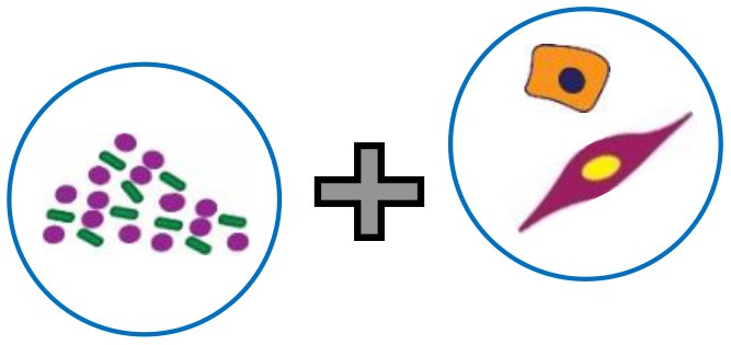
|
Human Skin cells with biofilm or biofilm-conditioned media
Study the effects of wound colonizing bacteria by co-culturing human skin cells such as keratinocytes and fibroblasts with biofilms. It recapitulates host-microbe interactions in the wound bed resulting in changes in host cell migration, proliferation, and gene expression. Human Skin Equivalents (HSEs) 3D structures that mimic human skin layers and recapitulate bacterial attachment and biofilm formation under conditions close to native architecture. |
Holland et al., 2008, 2009; Charles et al., 2009; Kirker et al., 2009, 2012; Secor et al., 2011; Haisma et al., 2013; Tankersley et al., 2014; Alves et al., 2018 |
Microbes+Immune Cells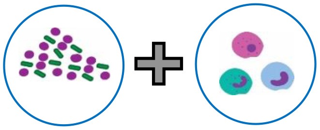
|
Infection-immunity interface on a microfluidic platform
Study interactions between the wound pathogen S. aureus (not specific for biofilms) and neutrophils across two compartments, enabling the study of neutrophil recruitment, migration, and engulfment. |
Brackman and Coenye, 2016 | |
Microbes+Extracellular Matrix
|
Polymer surface coated with gel-like collagen matrix
Study the role of matrix in biofilm formation and structure using comparisons between coated and uncoated surfaces. Collagen mold model with transwell inserts Biofilms embedded in collagen and structured as a void, recapitulating biomimetic effects such as antibiotic diffusion distance through the matrix. |
Werthén et al., 2010; Price et al., 2016 | |
Microbes+Wound fluid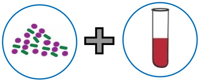
|
Lubbock model (Bolton broth) and its variants
Widely-used to mimic the wound infection state. It enables the study of biofilms and interspecies interactions and has been used to study the effects of antibiotics and other antimicrobial compounds on biofilms. Simulated sweat and serum media Enables the study of growth and biofilm formation under wound-relevant nutritional and chemical conditions. |
Sun et al., 2008, 2014; Dalton et al., 2011; DeLeon et al., 2014; Dowd et al., 2014; Sojka et al., 2016 | |
| Ex vivo | 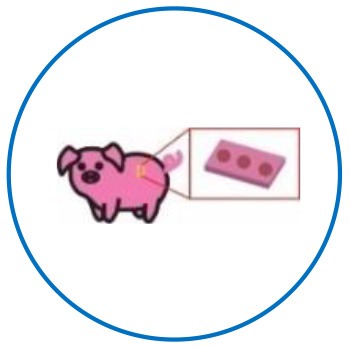 |
Biological skin tissue from pigs: A high degree of anatomic and physiological similarity to human skin and immune system. Enables the actual creation of a wound (thermal injuries, infected state). Biological tissue supports biofilm growth. Enables testing of immune parameters such as cytokine responses. Can be leveraged to test therapeutics under closely human-relevant conditions. |
Steinstraesser et al., 2010; Yang et al., 2013; Thet et al., 2016 |
| Porcine skin | |||
 |
Biological tissue from human skin:
Can faithfully recapitulate biomimetic features of the chronic wound infection state. Demonstration of biofilm formation and critical host immune factors including cellular and cytokine responses. Can be leveraged to test therapeutics under human-relevant conditions. |
Misic et al., 2014; Schaudinn et al., 2017; Ashrafi et al., 2018 | |
| Human skin |
The authors apologize for these errors and state that this does not change the scientific conclusions of the article in any way. The original article has been updated.
References
- Alves P. M., Al-Badi E., Withycombe C., Jones P. M., Purdy K. J., Maddocks S. E. (2018). Interaction between Staphylococcus aureus and Pseudomonas aeruginosa is beneficial for colonisation and pathogenicity in a mixed biofilm. Pathog Dis. 76:fty003. 10.1093/femspd/fty003 [DOI] [PubMed] [Google Scholar]
- Ashrafi M., Novak-Frazer L., Bates M., Baguneid M., Alonso-Rasgado T., Xia G., et al. (2018). Validation of biofilm formation on human skin wound models and demonstration of clinically translatable bacteria-specific volatile signatures. Sci. Rep. 8:9431. 10.1038/s41598-018-27504-z [DOI] [PMC free article] [PubMed] [Google Scholar]
- Brackman G., Coenye T. (2016). In vitro and in vivo biofilm wound models and their application. Adv. Exp. Med. Biol 15–32. 10.1007/5584_2015_5002 [DOI] [PubMed] [Google Scholar]
- Charles C. A., Ricotti C. A., Davis S. C., Mertz P. M., Kirsner R. S. (2009). Use of tissue-engineered skin to study in vitro biofilm development. Dermatol. Surg. 35, 1334–1341. 10.1111/j.1524-4725.2009.01238.x [DOI] [PubMed] [Google Scholar]
- Dalton T., Dowd S. E., Wolcott R. D., Sun Y., Watters C., Griswold J. A., et al. (2011). An in vivo polymicrobial biofilm wound infection model to study interspecies interactions. PLoS ONE 6:e27317. 10.1371/journal.pone.0027317 [DOI] [PMC free article] [PubMed] [Google Scholar]
- DeLeon S., Clinton A., Fowler H., Everett J., Horswill A. R., Rumbaugh K. P. (2014). Synergistic interactions of Pseudomonas aeruginosa and Staphylococcus aureus in an in vitro wound model. Infect. Immun. 82, 4718–4728. 10.1128/IAI.02198-14 [DOI] [PMC free article] [PubMed] [Google Scholar]
- Dowd S. E., Sun Y., Smith E., Kennedy J. P., Jones C. E., Wolcott R. (2014). Effects of biofilm treatments on the multi-species Lubbock chronic wound biofilm model. J. Wound Care 18, 508–512. 10.12968/jowc.2009.18.12.45608 [DOI] [PubMed] [Google Scholar]
- Haisma E. M., Rietveld M. H., Breij A., Van Dissel J. T., El Ghalbzouri A., Nibbering P. H. (2013). Inflammatory and antimicrobial responses to methicillin-resistant Staphylococcus aureus in an in vitro wound infection model. PLoS ONE 8:e82800. 10.1371/journal.pone.0082800 [DOI] [PMC free article] [PubMed] [Google Scholar]
- Holland D. B., Bojar R. A., Farrar M. D., Holland K. T. (2009). Differential innate immune responses of a living skin equivalent model colonized by Staphylococcus epidermidis or Staphylococcus aureus. FEMS Microbiol. Lett. 290, 149–155. 10.1111/j.1574-6968.2008.01402.x [DOI] [PubMed] [Google Scholar]
- Holland D. B., Bojar R. A., Jeremy A. H. T., Ingham E., Holland K. T. (2008). Microbial colonization of an in vitro model of a tissue engineered human skin equivalent–a novel approach. FEMS Microbiol. Lett. 279, 110–115. 10.1111/j.1574-6968.2007.01021.x [DOI] [PubMed] [Google Scholar]
- Kirker K. R., James G. A., Fleckman P., Olerud J. E., Stewart P. S. (2012). Differential effects of planktonic and biofilm MRSA on human fibroblasts. Wound Repair Regen. 20, 253–261. 10.1111/j.1524-475X.2012.00769.x [DOI] [PMC free article] [PubMed] [Google Scholar]
- Kirker K. R., Secor P. R., James G. A., Fleckman P., Olerud J. E., Stewart P. S. (2009). Loss of viability and induction of apoptosis in human keratinocytes exposed to Staphylococcus aureus biofilms in vitro. Wound Repair Regen. 17, 690–699. 10.1111/j.1524-475X.2009.00523.x [DOI] [PMC free article] [PubMed] [Google Scholar]
- Misic A. M., Gardner S. E., Grice E. A. (2014). The wound microbiome: modern approaches to examining the role of microorganisms in impaired chronic wound healing. Adv. Wound Care 3, 502–510. 10.1089/wound.2012.0397 [DOI] [PMC free article] [PubMed] [Google Scholar]
- Price B. L., Lovering A. M., Bowling F. L., Dobson C. B. (2016). Development of a novel collagen wound model to simulate the activity and distribution of antimicrobials in soft tissue during diabetic foot infection. Antimicrob. Agents Chemother. 60, 6880–6889. 10.1128/AAC.01064-16 [DOI] [PMC free article] [PubMed] [Google Scholar]
- Schaudinn C., Dittmann C., Jurisch J., Laue M., Günday-Türeli N., Blume-Peytavi U., et al. (2017). Development, standardization and testing of a bacterial wound infection model based on ex vivo human skin. PLoS ONE 12:e0186946. 10.1371/journal.pone.0186946 [DOI] [PMC free article] [PubMed] [Google Scholar]
- Secor P. R., James G. A., Fleckman P., Olerud J. E., McInnerney K., Stewart P. S. (2011). Staphylococcus aureus biofilm and planktonic cultures differentially impact gene expression, mapk phosphorylation, and cytokine production in human keratinocytes. BMC Microbiol. 11:143. 10.1186/1471-2180-11-143 [DOI] [PMC free article] [PubMed] [Google Scholar]
- Sojka M., Valachova I., Bucekova M., Majtan J. (2016). Antibiofilm efficacy of honey and bee-derived defensin-1 on multispecies wound biofilm. J. Med. Microbiol. 65, 337–344. 10.1099/jmm.0.000227 [DOI] [PubMed] [Google Scholar]
- Steinstraesser L., Sorkin M., Niederbichler A. D., Becerikli M., Stupka J., Daigeler A., et al. (2010). A novel human skin chamber model to study wound infection ex vivo. Arch Dermatol Res. 302, 357–365. 10.1007/s00403-009-1009-8 [DOI] [PMC free article] [PubMed] [Google Scholar]
- Sun Y., Dowd S. E., Smith E., Rhoads D. D., Wolcott R. D. (2008). In vitro multispecies Lubbock chronic wound biofilm model. Wound Repair Regen. 16, 805–813. 10.1111/j.1524-475X.2008.00434.x [DOI] [PubMed] [Google Scholar]
- Sun Y., Smith E., Wolcott R., Dowd S. E. (2014). Propagation of anaerobic bacteria within an aerobic multi-species chronic wound biofilm model. J. Wound Care 18, 426–431. 10.12968/jowc.2009.18.10.44604 [DOI] [PubMed] [Google Scholar]
- Tankersley A., Frank M. B., Bebak M., Brennan R. (2014). Early effects of Staphylococcus aureus biofilm secreted products on inflammatory responses of human epithelial keratinocytes. J. Inflamm. 11:17. 10.1186/1476-9255-11-17 [DOI] [PMC free article] [PubMed] [Google Scholar]
- Thet N. T., Alves D. R., Bean J. E., Booth S., Nzakizwanayo J., Young A. E. R., et al. (2016). Prototype development of the intelligent hydrogel wound dressing and its efficacy in the detection of model pathogenic wound biofilms. ACS Appl. Mater. Interfaces 8, 14909–14919. 10.1021/acsami.5b07372 [DOI] [PubMed] [Google Scholar]
- Werthén M., Henriksson L., Jensen P. Ø., Sternberg C., Givskov M., Bjarnsholt T. (2010). An in vitro model of bacterial infections in wounds and other soft tissues. APMIS 118, 156–164. 10.1111/j.1600-0463.2009.02580.x [DOI] [PubMed] [Google Scholar]
- Yang Q., Phillips P. L., Sampson E. M., Progulske-Fox A., Jin S., Antonelli P., et al. (2013). Development of a novel ex vivo porcine skin explant model for the assessment of mature bacterial biofilms. Wound Repair Regen. 21, 704–714. 10.1111/wrr.12074 [DOI] [PubMed] [Google Scholar]


