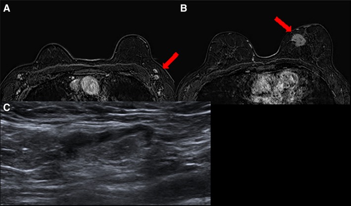
Figure 3.

MRI and sonographic characteristics of benign nodes. T1 fat‐saturated magnetic resonance imaging 2 minutes after administration of contrast media (A) in a woman aged 68 years with a suspicious mass in the median‐inner quadrant of the left breast (B, red arrow) showing bilateral enlarged nodes with central fatty hilum. (C): The targeted ultrasound demonstrated normal appearance of one of the nodes. A fine‐needle aspiration was performed with a cytologic diagnosis: polymorphous lymphoid population, negative for malignant cells.
