Abstract
Context: Angelica sinensis L. (Umbelliferae) has medicinal properties.
Objectives: The present study evaluates the haematopoietic effects of A. sinensis polysaccharides (ASP) against lisinopril-induced anaemia.
Materials and methods: Thirty healthy adult male albino rats were randomly divided into five groups (n = 6). Group I was control group. Group II was treated with angiotensin-converting enzyme inhibitor (ACEI, 20 mg/kg/day) to induce anaemia. In group III, erythropoietin (EPO, 100 IU/kg/each) was administered in combination with ACEI. Group IV was treated with ASP (1 g/kg/day), extracted from A. sinensis root caps. In Group V, ASP (1 g/kg/day) was treated with ACEI. After 28 days, blood and tissue samples were collected for haematological and histopathological analysis, respectively.
Results: The results showed that ACEI significantly reduced the haemoglobin (Hb, 10.0 g/dL), packed cell volume (PCV, 39.5%), red blood cells (RBCs, 6.2 million/mm3), mean corpuscular volume (MCV, 53.5 fL) and mean corpuscular haemoglobin (MCH, 16.2 pg/cell) values. In the group treated with ASP, the Hb (13.7 g/dL) and RBCs (7.8 million/mm3) increased significantly (p < 0.05). The combination of ASP and ACEI led to the significant (p < 0.05) reduction in Hb (10.7 g/dL), PCV (33.3%), RBCs (6.0 million/mm3), MCV (54.42 fL) and MCH (16.44 pg/cell) values. While histopathological examination of the liver and kidney cells showed a mild degree of toxicity in the ASP-treated group.
Conclusion: ASP has a potentiating effect on haematological parameters when given alone. However, when administered simultaneously with lisinopril, it showed an unfavourable effect with more complicated anaemia so it should not be used with ACEIs.
Keywords: ACE inhibitors, recombinant human erythropoietin, haemoglobin level, RBC count, PCV, MCV, MCH
Introduction
Angiotensin-converting enzyme inhibitors (ACEIs) are widely used for the treatment of congestive heart failure, diabetes mellitus and hypertension. However, prolonged use of these medications is reported to cause anaemia (Ajmal et al. 2013). ACEIs reduced erythropoietin (EPO) levels, circulating insulin-like growth factor (IGF-1) and haematocrit. ACE regulates a group of bioactive peptides, such as N-acetyl-ser-asp (Ac-SDKP), substance P and angiotensin 1-7. These peptides involve in the regulation of haematopoiesis pathways (Shen & Bernstein 2011). Angiotensin II enhances the proliferation of erythroid progenitor cells so erythropoiesis is inhibited by decreasing angiotensin II level (MacDougall 2001). Inhibition of the renin–angiotensin system with ACEIs is correlated with decreased haemoglobin levels and reduced EPO production (Tang & Katz 2006).
Herbal medicines are based on the principle that plants contain natural chemicals that promote health and cure diseases; therefore focus on plant research has increased worldwide (Alves & Rosa 2007). For centuries, various traditional Chinese medicine formulas have been used as thrombopoietic and haematopoietic agents (Liu et al. 2010). Angelica sinensis L. (Umbelliferae) is a famous Chinese herb utilized for the treatment of irregular menstruation, anaemia, and uterine bleeding with the dose rate of 1–2 g/kg (Liu et al. 2012). The root of A. sinensis contains various polysaccharides such as galactose, glucose, arabinose, rhamnose and xylose (Wang et al. 2003; Liu et al. 2012). The polysaccharides separated from A. sinensis increase haematopoiesis by enhancing haematopoietic cells, promoting the release of EPO (Liu et al. 2012) and regulation of iron homeostasis (Wang et al. 2007, 2011). The bioactivity analysis demonstrates that A. sinensis polysaccharides (ASP) involve in increasing erythrocyte and leucocyte count in peripheral blood and enhancing bone marrow haematopoiesis (Zhao et al. 2012; Zahra et al. 2016). ASP has been used as a health-supporting agent in cancer patients under chemo-radiation treatment and anaemic patients (Lee et al. 2012) and also enhances thrombopoiesis and haematopoiesis (Liu et al. 2010).
Keeping these facts in mind, the present experimental study was designed to elucidate the haematopoietic effects of ASP on haematological parameters in rats treated with ACEIs.
Materials and methods
Experimental animals
Thirty healthy adult male albino Wistar rats weighing about 120 ± 15 g were purchased from a local market of Faisalabad, Pakistan. The animals were housed at 25 ± 5 °C in a well-ventilated condition under 12 h light and dark cycles in animal room of the Department of Physiology and Pharmacology, University of Agriculture, Faisalabad. The rats were provided normal feed (rat chow) for 28 days twice a day and drinking water ad libitum. The experiments were carried out in accordance with the guidelines of the Directorate of Graduate Studies and Institutional Animal Ethical Committee with protocol approval number as DGS/20174-79.
Drugs and experimental protocols
ACEI; Tablet Zestril® (Lisinopril 5 mg), purchased from Searle Pakistan Limited Karachi-Pakistan, was used to induce anaemia. Synthetic anti-anaemic drug; Injection Eritrogen® (Recombinant human EPO 4000 IU/mL subcutaneously) was used to cure anaemia. After 7 days of acclimatization, 30 albino rats were randomly divided into five equal groups each group carried six animals. Group I was the control group and given normal feed and water ad libitum. Group II was treated with ACEI (20 mg/kg/day) orally to induce the anaemia for 28 days (Dodiya et al. 2013; Albarwani et al. 2015). In group III, EPO (100 IU/kg/each) was administered in combination with ACEI (20 mg/kg/day) on alternate day subcutaneously for 28 days (Lebel et al. 2006). Group IV was treated with ASP at the dose of 1 g/kg/day intragastrically for 28 days. In group V, ASP (1 g/kg/day) was administered in combination with ACEI (20 mg/kg/day) intragastrically for 28 days (Wang et al. 2011).
Plant material
The dry roots of A. sinensis were purchased from the local herbal market of Faisalabad, Pakistan. Identified and authenticated by Dr Mansoor Hameed, Department of Botany, Faculty of Sciences, University of Agriculture, Faisalabad, Pakistan. Angelica sinensis root caps were washed thoroughly with distilled water to remove extraneous material. The root caps were dried in shade and powdered with an electrical grinder. The powdered material was passed through a mesh sieve and then stored in air tight container.
Preparation of ASP
Aqueous extraction of A. sinensis root caps was carried out in the Department of Physiology and Pharmacology, UAF. Polysaccharide fraction was isolated by the ethanol precipitation method as described by Cho et al. (2000) and modified by Ye et al. (2003). Angelica sinensis root caps (100 g) were boiled for 3–4 h periods with water for a total of 12 h. After each 4 h period of boiling, the water extract was collected and the residue was boiled again with water for another 4 h period. All extracts were finally pooled and mixed with a concentrated ethanol solution (final concentration 75% v/v) to precipitate the polysaccharide-enriched fraction. The precipitates were dried to a thick semi-solid mass of brown colour. It was stored at −20 °C until further use.
Sampling and analysis
The body weight of each group was determined. After the treatment period, blood was collected from the jugular vein of rats into tubes containing ethylene diamine tetra-acetic acid (EDTA) for haematological parameters. Estimation of total red blood cell (RBC), white blood cell (WBC) and platelet count was done using Neubauer haemocytometer (Feinoptik, Germany). Haemoglobin (Hb) content and packed cell volume (PCV) was estimated by Sahli’s haemoglobinometer and centrifuging blood in haematocrit tube and reading % RBC in a hematocrit reader, respectively. Erythrocyte indices including mean corpuscular volume (MCV), which indicates the volume of average red cell in a sample expressed in femtolitres (fL) and calculated using the formula: MCV = PCV/RBC × 10. While, mean corpuscular haemoglobin (MCH), which represents the absolute amount of Hb in the average red cell in a sample in units of picogram (pg) per cell. The MCH was calculated from Hb and RBC using the following formula: MCH = (Hb × 10)/RBC. The weight of liver, kidney and spleen was measured at the 28th day after sacrificing.
Histopathological analysis
Formalin-fixed kidney and liver biopsies were processed in graded ethanol concentrations and fixed in paraffin blocks. Their fragments were arranged perpendicular to the plane of the section in the block and 6 μm thick transverse fragments were cut and mounted on glass slides and stained with haematoxylin and eosin (H and E stain). Microscopy was completed on an Olympus PM-10 ADS automatic light microscope (Olympus optical Co., Tokyo, Japan) with a 20 × objective.
Statistical analysis
The values were expressed as mean ± SE. The difference between control and treated values were tested by performing one-way analysis of variance (ANOVA) and Dunnett’s t-test at 5% level of significance.
Results
The effects on RBCs count (million/mm3), WBC count (thousand/mm3), platelet count (109/L), PCV (%), Hb (g/dL), MCV (fL) and MCH (pg/cell) values of control and treated groups of albino rats after 28 days are presented in Table 1. ACEIs significantly (p < 0.05) lowered the Hb value (10.0 ± 0.46 g/dL), PCV (39.5 ± 1.73%), RBCs count (6.2 ± 0.25 million/mm3), MCV (53.5 ± 0.46 fL) and MCH value (16.2 ± 0.02 pg/cell). Group IV, which was administered ASP, significantly (P < 0.05) increased the Hb value (13.7 ± 1.91 g/dL), RBCs count (7.8 ± 1.02 million/mm3) but significantly (p < 0.01) reduced the MCV value (56.2 ± 0.289 fL) while the effect on MCH value (17.5 ± 0.12 pg/cell) remained non-significant (p > 0.05) compared with control group. Unexpectedly, Hb (10.7 ± 0.10 g/dL), PCV (33.3 ± 2.79%), RBCs count (6.0 ± 0.43 million/mm3), MCV (54.42 ± 1.21 fL) and MCH value (16.44 ± 0.34 pg/cell) decreased significantly (p < 0.05) in group V treated with ASP + ACEI as compared to control group after 28 days. There was no significant increase in WBC and platelet count in these groups as compared to control group as presented in Table 1.
Table 1.
Mean ± SEM values of haematological parameters in control and treated groups of adult male albino rats (n = 6) after 28 days.
| Groups |
|||||
|---|---|---|---|---|---|
| Parameters | I | II | III | IV | V |
| Hb (g/dL) | 11.9 ± 0.59 | 10.0 ± 0.46a | 17.9 ± 0.25a | 13.7 ± 1.91a | 10.7 ± 0.10a |
| PCV (%) | 42.1 ± 1.95 | 39.5 ± 1.73a | 61.0 ± 0.95a | 44.1 ± 6.02NS | 33.3 ± 2.79a |
| RBC count (million/mm3) | 6.8 ± 0.34 | 6.2 ± 0.25a | 10.0 ± 0.12a | 7.8 ± 1.02a | 6.0 ± 0.43a |
| WBC count (thousand/mm3) | 9.6 ± 0.8 | 7.5 ± 0.31NS | 7.3 ± 0.93NS | 11.47 ± 1.0NS | 6.1 ± 0.38NS |
| Platelets count (109/L) | 0.87 ± 0.13 | 0.98 ± 0.63NS | 0.67 ± 0.76NS | 1.05 ± 0.15NS | 0. 62 ± 0.17NS |
| MCV (fL) | 61.5 ± 0.73 | 53.5 ± 0 .46b | 60.6 ± 0.50NS | 56.2 ± 0.289** | 54.42 ± 1.21b |
| MCH (pg/cell) | 17.5 ± 0.22 | 16.2 ± 0.02b | 17.7 ± 0.04NS | 17.5 ± 0.12NS | 16.44 ± 0.34b |
Significance level is shown by
(p < 0.05),
(p < 0.01).
Non-significant.
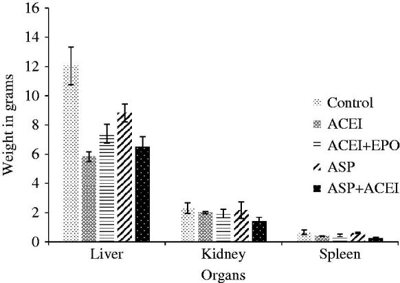
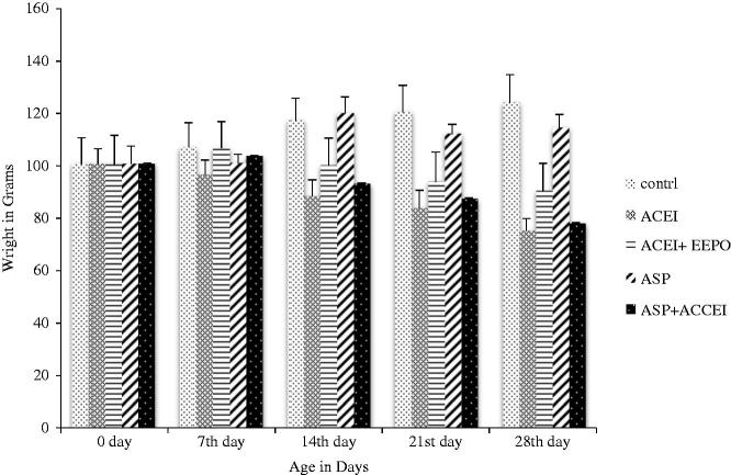
The mean values for body weight of ACEI group and ASP + ACEI groups were 74.57 ± 5.3 and 77.41 ± 8.04 g, respectively, that showed significant reduction in body weight in these groups as compared to control group after 28 days. Mean values for body weight of EPO + ACEI and ASP groups were 89.785 ± 11.1 and 113.77 ± 5.9 g that showed non-significant reduction in body weight in these groups against control group after 28 days presented in Figure 1. The mean values for the liver, kidney and spleen weight showed non-significant difference in weight presented in Figure 2.
Figure 1.
Body weight of control and treated rats during the experimental period of 28 days.
Figure 2.

Weight of different organs of control and treated groups of albino rats after 28 days.
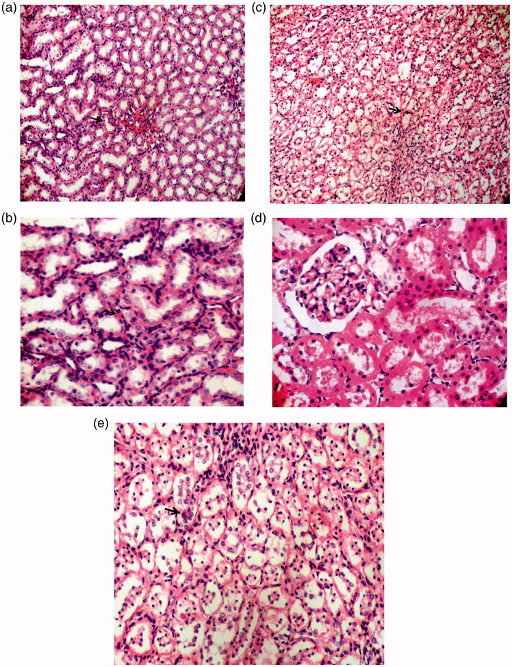
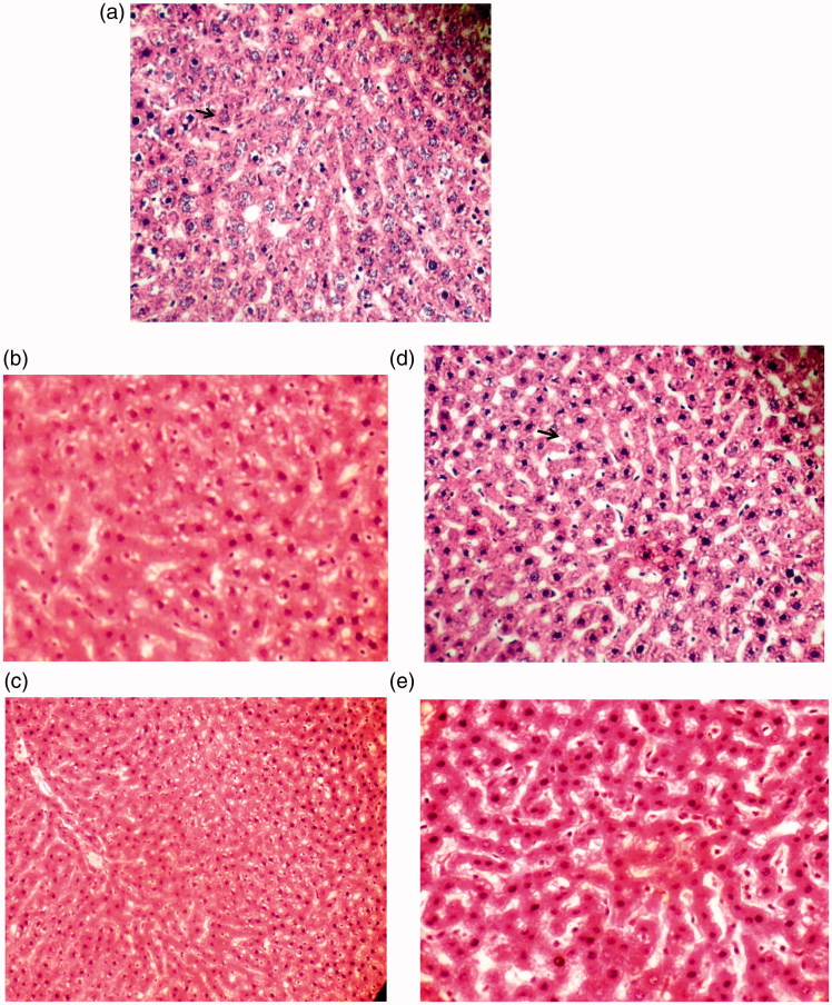
In EPO + ACEI group showing mild degree of condensation pyknotic changes in the nuclei of tubular epithelial cells (Figure 3(c)) while ASP + ACEI group showing mild degree of pyknotic changes in nuclei of tubular epithelial cells along with normal tubular epithelium (Figure 3(e)) in comparison with the kidney of control group (Figure 3(a)) presented with arrows. Groups treated with ACEI (Figure 3(b)) and ASP (Figure 3(d)) showing mild degree of condensation and normal tubular epithelium and glomeruli with clear urinary space in renal parenchyma, respectively. ASP-treated group showing individual cell necrosis in hepatic parenchyma (Figure 4(d)) while ACEI-treated group (Figure 4(b)) showing mild degree of vacuolar degeneration as compared to liver of control group (Figure 4(a)) presented with arrows. In EPO + ACEI group (Figure 4(c)) showing mild degree of vacuolar degeneration in the cytoplasm of hepatocytes, whereas group treated with ASP + ACEI (Figure 4(e)) showing mild to moderate degree of vacuolar degeneration condensed and pyknotic nuclei at some places.
Figure 3.
Photomicrograph of kidney (a) control group (b) ACEI group (c) EPO + ACEI group (d) ASP group (e) ASP + ACEI group (200 and 400X, H&E Staining).
Figure 4.
Photomicrograph of liver (a) control group (b) ACEI group (c) EPO + ACEI group (d) ASP group (e) ASP + ACEI group (200 and 400X, H&E Staining).
Discussion
Use of an ACEI is correlated with decreased Hb levels and reduced EPO production. This reduction in the EPO level increases the hepicidin (antimicrobial peptide produced by hepatocytes) expression which causes systemic iron deficiency and anaemia (Handelman & Levin, 2008). Another action of ACEI is the pharmacological inhibition of the renin-angiotensin system in clinical trials, which is correlated with minute but statistically significant reduction in Hb levels (Tang & Katz 2006). After the initiation of the treatment with high doses of ACEIs, isolated normochrome anaemia in ACE knock-out mice while leucopenia and anaemia in humans have been reported (Savary et al. 2005). ACEIs may aggravate anaemia by decreasing Ang II levels that have an association with clinical management of chronically ill patients (Cole et al. 2000; Hayashi et al. 2001). Anaemia contributes to the worsening of cardiovascular diseases in many cases, therefore, the management of anaemia is necessary for controlling the patient’s overall condition (Ajmal et al. 2013). The link between chronic heart failure and anaemia may be associated with high mortality (Anand et al. 2004). Asinensis sinensis was found to be effective for enhancing haematopoiesis by increasing the secretion of some haematopoietic growth factors (EPO) via stimulating haematopoietic cells and muscle tissues (Sarker & Nahar 2004). Roots of A. sinensis-containing ASP are reported to reduce anaemia (Liu et al. 2012; Zahra et al. 2016).
In the present study, group II treated with ACEI showed decreased Hb, PCV, RBCs count, MCV and MCH values as compared to group I (control group) that indicate anaemia in rats while there was non-significant difference in WBC and platelet count. This effect on haematological parameters might be due to the depression of renin–angiotensin system by ACEIs, as this system is involved in the regulation of haematopoiesis (Cole et al. 2000). This depression of renin–angiotensin system down regulates EPO which might affect the hepcidin expression and cause iron deficiency with anaemia (Handelman & Levin 2008; Liu et al. 2012). In our study, the EPO-treated group showed improvement in the haematological parameters and anaemia induced by lisinopril (ACEI). This could be due to direct combination of EPO to its receptor on the cell surface and intracellular signal transduction to suppress the expression and DNA-binding activity of transcription factor CAAT (controlled amino acid therapy)/enhancer-binding proteins α (C/EBPα) and phospho-STAT3/5, thereby inhibiting the expression of hepcidin to improve the anaemic conditions (Pinto et al. 2008). ASPs from the root cap were selected in the present study because of medicinal importance of root mentioned in the literature (Liu et al. 2012; Lim & Yun 2014). Recently, ASPs have also been reported to decreased hepcidin expression by suppressing the expression of JAK1/2, phospho-JAK1/2, phospho-SMAD1/5/8, phospho-ERK1/2 and promoting the expression of SMAD7 in the liver (Zhang et al. 2014). This decreased hepicidin expression might be due to increased haematopoiesis by ASP consequently reduced the anaemia. Present study reported that Hb and RBCs count increased and the MCV value decreased in ASP-treated group compared to ACEI-treated group, while change in MCH value was non-significant; these results were in line with our previous study (Zahra et al. 2016). Increase in RBCs count and decrease in MCV value resulted in the compensatory effect on the change in haematocrit value, so it remained non-significant. In this situation RBCs were referred as normochromic. As various studies have also shown, ASP can be used as a health-supporting agent in anaemic patients by increasing plasma iron levels through inhibition of hepcidin expression (Wang et al. 2011; Lee et al. 2012). ASP also increases haematopoiesis by promoting the release of haematopoietic growth factors such as EPO by enhancing haematopoietic cells along with blood circulation in the body (Wang et al. 2007; Liu et al. 2012). The bioactivity analysis demonstrates that ASP involve in increasing erythrocyte and leucocyte count in peripheral blood and enhancing bone marrow haematopoiesis (Zhao et al. 2012).
In group V, which was treated with ASP along with ACEI showed that Hb level, PCV level, RBC count, MCV and MCH level decreased significantly (p < 0.05) as compared to control group that indicated the ineffectiveness of combination of ACEI with ASP. It showed that when ASP is given alone then it will exert its anti-anaemic effect but when given in combination with ACEI, it will produce detrimental effect with and will lead towards severe anaemia. This might be due to the interaction between ACEI and ASP or with some other component present in A. sinensis, which might have effect on haematological parameters in combination with ACEIs. There was no significant effect on body and organ weight in all the treated groups. In a study, it has been shown that long-term ACEI treatment reduces serum leptin concentration, food intake and body weight gain. In adipose tissue, ACEI treatment reduces ACE activity that increases the expression of peroxisome proliferator-activated receptor gamma, hormone-sensitive lipase, adiponectin, fatty acid synthase, superoxide dismutase and catalase resulting in body weight reduction (Santos et al. 2009).
In case of histopathological examinations of liver and kidney, ASP showed necrotic changes at the cellular level that showed a mild degree of toxicity on both the kidney and liver. This toxicity might be due to variations in the hepcidin and EPO levels in kidney and liver tissues. The toxicity problem can be reduced by reducing the intervention time to 2 or 3 weeks because more prolonged treatment may cause severe damage to kidney and liver. It is a common belief that herbal medicines are free of any side effects because of their natural origins. But dose, frequency and manner of administration are not fully controlled and reviewed as these are usually self-prescribed. Herbs containing chemicals may be natural to the plants but for the human body, they are not natural (Arsad et al. 2014).
Conclusions
In case of ASP, Hb level and RBCs count increased. In rats treated with ASP + ACEI, there was not only reduction in Hb value, PCV value and RBCs count but there was also microcytic and hypochromic anaemia. It means ASP may have effect against anaemia but when it is administered simultaneously with ACEI to cure anaemia then it showed unfavourable effect with more complicated anaemia so it should not be used with ACEIs. Histopathological examination of liver and kidney revealed that ASP showed toxicity within 28 days. These results end up with a conclusion that ASP will be toxic when it will be given for a longer period of time. So it is not safe for the patients with cardiovascular diseases for longer period of time.
Disclosure statement
The authors report no conflicts of interest. The authors alone are responsible for the content and writing of this article.
References
- Ajmal A, Gessert CE, Johnson BP, Renier CM, Palcher JA.. 2013. Effect of angiotensin converting enzyme inhibitors and angiotensin receptor blockers on hemoglobin levels. BMC Res Notes. 6:443–460. [DOI] [PMC free article] [PubMed] [Google Scholar]
- Albarwani S, Al-Siyabi S, Al-Husseini I, Al-Ismail A, Al-Lawati I, Al-Bahrani I, Tanira MO.. 2015. Lisinopril alters contribution of nitric oxide and K(Ca) channels to vasodilatation in small mesenteric arteries of spontaneously hypertensive rats. Physiol Res. 64:39–49. [DOI] [PubMed] [Google Scholar]
- Alves RR, Rosa IM.. 2007. Biodiversity, traditional medicine and public health: where do they meet? J Ethnobiol Ethnomed. 3:14–46. [DOI] [PMC free article] [PubMed] [Google Scholar]
- Anand I, McMurray JJ, Whitmore J, Warren M, Pham A, McCamish MA, Burton PB.. 2004. Anemia and its relationship to clinical outcome in heart failure. Circulation. 110:149–154. [DOI] [PubMed] [Google Scholar]
- Arsad SS, Esa NM, Hamzah H.. 2014. Histopathologic changes in liver and kidney tissues from male Sprague Dawley rats treated with Rhaphidophora decursiva (Roxb.) Schott extract. J Cytol Histo. S4:001. [Google Scholar]
- Cho CH, Mei QB, Shang P, Lee SS, So HL, Guo X, Li Y.. 2000. Study of the gastrointestinal protective effects of polysaccharides from Angelica sinensis in rats. Planta Med. 66:348–351. [DOI] [PubMed] [Google Scholar]
- Cole J, Ertoy D, Lin H, Sutliff RL, Ezan E, Guyene TT, Capecchi M, Corvol P, Bernstein KE.. 2000. . Lack of angiotensin II-facilitated erythropoiesis causes anemia in angiotensin-converting enzyme-deficient mice. J Clin Invest. 106:1391–1398. [DOI] [PMC free article] [PubMed] [Google Scholar]
- Dodiya H, Kale V, Goswami S, Sundar R, Jain M.. 2013. Evaluation of adverse effects of lisinopril and rosuvastatin on hematological and biochemical analytes in Wistar rats. Toxicol Int. 20:170–176. [DOI] [PMC free article] [PubMed] [Google Scholar]
- Handelman GJ, Levin NW.. 2008. Iron and anemia in human biology: a review of mechanisms. Heart Fail Rev. 13:393–404. [DOI] [PubMed] [Google Scholar]
- Hayashi K, Hasegawa K, Kobayashi S.. 2001. Effects of angiotensin-converting enzyme inhibitors on the treatment of anemia with erythropoietin. Kidney Int. 60:1910–1916. [DOI] [PubMed] [Google Scholar]
- Lebel M, Rodrigue ME, Agharazii M, Larivière R.. 2006. Antihypertensive and renal protective effects of renin-angiotensin system blockade in uremic rats treated with erythropoietin. Am J Hypertens. 19:1286–1292. [DOI] [PubMed] [Google Scholar]
- Lee JG, Hsieh WT, Chen SU, Chiang BH.. 2012. Hematopoietic and myeloprotective activities of an acidic Angelica sinensis polysaccharide on human CD34+ stem cells. J Ethnopharmacol. 15:739–745. [DOI] [PubMed] [Google Scholar]
- Lim DW, Yun TK.. 2014. Anti-osteoporotic effects of Angelica sinensis (Oliv.) Diels extract on ovariectomized rats and its oral toxicity in rats. Nutrients. 6:4362–4372. [DOI] [PMC free article] [PubMed] [Google Scholar]
- Liu C, Li J, Meng FY, Liang SX, Deng R, Li CK, Pong NH, Lau CP, Cheng SW, Ye JY, et al. 2010. Polysaccharides from the root of Angelica sinensis promotes hematopoiesis and thrombopoiesis through the PI3K/AKT pathway. BMC Complement Altern Med. 10:79. [DOI] [PMC free article] [PubMed] [Google Scholar]
- Liu JY, Zhang Y, You RX, Zeng F, Guo D, Wang KP.. 2012. Polysaccharide isolated from Angelica sinensis inhibits hepcidin expression in rats with iron deficiency anemia. J Med Food. 15:923–929. [DOI] [PMC free article] [PubMed] [Google Scholar]
- MacDougall IC. 2001. Ace inhibitors and erythropoietin responsiveness. Am J Kidney Dis. 38:649–651. [DOI] [PubMed] [Google Scholar]
- Pinto JP, Ribeiro S, Pontes H, Thowfeequ S, Tosh D, Carvalho F, Porto G.. 2008. Erythropoietin mediates hepcidin expression in hepatocytes through EPOR signaling and regulation of C/EBPalpha. Blood. 111:5727–5733. [DOI] [PMC free article] [PubMed] [Google Scholar]
- Santos EL, de Picoli SK, da Silva ED, Batista EC, Martins PJ, D'Almeida V, Pesquero JB.. 2009. Long term treatment with ACE inhibitor enalapril decreases body weight gain and increases life span in rats. Biochem Pharmacol. 78:951–958. [DOI] [PubMed] [Google Scholar]
- Sarker SD, Nahar L.. 2004. Natural medicine: the genus Angelica. Curr Med Chem. 11:1479–1500. [DOI] [PubMed] [Google Scholar]
- Savary K, Michaud A, Favier J, Larger E, Corvol P, Gasc JM.. 2005. Role of the renin-angiotensin system in primitive erythropoiesis in the chick embryo. Blood. 105:103–110. [DOI] [PubMed] [Google Scholar]
- Shen XZ, Bernstein KE.. 2011. The peptide network regulated by angiotensin converting enzyme (ACE) in hematopoiesis. Cell Cycle. 10:1363–1369. [DOI] [PMC free article] [PubMed] [Google Scholar]
- Tang YD, Katz SD.. 2006. Contemporary reviews in cardiovascular medicine: anemia in chronic heart failure; prevalence, etiology, clinical correlates, and treatment options. Circulation. 113:2454–2461. [DOI] [PubMed] [Google Scholar]
- Wang KP, Zeng F, Liu JY, Guo D, Zhang Y.. 2011. Inhibitory effect of polysaccharides isolated from Angelica sinensis on hepcidin expression. J Ethnopharmacol. 134:944–948. [DOI] [PubMed] [Google Scholar]
- Wang PP, Zhang Y, Dai LQ, Wang KP.. 2007. Effect of Angelica sinensis polysaccharide-iron complex on iron deficiency anemia in rats. Chin J Integr Med. 13:297–300. [DOI] [PubMed] [Google Scholar]
- Wang QJ, Ding F, Zhu NN, He P, Fang Y.. 2003. Determination of the compositions of polysaccharides from Chinese herbs by capillary zone electrophoresis with amperometric detection. Biomed Chromatogr. 17:483–488. [DOI] [PubMed] [Google Scholar]
- Ye YN, So HL, Liu ES, Shin VY, Cho CH.. 2003. Effect of polysaccharides from Angelica sinensis on gastric ulcer healing. Life Sci. 72:925–932. [DOI] [PubMed] [Google Scholar]
- Zahra SK, Aslam B, Javed I, Khaliq T, Khan JA, Raza A.. 2016. Hematopoietic potential of polysaccharides isolated from Angelica sinensis against ACE inhibitor induced anemia in albino rats. Pak Vet J. 36:11–15. [Google Scholar]
- Zhang Y, Cheng Y, Wang N, Zhang Q, Wang K.. 2014. The action of JAK, SMAD and ERK signal pathways on hepcidin suppression by polysaccharides from Angelica sinensis in rats with iron deficiency anemia. Food Funct. 5:1381–1388. [DOI] [PubMed] [Google Scholar]
- Zhao L, Wang Y, Shen HL, Shen XD, Nie Y, Wang Y, Han T, Yin M, Zhang QY.. 2012. Structural characterization and radioprotection of bone marrow hematopoiesis of two novel polysaccharides from the root of Angelica sinensis (Oliv.) Diels. Fitoterapia. 83:1712–1720. [DOI] [PubMed] [Google Scholar]


