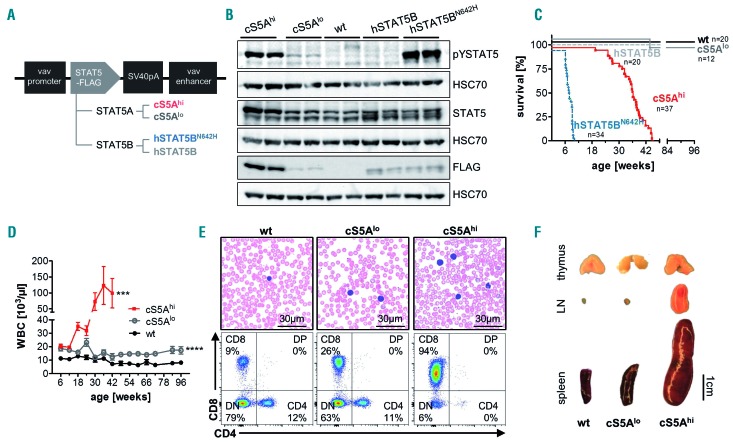
Figure 1.
Expression of a gain-of-function Stat5a or STAT5B variant leads to a polyclonal CD8+ T-cell disease. (A) Schematic representation of the FLAG-tagged STAT5 constructs for generation of transgenic mouse lines expressing hyperactive Stat5a (cS5Alo and cS5Ahi) or human STAT5B (hSTAT5B and hSTAT5BN642H). (B) Immunoblot on lymph node lysates from cS5Ahi, cS5Alo, wildtype (wt), hSTAT5B, and hSTAT5BN642H mice (n=2/genotype) using antibodies to FLAG, phosphotyrosine(Y694)-STAT5 (pYSTAT5) and STAT5. HSC70 was used as a loading control. Representative blot of four experiments. (C) Kaplan-Meier disease-free survival plot of wt (n=20), cS5Alo (n=12), cS5Ahi (n=37), hSTAT5B (n=20) and hSTAT5BN642H (n=34) mice; P≤0.0001 with the log-rank (Mantel-Cox) test. (D) White blood cell (WBC) count measured at 6-week intervals from wt (n≥6), cS5Alo (n≥8) and cS5Ahi (n≥10) mice for 66 weeks (cS5Ahi until 42 weeks, unpaired t-test wt vs. cS5Alo P≤0.0001, wt vs. cS5Ahi P=0.003). (E) Representative blood smears of 32-week old mice, scale bar 30 mm, and representative flow cytometry dot plots of CD4 and CD8 cells in peripheral blood of 32-week old wt, cS5Alo and cS5Ahi mice. (F) Macroscopic appearance of thymi, lymph nodes (LN), and spleens of representative age-matched wt, cS5Alo and diseased cS5Ahi mice.

