Key Points
Treatment of relapsed refractory T-PLL with venetoclax monotherapy results in only transient and minor clinical responses.
In vitro analyses pre- and postvenetoclax indicate dual dependence on BCL2 and MCL1; combined BCL2 and MCL1 inhibition are synergistic.
Introduction
T-prolymphocytic leukemia (T-PLL) is a rare mature T-cell malignancy that pursues an aggressive clinical course. Established treatment options are limited to the CD52 monoclonal antibody alemtuzumab1 or purine analog-based chemotherapy. Nonresponders to alemtuzumab exhibit a median overall survival of 4 months, whereas for responders, the 5-year overall survival is 20%.2
Venetoclax is a potent selective oral inhibitor of the BCL2 antiapoptotic protein, with demonstrated efficacy in relapsed or refractory (R/R) chronic lymphocytic leukemia (CLL)3,4 and mantle cell lymphoma.5,6 In contrast, 2 cases of R/R T-PLL treated with venetoclax monotherapy demonstrated only moderate and transient activity.7 There are no other published in vivo data on the use of venetoclax in T-PLL. We report an in vitro analysis of a patient with refractory T-PLL who received venetoclax monotherapy and show dual dependence on BCL2 and MCL1. This synergistic combination was observed in 10 other T-PLL patient samples, suggesting that concurrent inhibition of antiapoptotic proteins may provide more effective therapy.
Methods
In vitro analysis of drug sensitivity
T-PLL cells were maintained in RPMI 1640 media, with or without the addition of interleukin-2 (IL-2; 10 ng/mL), IL-4 (10 ng/mL), and CD154 cross-linking antibody (10 ng/mL), as described previously.8 IL-4 (204-IL) and CD154 (2706-CL)/anti-his (MAB050) were purchased from R&D Systems (Minneapolis, MN), and IL-2 (200-02) was purchased from PeproTech (Rocky Hill, NJ). The SUP-T11 cell line was obtained from DSMZ (Braunschweig, Germany). Cells were treated with venetoclax,9 A1331852,10 and S6384511 (Selleck Chemicals, Houston, TX) for 24 hours before analysis using CellTiter-Glo.12
Statistical analysis
Combinations of drugs were assessed by calculating combination indexes (CIs) using CalcuSyn.13
Ethical approval.
Samples were collected after informed written patient consent in accordance with the Declaration of Helsinki and appropriate institutional ethical approvals from the Oxford Radcliffe Biobank (REC: 09/H0606/5+5) and the University of Leicester Haematological Malignancies Tissue Bank (Leicestershire, Northamptonshire and Rutland REC06/Q2501/122).
Results and discussion
Clinical response to venetoclax in a patient with refractory T-PLL
A 48-year-old woman presented with weight loss and fatigue. Her white cell count (WCC) was 40 × 109/L, with peripheral blood (PB) morphology, immunophenotyping, and fluorescent in situ hybridization consistent with T-PLL. IV alemtuzumab (3 and, subsequently, 5 times weekly), pentostatin, and, finally, fludarabine, mitoxantrone, and dexamethasone were all ineffective (summarized in detail in supplemental Table 1). At this juncture, a trial of self-funded venetoclax monotherapy was commenced.
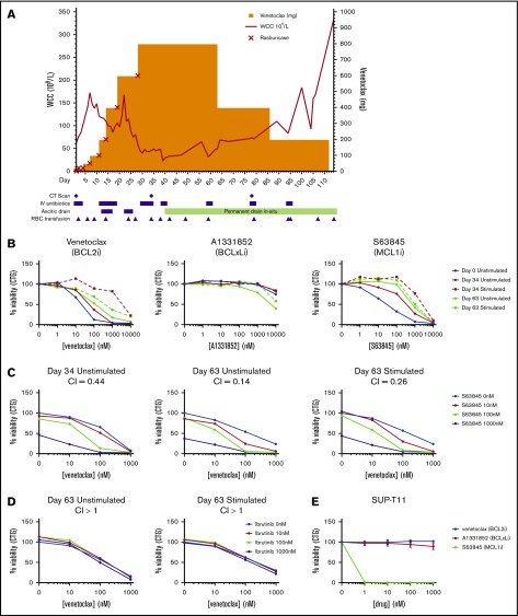
At the commencement of venetoclax, the patient had a WCC of 61 × 109/L, was anemic and thrombocytopenic, and had splenomegaly (22 cm by computerized tomography), ascites, and an Eastern Cooperative Oncology Group performance status of 3. Venetoclax was commenced at 20 mg daily.14 Dose escalation to 50, 100, 200, 400, and 600 mg daily proceeded every ∼3 days (supplemental Table 2). Rasburicase (flat dose 7.5 mg IV) and IV saline hydration were given as tumor lysis syndrome prophylaxis at each escalation. No biochemical or clinical tumor lysis syndrome was noted (Figure 1). In light of the aggressive nature of the disease, a decision was made to escalate to a maximum tolerable dose. During dose escalation to 600 mg, an initial WCC doubling time of ∼5 days was observed. The final escalation to 800 mg was reached at day (D)29 after excellent initial tolerance and a limited WCC response (Figure 1). No initial dose-related response was seen with a peak WCC ∼170 × 109/L observed. The WCC eventually decreased to a nadir of 22 × 109/L by D39. Computed tomography imaging at D77 showed a minor reduction in splenomegaly (19 cm).
Figure 1.
Clinical course and in vitro analysis. (A) Venetoclax monotherapy dose escalation, WCC level, and clinical course. (B) T-PLL cells isolated at D0, D34, and D63 were incubated with different concentrations of venetoclax (left panel), S63845 (middle panel), or A1331852 (right panel), with and without stimulation with IL-2, IL-4, and CD40L for 24 hours before analysis of cell viability using CellTiter-Glo. (C) Unstimulated and stimulated patient cells were exposed to different concentrations of venetoclax and S63845 in combination before analysis of cell viability using CellTiter-Glo at 24 hours. (D) Unstimulated and stimulated patient cells were exposed to different concentrations of venetoclax and ibrutinib in combination before analysis of cell viability using CellTiter-Glo at 48 hours. Experiments with primary samples (B-D) were performed in triplicate, and data shown are mean values. CI values < 1 indicate synergy. (E) SUP-T11 cell line was exposed to different concentrations of venetoclax, S63845, or A1331852 before analysis of cell viability using CellTiter-Glo. Data are shown as mean ± standard deviation. n = 3.
Despite an initial minor reduction in the lymphocytosis, there was a minimal response in other disease parameters. The patient required ongoing transfusion support and a semipermanent ascitic drain because of rapid reaccumulation of ascites. Diarrhea (grade 2) and neutropenia (grade 4) both occurred at 800 mg daily. In light of the rapid disease progression, the patient was subsequently managed with palliative intent and died on D117.
Sensitivity of T-PLL cells to BCL2 inhibition and MCL1 inhibition in vitro
T-PLL cells from the patient were assessed for sensitivity to venetoclax and other specific BH3 mimetics targeting BCLxL (A1331852) and MCL1 (S63845) in vitro. Patient samples taken prevenetoclax (D0), at 800 mg daily (D34), and after dose reduction (D63) were treated with BH3 mimetics for 24 hours. Pretreatment, the cells were sensitive to venetoclax and the MCL1 inhibitor S63845 (50% effective concentration [EC50], 18 nM and 36 nM, respectively) (Figure 1B). In contrast, the cells were completely resistant to BCLxL inhibition with A1331852 (Figure 1B). Cells taken from the later time points exhibited a slight decrease in response to venetoclax but remained sensitive (EC50, 58 nM and 114 nM). Stimulation with IL-2, IL-4, and CD40L was undertaken to mimic the effects of the lymph node microenvironment.8 Stimulation inhibited responses to BCL2 inhibition (BCL2i) and MCL1 inhibition (MCL1i) (Figure 1B), suggesting that disease progression may have been due to microenvironmental signaling. Because of the joint dependence upon BCL2 and MCL1, the specific mimetics were also tested in combination (Figure 1C). The combination of venetoclax and S63845 was synergistic with a CI of 0.44, indicative of a strong synergistic interaction. Moreover, this synergistic interaction was also observed in the later time point sample under unstimulated and stimulated conditions. A clinical trial of T-PLL with venetoclax and ibrutinib is recruiting (NCT03873493); therefore, this combination was also assessed. However, for this sample, the addition of ibrutinib did not enhance the effect of venetoclax (Figure 1D). In contrast to the patient samples, the SUP-T11 cell line showed picomolar sensitivity to MCL1i (EC50, 10 pM) but was resistant to BCL2i and BCLxL inhibition (Figure 1E).
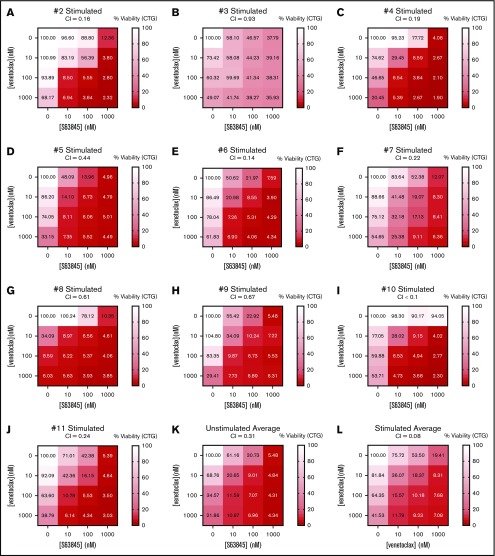
To interrogate whether this response was patient specific or could be applied more broadly, the sensitivity of 10 other T-PLL patient samples to BH3 mimetics were also assessed in vitro (supplemental Figure 1). Nine of 10 patients responded to venetoclax and/or S63845 within the nanomolar range, whereas only 1 patient was sensitive to A1331852. Stimulation decreased sensitivity to the mimetics. The combination of venetoclax plus S63845 was also tested (Figure 2; supplemental Figure 2). Despite the decreased sensitivity to the mimetics under stimulated conditions, the combination was synergistic in all samples. The average response to the combination across all samples had CI values of 0.31 and 0.08 in in unstimulated and stimulated samples, respectively, indicating that dual targeting of these proteins might be effective, irrespective of microenvironment signaling.
Figure 2.
Synergistic interaction between BCL2i and MCL1i. (A-J) Cells isolated from 10 T-PLL patients were stimulated with IL-2, IL-4, and CD40L and treated with different concentrations of venetoclax and S63845 in combination for 24 hours before analysis of cell viability using CellTiter-Glo. All unstimulated (K) and stimulated (L) responses across all samples averaged. Experiments were performed in triplicate, and data shown are mean values. CI values <1 indicate synergy.
Boidol et al7 demonstrated variable sensitivity of T-PLL to venetoclax in vitro; T-PLL was less sensitive than CLL but more sensitive than acute myeloid leukemia. In their study, EC50 of venetoclax in T-PLL varied from 36 nM to 1040 nM. Our data, from the patient samples and the SUP-T11 cell line (which, unusually for T-PLL, exhibits biallelic TP53 mutations), highlight the functional importance of MCL1, as well as BCL2. Boidol et al7 also showed that venetoclax monotherapy at 800 to 1200 mg daily was associated with only transient responses in 2 patients with T-PLL. We describe the third patient with R/R T-PLL in the literature to receive venetoclax monotherapy and, again, demonstrate a short-lived response, limited to the PB. The in vitro sensitivity of T-PLL contrasts with the lack of overall clinical efficacy. Given the response in the PB alone, it is possible that only subclonal PB T-PLL populations were sensitive to BCL2i. This is supported by the impaired in vitro response to treatment following cytokine stimulation, which has previously been demonstrated to induce resistance to navitoclax (BCL2, BCLxL, BCL-w inhibitor) in CLL.8 Collectively, these data suggest that, in contrast to CLL, BCL2i alone is not adequate to treat T-PLL satisfactorily. Our data indicate a strong dependence on MCL1, as well as BCL2, and in vitro analysis of 11 patient samples also unveiled a strong synergy between BCL2i and MCL1i. Previous studies have shown SNS-032, a cyclin-dependent kinase inhibitor that downregulates the short-lived MCL1 protein, to be extremely effective, in a T-PLL–specific manner, in combination with venetoclax.15 MCL1i is currently being evaluated in clinical trials (NCT02979366, NCT02992483, NCT03672695) and may provide more effective options for patients with T-PLL as monotherapy or in combination with BCL2i.
Supplementary Material
The full-text version of this article contains a data supplement.
Acknowledgments
This work was supported by Cancer Research UK in conjunction with the UK Department of Health and an Experimental Cancer Medicine Centre grant (C10604/A25151). T.A.E. and A.H.S. acknowledge support from the Oxford National Institute for Health Research Biomedical Research Centre.
Footnotes
Data sharing requests should be sent to Toby A. Eyre (toby.eyre@ouh.nhs.uk).
Authorship
Contribution: V.M.S. performed research, analyzed data, and wrote the manuscript; O.L. analyzed data and wrote the manuscript; D.C. contributed analytical tools and analyzed data; L.P., A.H.S., O.G., and S.M. performed research; D.B. contributed analytical tools; S.J. performed research and analyzed data; and M.J.S.D. and T.A.E. designed and performed research, supervised the study, analyzed data, and wrote the manuscript.
Conflict-of-interest disclosure: T.A.E. has received speaker fees, honorarium, and travel grants from AbbVie. M.J.S.D. has received travel grants from AbbVie. A.H.S. has received speaker bureau fees from AbbVie. The remaining authors declare no competing financial interests.
Correspondence: Toby A. Eyre, Department of Haematology, Cancer and Haematology Centre, Oxford University Hospitals NHS Trust, Oxford OX3 7LE, United Kingdom; e-mail: toby.eyre@ouh.nhs.uk.
References
- 1.Dearden CE, Matutes E, Cazin B, et al. . High remission rate in T-cell prolymphocytic leukemia with CAMPATH-1H. Blood. 2001;98(6):1721-1726. [DOI] [PubMed] [Google Scholar]
- 2.Herling M, Patel KA, Teitell MA, et al. . High TCL1 expression and intact T-cell receptor signaling define a hyperproliferative subset of T-cell prolymphocytic leukemia. Blood. 2008;111(1):328-337. [DOI] [PMC free article] [PubMed] [Google Scholar]
- 3.Stilgenbauer S, Eichhorst B, Schetelig J, et al. . Venetoclax in relapsed or refractory chronic lymphocytic leukaemia with 17p deletion: a multicentre, open-label, phase 2 study. Lancet Oncol. 2016;17(6):768-778. [DOI] [PubMed] [Google Scholar]
- 4.Roberts AW, Davids MS, Pagel JM, et al. . Targeting BCL2 with venetoclax in relapsed chronic lymphocytic leukemia. N Engl J Med. 2016;374(4):311-322. [DOI] [PMC free article] [PubMed] [Google Scholar]
- 5.Davids MS, Roberts AW, Seymour JF, et al. . Phase I first-in-human study of venetoclax in patients with relapsed or refractory non-Hodgkin lymphoma. J Clin Oncol. 2017;35(8):826-833. [DOI] [PMC free article] [PubMed] [Google Scholar]
- 6.Eyre TA, Walter HS, Iyengar S, et al. . Efficacy of venetoclax monotherapy in patients with relapsed, refractory mantle cell lymphoma after bruton tyrosine kinase inhibitor therapy. Haematologica. 2019;104(2):e68-e71. [DOI] [PMC free article] [PubMed] [Google Scholar]
- 7.Boidol B, Kornauth C, van der Kouwe E, et al. . First-in-human response of BCL-2 inhibitor venetoclax in T-cell prolymphocytic leukemia. Blood. 2017;130(23):2499-2503. [DOI] [PubMed] [Google Scholar]
- 8.Vogler M, Butterworth M, Majid A, et al. . Concurrent up-regulation of BCL-XL and BCL2A1 induces approximately 1000-fold resistance to ABT-737 in chronic lymphocytic leukemia. Blood. 2009;113(18):4403-4413. [DOI] [PubMed] [Google Scholar]
- 9.Souers AJ, Leverson JD, Boghaert ER, et al. . ABT-199, a potent and selective BCL-2 inhibitor, achieves antitumor activity while sparing platelets. Nat Med. 2013;19(2):202-208. [DOI] [PubMed] [Google Scholar]
- 10.Leverson JD, Phillips DC, Mitten MJ, et al. . Exploiting selective BCL-2 family inhibitors to dissect cell survival dependencies and define improved strategies for cancer therapy. Sci Transl Med. 2015;7(279):279ra40. [DOI] [PubMed] [Google Scholar]
- 11.Kotschy A, Szlavik Z, Murray J, et al. . The MCL1 inhibitor S63845 is tolerable and effective in diverse cancer models. Nature. 2016;538(7626):477-482. [DOI] [PubMed] [Google Scholar]
- 12.Kozaki R, Vogler M, Walter HS, et al. . Responses to the selective Bruton’s tyrosine kinase (BTK) inhibitor tirabrutinib (ONO/GS-4059) in diffuse large B-cell lymphoma cell lines. Cancers (Basel). 2018;10(4):E127. [DOI] [PMC free article] [PubMed] [Google Scholar]
- 13.Chou TC. Drug combination studies and their synergy quantification using the Chou-Talalay method. Cancer Res. 2010;70(2):440-446. [DOI] [PubMed] [Google Scholar]
- 14.AbbVie Venclyxto. Summary of product characteristics. https://www.ema.europa.eu/en/documents/product-information/venclyxto-epar-product-information_en.pdf. Accessed 5 January 2020.
- 15.Andersson EI, Pützer S, Yadav B, et al. . Discovery of novel drug sensitivities in T-PLL by high-throughput ex vivo drug testing and mutation profiling. Leukemia. 2018;32(3):774-787. [DOI] [PubMed] [Google Scholar]
Associated Data
This section collects any data citations, data availability statements, or supplementary materials included in this article.


