Abstract
Background
This study investigated the remineralisation effect of bioactive glass on artificial dentine caries.
Methods
Dentine disks with artificial caries were treated with bioactive glass (group BAG), casein phosphopeptide–amorphous calcium phosphate (CPP-ACP) (group CPP-ACP), sodium fluoride glycerol (group F) or deionized water (group W). All disks were subjected to pH cycling for 28 days subsequently. The topography, microhardness and remineralisation depth of the dentine carious lesion were assessed by atomic force microscopy (AFM), microhardness testing and confocal laser scanning microscope (CLSM), respectively.
Results
AFM images indicated mineral depositions on the surface of the carious lesion in group BAG. The changes of Vickers hardness number (ΔVHN, mean ± SD) after pH cycling were 9.67 ± 3.60, 6.06 ± 3.83, 5.00 ± 2.19 and − 1.90 ± 2.09 (p < 0.001) in group BAG, group CPP-ACP, group F and group W, respectively. The remineralisation depth (mean ± SD) of the carious lesion in group BAG, group CPP-ACP, group F and group W were 165 ± 11 μm, 111 ± 11 μm, 75 ± 6 μm and 0 μm (p < 0.001), respectively.
Conclusion
Bioactive glass possessed a promising remineralisation effect on artificial dentine caries and could be a therapeutic choice for caries management.
Keywords: Artificial caries, Dentine, Remineralisation, Bioactive glass, CPP-ACP
Background
Dental caries (tooth decay) is one of the most prevalent chronic disease [1]. Dentine caries refers to the situation in which caries has progressed into dentine and caused significant lesion depth, it can progress rapidly since dentine is a porous organic-inorganic composite material. The traditional management of dentine caries has focused primarily on treatment via the excision of diseased tissues and the subsequent restoration of the defect [2]. The primary goal of contemporary mineral invasive dentistry is to respect tooth structure, retaining viable and biologically repairable tissues to maintain tooth vitality. Therefore, retaining demineralised dentine which has no bacteria invasion and restored it with bioactive materials which has remineralization capability is the trend of caries treatment. This procedure can not only prevent further bacterial infection, but also preserve dental hard tissues as much as possible, which is beneficial to protect dental pulp tissues, and increase the retention ability and resistance performance of restoration materials [3]. Bioactive materials play an important role in treatment of the partial removal of caries.
Bioactive materials are therefore been introduced since whey will be intended to interact in some positive way with the oral environment. 45S5 bioactive glass (BAG) was initially introduced in 1970s, it is a glass in Na2O-CaO-SiO2-P2O5 system, high in calcium content [4]. It was found to be able to bond with bone rapidly and strongly, it stimulates bone growth away from the bone–implant interface [5]. The mechanism for bone bonding is attributed to a hydroxycarbonate apatite (HCA) layer on the surface of the glass, following initial glass dissolution. BAG has been introduced into dentistry to treat dentine hypersensitivity in 2004 [6]. In vitro studies showed that the BAGs particles can adhere to dentine and form an HCA layer which is similar in composition to dentine, therefore block the dentinal tubules [7]. This indicates that BAG seems to work by stimulating mineralization (calcium phosphate deposition over the dentine tubules) [8, 9].
Apart from treating dentine hypersensitivity, BAG has been used in different areas in dentistry. A.S. Bakry’s studies showed that BAG can be used to treat enamel leukoplakia caused by orthodontic treatment and as a temporary filling material for remineralization [10, 11]. BAG also can be used as an auxiliary material for tooth bleaching to prevent/repair the damage caused by enamel bleaching agent [12]. Research shows that a novel BAG has been developed to as a viable alternative to adhesive removal with a TC bur [9]. A combined dentine pre-treatment using BAG followed by polyacrylic acid may increase the bond strength and maintain it stable over time [13]. Increasing BAG filler content in pit and fissure sealants may prevent secondary caries at enamel edge [14]. However, the effect and mechanisms of BAG on dentine caries is still unclear.
It had also been reported that several other materials could remineralize dentin, including casein phosphopeptide-amorphous calcium phosphate (CPP-ACP) and fluoride compounds [1, 15, 16]. CPP-ACP enhances remineralization by stabilizing calcium phosphate so that high concentrations of calcium ions and phosphate ions exist in the solution. Fluoride has been shown to enhance the remineralisation of caries [17]. Fluoride is mainly combined with supersaturated calcium and phosphorus ions to further promote the deposition of calcium and phosphorus, forming new antacid fluorapatite crystals and realizing remineralization. These studies have proclaimed sufficient observations to prove the formation of mineral depositions on dentin surface after treatment. In this study, CPP-ACP and sodium fluoride are used as positive controls, pH-cycling model was used to simulate the dynamic variation in mineral saturation and the pH altering with the natural caries process, which refers to in vitro experimental protocols including exposure of dentin to combinations of demineralization and remineralization. The null hypothesis of the study is that BAG does not have reminerlisation effect on artificial dentine caries.
Methods
Dentine disks preparation
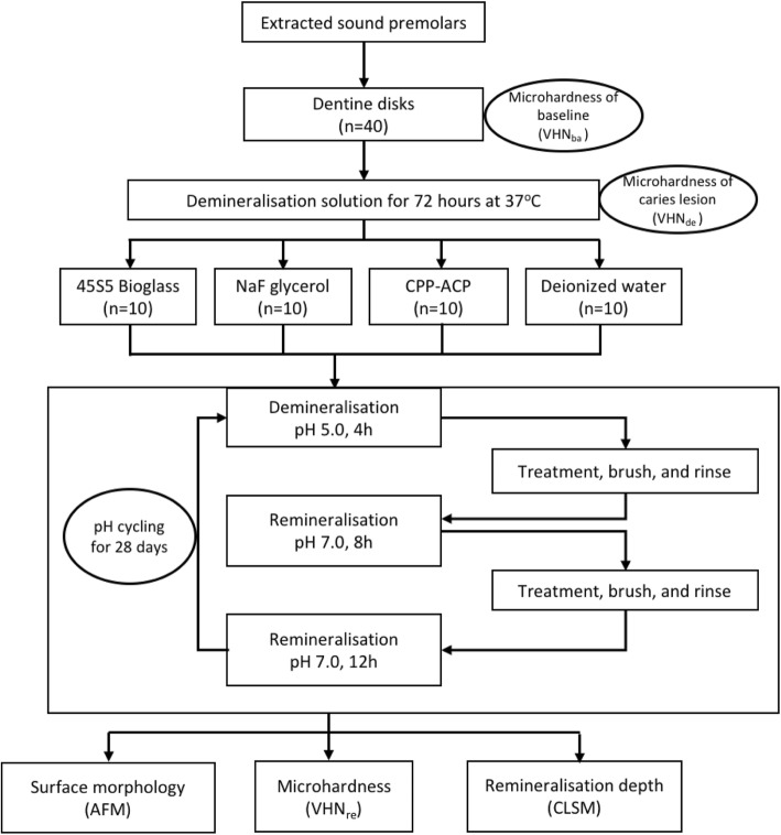
Ethical approval was obtained from Ethics Committee of the School and Hospital of Stomatology, Nanjing Medical University (2019–284). This study was conducted in full accordance with the Declaration of Helsinki of the World Medical Association. All participants received dental treatment at the Hospital of Stomatology of Nanjing Medical University and provided written informed consent. The written consents were obtained from the parents/guardians of the teenagers who were under 16 years of age. Forty human premolars extracted within one month for orthodontic reasons were collected and stored in deionized water containing 0.1% thymol at 4 °C prior to the experiment. Crowns with caries, restorations, or fractures were abandoned. The flow chart in Fig. 1 summarises the protocol of this study.
Fig. 1.
Flowchart of experimental design
Forty dentine disks with a thickness of 1.0 mm, perpendicular to the long axis of the tooth above the cemento-enamel junction, were prepared by low-speed water cooled diamond saw (Isomet, Buehler Ltd., Lake Bluff, IL, USA). All disks were free of coronal enamel or pulpal exposures. A standard smear layer was created on the coronal side of the dentin surface using silicon carbide papers of 600-grit, 800-grit, 1200-grit and ultrasonically washed in deionized water 3 times each for 60s, while the opposite sides were coated with acid-resistant nail polish.
Demineralisation and remineralisation solutions
The demineralisation solution contains was 0.05 M acetic acid containing 2.2 mM CaCl2·2H2O (Shanghai Ling Feng Chemical Reagents Co., Ltd.,) and 2.20 mM KH2PO4 (Shanghai Ling Feng Chemical Reagents Co., Ltd.,) and was adjusted to pH 5.0.
The remineralisation solution contained 1.5 mM CaCl2·2H2O, 0.90 mM KH2PO4 and 130 mM KCl (Shanghai Ling Feng Chemical Reagents Co., Ltd.,), and was adjusted to pH 7.0. Both of them were freshly prepared [18].
Preparation of artificial lesions
All disks were immersed into deminerlisation solution for 72 h at 37 °C. The surface hardness of the disks was characterized by Vicks microhardness number (VHN).
Experimental procedure
The demineralized dentine disks were randomly assigned into four groups (n = 10). The treatments were applied twice a day using electric toothbrush (Colgate 360°, Colgate- Palmolive Co.), the disks were rinsed thoroughly after brushing to mimic the real situation.
Group 1: 0.075 g/mL BAG paste (Actimins Paste, Datsing Bio-Tech Co. Ltd., Beijing, China), (Na2O2 4.5 wt%, CaO2 4.5 wt%, P2O5 6.0 wt%, SiO2 45 wt%).
Group 2: Sodium fluoride and Glycerin Paste (75% sodium fluoride and 25% Glycerol).
Group 3: 10% CPP-ACP (Recaldent™, Japan GC Co.,Ltd) (CPP–ACP:10%; Ca content: 13 mg/g; P content: 5.6 mg/g).
Group 4: Deionized water.
All disks were subjected to 28 days’ pH cycles, which consisted of 4 h demineralization solution followed by a 20 h remineralisation solution. Each disk was placed in a 15 mL container. All the solutions were freshly made prior to use. All the disks were collected for testing after pH cycling.
Surface roughness test
Three disks from each group embedded in epoxy resin were imaged using an atomic force microscope (AFM; CSPM 5000, Ben Yuan Ltd., Beijing, China) in order to analyze surface morphology changes. The dentine disks were polished with silicon carbide paper (2000 grit), then 1.0, 0.3 and 0.05 μm diamond mask alumina suspensions sequentially, followed by ultrasonically cleaning in deionized water for 15 min to remove the residues [19].
Topographical images of the surface were performed in the tapping mode using a silicon nitride scanning probe in admosphere, in which the probe periodically touches the sample surface, producing higher quality images [15]. Each dentine disk was observed in 4 different sites and obtained three-dimensional images of the dentin surface. In each image, a field of view at 50 μm × 50 μm scan size, 1.5 Hz scan rate and a resolution of 512 by 512 pixels were employed on the entire surface.
Surface microhardness test
Seven disks from each group were randomly selected to measure the microhardness respectively of baseline (VHNba), before pH cycling (VHNde) and after pH cycling (VHNre). The microhardness value of each disk was measured with a Vickers indenter on a Hardness Tester (DHV-1000, Shangcai testermachine Co., LTD, China).
Indentations were made with a Vickers diamond indenter of three widely similarly positioned locations. The indentations with loads of 0.98 N and time for 15 s were considered to be suitable for dentin measurement of the long and short indentation diagonals and resulted in minimum surface damage. As the apexes of the diagonals were estimated on the surface, Vickers number could be converted by the size of the indentation. Three values were averaged to produce one hardness value for each specimen. The change in Vickers hardness number (ΔVHN) was determined as the difference of the caries lesion before and after pH cycling (ΔVHN = VHNre - VHNde).
Confocal laser scanning microscopy (CLSM)
The disks from microhardness study were cut to thin sections with thickness of 500 μm along the treatment surface, and then stained with a freshly prepared 0.1% Rhodamine B solution (Aldrich Chem. Co., Milwaukee, WI, USA) for 1 h, and rinsed for 3 times with deionized water. Samples were analyzed with a confocal laser scanning microscopy (CLSM, CarlZeiss LSM 710, Carl Zeiss, Inc., Germany). The reflection imaging was performed using the laser. Standard settings for contrast, brightness and laser power were used for all images. The remineralisation depths (H) were quantitatively analyzed with an image-analysis system (Image Pro-Plus, 6.0).
Statistical analysis
All of the data were assessed for a normal distribution using the Shapiro–Wilk test for normality (p > 0.05). A one-way ANOVA was used to compare the VHN and remineralisation depth across the four treatment groups, followed by LSD multiple comparison was used to compare among groups. All of the analyses were conducted using IBM SPSS Version 2.0 software (IBM Corporation, Armonk, New York, USA). The cut-off level for significance was taken as 5% for all of the analyses.
Results
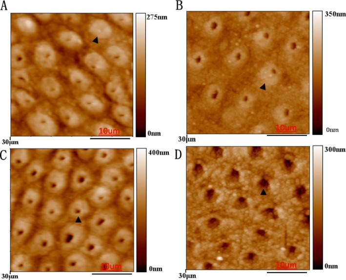
Figure 2 showed the surfaces of the dentine disks after treatments and pH cycling. We observed that dentine collagen fibres were not exposed on the relatively smooth surface of the BAG, fluoride and CPP-ACP treated dentine (Fig. 2 a, 12B and 2C). In particular, parcipatation on the peritubular dentine, and little space remained in both inter-tubular and intra-tubular areas. Figure 2 d is the negative control which received water, enlarged dentinal tubulars when comparing with other groups, indicating partial demineralisation.
Fig. 2.
AFM micrographs in the tapping mode of specimen surfaces after 28-day treatment by bioactive glass a, sodium fluoride glycerin b, CPP-ACP c and deionized water d
The means and standard deviations of VHN of the dentine of 4 groups of the baseline, demineralized and after pH cycling are summarized in Table 1. Group BAG, Group CPP-ACP and Group F showed higher VHN when comparing Group W after 28 days pH cycling (p = 0.020). There was no significant difference in VHN among different groups in the baseline (p = 0.919), as well as after 72 h demineralization (p = 0.290). Group BAG and Group CPP-ACP presented larger ΔVHN when comparing with Group F (p < 0.001).
Table 1.
Mean VHN and SD of dentin surface in sound dentine, after demineralisation and after pH cycling. VHN, Vickers microhardness numbers
| Group (n = 7 each) | VHNba Mean ± SD | VHNde Mean ± SD | VHNre Mean ± SD | ΔVHN (VHNre - VHNde) Mean ± SD |
|---|---|---|---|---|
| Group BAG | 60.44 ± 6.41 | 13.05 ± 2.90 | 22.72 ± 3.91a | 9.67 ± 3.60a |
| Group CPP-ACP | 60.67 ± 3.53 | 15.60 ± 4.62 | 21.66 ± 3.92a | 6.06 ± 3.83a |
| Group F | 60.07 ± 9.95 | 13.35 ± 4.26 | 18.35 ± 5.62a | 5.00 ± 2.19b |
| Group W | 58.36 ± 2.82 | 16.82 ± 3.31 | 14.92 ± 3.23b | − 1.90 ± 2.09c |
| p value | 0.919 | 0.290 | 0.020 | < 0.001 |
| LSD comparison | N/A | N/A | a > b | a > b > c |
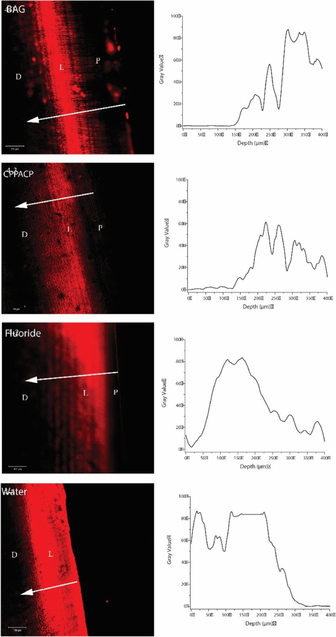
CLSM observation showed a red fluorescent band representing caries lesion. The remineralization is evidenced by the decrease in fluorescence on the superficial layer of the lesion (Fig. 3). The precipitation band was wider in Group BAG when compared to the fluoride treated and the control group. Correspondingly, Table 2 show the depth of remineralisation zone after 28 days pH cycling in the four experimental groups. The depth of remineralisation zone of Group BAG is 165.40 ± 11.09 μm, which is significantly higher (p < 0.001) than those in other groups, demonstrating a promising capability in remineralising dentine caries. Combined with the CLSM images, BAG promoted mineral deposition on the superficial layer of the lesion.
Fig. 3.
Confocal Laser Scanning Microscopy representative image of artificial dentin caries treated by bioactive glass (4–1), sodium fluoride glycerin (4–2), CPP-ACP (4–3) and deionized water (4–4). (L, lesion; D, sound dentin; P, precipitation band)
Table 2.
The depth of dentine remineralisation zone in 4 experimental groups (n = 7)
| Group | Depth (μm) |
|---|---|
| Group BAG | 165.40 ± 11.09a |
| Group CPP-ACP | 110.86 ± 10.55b |
| Group F | 75.18 ± 6.47c |
| Group Water | 0 |
| p value | < 0.001 |
| LSD comparison | a > b > c |
Discussion
This study investigated the remineralization effect of BAG on artificial dentine caries. It provides useful information about the microstructure changes in dentine caries after BAG application. According to the result of the study, the null hypothesis was rejected. BAG showed a promising remineralization effect on artificial dentine caries with increasing microhardness by forming a remineralization zone on the lesion surface. Hardness testing is an indirect method of tracking changes in the mineral content of dentine, and several microhardness studies on dentine in arrested carious lesions have been published [20, 21]. A limitation of the study is the chemical system used is the lack of biological component, in which antimicrobial of the treatment could be underestimated. A biological model can be employed in the next step to assess antimicrobial effect. In addition, the results cannot be extrapolated to the in vivo situation and caution should be exercised in their interpretation. In AFM study, the specimens require high quality polished surface. Polishing teeth could remove some attachments on the surface, but according to the AFM results, the BAG mainly embedded into dentin tubules to form deposits.
Two perspectives have been focus to achieve dentine caries remineralisation: coating nucleation templates on demineralized dentin or creating a local environment with high calcium and phosphorus concentration [22–24]. The process of remineralising dentine caries using BAG includes exchanging ions (Na+, Ca2+, PO43−, F−) in the silicate network of BAG with the surrounding oral liquid to supersaturate the ions in the fluid, which are then reprecipitated on the silicate network of BAG in the tissue [25]. BAG can make materials and tissues bond tightly, which is conducive to promoting the remineralization of calcium phosphate on the surface of teeth in vivo [26]. It can promote the formation of stable crystalline hydroxyapatite crystals on the surface of demineralized teeth in salivary environment, thus promoting dentine caries remineralisation. In current study, a very fine BAG powder (Actimins Paste, Datsing Bio-Tech Co. Ltd., Beijing, China) with the maximum grain size is less than 90 nm was used [27]. Small size particles facilitate the penetration into dentine caries, they also provide large surface area for reaction.
It had been demonstrated that dentine remineralisation occurs neither by spontaneous precipitation nor by nucleation of mineral on the organic matrix but by growth of residual crystals in the lesions [28]. And as it has been discovered by researchers that remineralization was possible even at a high degree of initial mineral loss, where it might have been considered that the caries process had happened [29]. It is advantageous to save softened but not the bacterial invasion demineralization dentin, which is consistent with minimum damage strategy for dentin caries treatment. Therefore, various active researches are currently being carried out to seal the exposed dentin tubules with some effective materials and improve the bonding at the dentin interface so as to repair demineralized dentin through remineralization.
Fluoride ions promote the formation of fluorapatite in enamel in the presence of calcium and phosphate ions produced during enamel demineralization by plaque bacterial organic acids. This is now believed to be the major mechanism of fluoride ion’s action in preventing enamel demineralization [30, 31]. It was documented that the anti-cariogenic effects of fluoride mainly through two principal mechanisms: inhibiting demineralization when fluoride is present on crystal surface during an acid challenge; and enhancing remineralization by forming a low soluble substance similar to acid-resistant mineral fluorapatite covering the crystal surface [9, 32]. Some scholars have also found that when the demineralized dentin does not contain hydroxyapatite, no new hydroxyapatite crystals will nucleate after immersion in the remineralized solution. Research has shown that fluoride has limited ability to remineralize dentin when the residual crystals of the lesion are insufficient [33]. CPP-ACP, which has been consider to promote remineralization of the carious lesions by maintaining a supersaturated state of enamel mineral, plays a key role in the biomineralization of dentin [15, 34]. It has also been suggested that CPP-ACP has a multifactorial anticariogenic mechanism. A vitro study showed that the presence of CPP-ACP prevents demineralization of dentin surface and promotes the remineralization of artificial caries-like dentin lesions.
In current study, the treatments were applied on the dentine disks via brushing by an electric toothbrush for 2 min, to mimic the real situation. The mineral was shown to deposit on the surface of the caries lesion in all treatment groups due to the AFM results (Fig. 2), which indicate that daily brush will not remove the deposit. We found that BAG group has the largest remineralisation depth when compared to other groups (Table 2). Ten Cate summarized the factors that enhance the remineralisation of deep lesions, and proposed that calcium may be rate limiting in remineralization [35]. Pronounced bonding capacity to tooth structure of BAG can be a major reason for this improved remineralisation effect. Based on results of this in vitro study, we believe that BAG inhibits the demineralization and/or promotes the remineralisation of artificial dentine caries under dynamic pH-cycling conditions. BAG has potential to a promising alternative to fluoride in the treatment of caries.
Conclusions
BAG possessed a promising remineralisation effect on artificial dentine caries and could be a therapeutic choice for caries management.
Acknowledgments
We would like to thank Professor Jianwei Zhao from Nanjing University for assisting with the AFM and VHN measurements.
Abbreviations
- AFM
Atomic force microscopy
- BAG
Bioactive glass
- CLSM
Confocal laser scanning microscope
- CPP-ACP
Casein phosphopeptide–amorphous calcium phosphate
- VHN
Vickers hardness number
Authors’ contributions
QW, MLM and YMC conceived and designed the experiments. QW performed the experiments, analysed the data and wrote the original paper. XW, SS and YX contributed to the methodology. MLM, XW, CHC and YMC revised the paper. All authors read and approved the final manuscript.
Funding
This study was financially supported by the general project of the Science and Technology Development Fund of Nanjing Medical University (Project number: NMUB2018171), and project of dentine allergy and bioactive glass research (Project number: 22011–001). These helped with sample collection and preparation, and analysis, and interpretation of the data.
Availability of data and materials
The datasets used and/or analysed during the current study available from the corresponding author on reasonable request.
Ethics approval and consent to participate
Ethical approval was obtained from Ethics Committee of the School and Hospital of Stomatology, Nanjing Medical University (2019–284). This study was conducted in full accordance with the Declaration of Helsinki of the World Medical Association. All participants received dental treatment at the Hospital of Stomatology of Nanjing Medical University and provided written informed consent. The written consents were obtained from the parents/guardians of the teenagers who were under 16 years of age.
Consent for publication
Not applicable.
Competing interests
The authors declare that they have no competing interests.
Footnotes
Publisher’s Note
Springer Nature remains neutral with regard to jurisdictional claims in published maps and institutional affiliations.
Contributor Information
May Lei Mei, Email: may.mei@otago.ac.nz.
Yaming Chen, Email: yaming_chen@njmu.edu.cn.
References
- 1.Rocha Gomes Torres C, Borges AB, Torres LM, Gomes IS, de Oliveira RS. Effect of caries infiltration technique and fluoride therapy on the colour masking of white spot lesions. J Dent. 2011;39(3):202–207. doi: 10.1016/j.jdent.2010.12.004. [DOI] [PubMed] [Google Scholar]
- 2.Weir MD, Ruan J, Zhang N, Chow LC, Zhang K, Chang X, Bai Y, Xu HHK. Effect of calcium phosphate nanocomposite on in vitro remineralization of human dentin lesions. Dent Mater. 2017;33(9):1033–1044. doi: 10.1016/j.dental.2017.06.015. [DOI] [PubMed] [Google Scholar]
- 3.Innes NPT, Chu CH, Fontana M, Lo ECM, Thomson WM, Uribe S, Heiland M, Jepsen S, Schwendicke F. A century of change towards prevention and minimal intervention in Cariology. J Dent Res. 2019;98(6):611–617. doi: 10.1177/0022034519837252. [DOI] [PubMed] [Google Scholar]
- 4.Jones JR. Review of bioactive glass: from Hench to hybrids. Acta Biomater. 2013;9(1):4457–4486. doi: 10.1016/j.actbio.2012.08.023. [DOI] [PubMed] [Google Scholar]
- 5.Rizwan M, Hamdi M, Basirun WJ. Bioglass® 45S5-based composites for bone tissue engineering and functional applications. Journal of biomedical materials research. Part A. 2017;105(11):3197–3223. doi: 10.1002/jbm.a.36156. [DOI] [PubMed] [Google Scholar]
- 6.Forsback AP, Areva S, Salonen JI. Mineralization of dentin induced by treatment with bioactive glass S53P4 in vitro. Acta Odontol Scand. 2004;62(1):14–20. doi: 10.1080/00016350310008012. [DOI] [PubMed] [Google Scholar]
- 7.Gillam DG, Tang JY, Mordan NJ, Newman HN. The effects of a novel bioglass dentifrice on dentine sensitivity: a scanning electron microscopy investigation. J Oral Rehabil. 2002;29(4):305–313. doi: 10.1046/j.1365-2842.2002.00824.x. [DOI] [PubMed] [Google Scholar]
- 8.Bakry AS, Takahashi H, Otsuki M, Tagami J. The durability of phosphoric acid promoted bioglass-dentin interaction layer. Dent Mater. 2013;29(4):357–364. doi: 10.1016/j.dental.2012.12.002. [DOI] [PubMed] [Google Scholar]
- 9.Taha AA, Patel MP, Hill RG, Fleming PS. The effect of bioactive glasses on enamel remineralization: A systematic review. J Dent. 2017;67(undefined):9–17. doi: 10.1016/j.jdent.2017.09.007. [DOI] [PubMed] [Google Scholar]
- 10.Bakry AS, Takahashi H, Otsuki M, Tagami J %J Dental materials : official publication of the Academy of Dental Materials: Evaluation of new treatment for incipient enamel demineralization using 45S5 bioglass. 2014, 30(3):314–320. [DOI] [PubMed]
- 11.Bakry AS, Abbassy MA %J Journal of dentistry: The efficacy of a bioglass (45S5) paste temporary filling used to remineralize enamel surfaces prior to bonding procedures. 2019, 85:33–38. [DOI] [PubMed]
- 12.Deng M, Wen HL, Dong XL, Li F, Xu X, Li H, Li JY, Zhou XD %J International journal of oral science: Effects of 45S5 bioglass on surface properties of dental enamel subjected to 35% hydrogen peroxide. 2013, 5(2):103–110. [DOI] [PMC free article] [PubMed]
- 13.Sauro S, Watson T, Moscardó AP, Luzi A, Feitosa VP, Banerjee A %J Journal of dentistry: The effect of dentine pre-treatment using bioglass and/or polyacrylic acid on the interfacial characteristics of resin-modified glass ionomer cements. 2018, 73:32–39. [DOI] [PubMed]
- 14.Yang SY, Kwon JS, Kim KN, Kim KM. %J Journal of dental research: Enamel Surface with Pit and Fissure Sealant Containing 45S5 Bioactive Glass. 2016, 95(5):550–557. [DOI] [PubMed]
- 15.Ahmadian E, Shahi S, Yazdani J, Maleki Dizaj S, Sharifi S. Local treatment of the dental caries using nanomaterials. Biomedicine & pharmacotherapy. Biomed pharmacother. 2018;108(undefined):443–447. doi: 10.1016/j.biopha.2018.09.026. [DOI] [PubMed] [Google Scholar]
- 16.Llena C; Esteve I; Rodríguez-Lozano FJ; Forner L, The application of casein phosphopeptide and amorphous calcium phosphate with fluoride (CPP-ACPF) for restoring mineral loss after dental bleaching with hydrogen or carbamide peroxide: An in-vitro study. Annals of anatomy = Anatomischer Anzeiger : official organ of the Anatomische Gesellschaft 2019, undefined, (undefined), undefined. [DOI] [PubMed]
- 17.Mei ML, Ito L, Cao Y, Li QL, Lo EC, Chu CH. Inhibitory effect of silver diamine fluoride on dentine demineralisation and collagen degradation. J Dent. 2013;41(9):809–817. doi: 10.1016/j.jdent.2013.06.009. [DOI] [PubMed] [Google Scholar]
- 18.ten Cate JM, Duijsters PP. Alternating demineralization and remineralization of artificial enamel lesions. Caries Res. 1982;16(3):201–210. doi: 10.1159/000260599. [DOI] [PubMed] [Google Scholar]
- 19.Sereda G, VanLaecken A, Turner JA. Monitoring demineralization and remineralization of human dentin by characterization of its structure with resonance-enhanced AFM-IR chemical mapping, nanoindentation, and SEM. Dent Mater. 2019;35(4):617–626. doi: 10.1016/j.dental.2019.02.007. [DOI] [PubMed] [Google Scholar]
- 20.Chu CH, Lo EC. Microhardness of dentine in primary teeth after topical fluoride applications. J Dent. 2008;36(6):387–391. doi: 10.1016/j.jdent.2008.02.013. [DOI] [PubMed] [Google Scholar]
- 21.Hosoya Y, Marshall SJ, Watanabe LG, Marshall GW. Microhardness of carious deciduous dentin. Oper Dent. 2000;25:81–89. [PubMed] [Google Scholar]
- 22.Liang K, Weir MD, Xie X, Wang L, Reynolds MA, Li J, Xu HH. Dentin remineralization in acid challenge environment via PAMAM and calcium phosphate composite. Dent Mater. 2016;32(11):1429–1440. doi: 10.1016/j.dental.2016.09.013. [DOI] [PubMed] [Google Scholar]
- 23.Yang Y, Lv XP, Shi W, Li JY, Li DX, Zhou XD, Zhang LL. 8DSS-promoted remineralization of initial enamel caries in vitro. J Dent Res. 2014;93(5):520–524. doi: 10.1177/0022034514522815. [DOI] [PubMed] [Google Scholar]
- 24.Tay FR, Pashley DH. Guided tissue remineralisation of partially demineralised human dentine. Biomaterials. 2008;29(8):1127–1137. doi: 10.1016/j.biomaterials.2007.11.001. [DOI] [PubMed] [Google Scholar]
- 25.Fernando D; Attik N; Pradelle-Plasse N; Jackson P; Grosgogeat B; Colon P, Bioactive glass for dentin remineralization: A systematic review. Materials science & engineering. C, Materials for biological applications 2017, 76, (undefined), 1369–1377. [DOI] [PubMed]
- 26.Curtis AR, West NX, Su B. Synthesis of nanobioglass and formation of apatite rods to occlude exposed dentine tubules and eliminate hypersensitivity. Acta Biomater. 2010;6(9):3740–3746. doi: 10.1016/j.actbio.2010.02.045. [DOI] [PubMed] [Google Scholar]
- 27.Xu YT, Wu Q, Chen YM, Smales RJ, Shi SY, Wang MT. Antimicrobial effects of a bioactive glass combined with fluoride or triclosan on Streptococcus mutans biofilm. Arch Oral Biol. 2015;60(7):1059–1065. doi: 10.1016/j.archoralbio.2015.03.007. [DOI] [PubMed] [Google Scholar]
- 28.Schwendicke F, Al-Abdi A, Pascual Moscardó A, Ferrando Cascales A, Sauro S. Remineralization effects of conventional and experimental ion-releasing materials in chemically or bacterially-induced dentin caries lesions. Dent Mater. 2019;35(5):772–779. doi: 10.1016/j.dental.2019.02.021. [DOI] [PubMed] [Google Scholar]
- 29.Cao CY, Mei ML, Li QL, Lo EC, Chu CH. Methods for biomimetic remineralization of human dentine: a systematic review. Int J Mol Sci. 2015;16(3):4615–4627. doi: 10.3390/ijms16034615. [DOI] [PMC free article] [PubMed] [Google Scholar]
- 30.Lynch RJ, Navada R, Walia R. Low-levels of fluoride in plaque and saliva and their effects on the demineralisation and remineralisation of enamel; role of fluoride toothpastes. Int Dent J. 2004;54(null):304–309. doi: 10.1111/j.1875-595X.2004.tb00003.x. [DOI] [PubMed] [Google Scholar]
- 31.Reynolds EC, Cai F, Cochrane NJ, Shen P, Walker GD, Morgan MV, Reynolds C. Fluoride and casein phosphopeptide-amorphous calcium phosphate. J Dent Res. 2008;87(4):344–348. doi: 10.1177/154405910808700420. [DOI] [PubMed] [Google Scholar]
- 32.Vieira AE, Delbem AC, Sassaki KT, Rodrigues E, Cury JA, Cunha RF. Fluoride dose response in pH-cycling models using bovine enamel. Caries Res. 2005;39(6):514–520. doi: 10.1159/000088189. [DOI] [PubMed] [Google Scholar]
- 33.Zhang X, Neoh KG, Lin CC, Kishen A. Remineralization of partially demineralized dentine substrate based on a biomimetic strategy. Journal of materials science. Mater Med. 2012;23(3):733–742. doi: 10.1007/s10856-012-4550-5. [DOI] [PubMed] [Google Scholar]
- 34.Rahiotis C, Vougiouklakis G. Effect of a CPP-ACP agent on the demineralization and remineralization of dentine in vitro. J Dent. 2007;35(8):695–698. doi: 10.1016/j.jdent.2007.05.008. [DOI] [PubMed] [Google Scholar]
- 35.ten Cate JM. Remineralization of deep enamel dentine caries lesions. Aust Dent J. 2008;53(3):281–285. doi: 10.1111/j.1834-7819.2008.00063.x. [DOI] [PubMed] [Google Scholar]
Associated Data
This section collects any data citations, data availability statements, or supplementary materials included in this article.
Data Availability Statement
The datasets used and/or analysed during the current study available from the corresponding author on reasonable request.


