Abstract
Background
Strains of the adductor muscle group of the hip are common amongst ice hockey players. The ratio of isometric strengths between the hip adductors and abductors has been offered as a risk factor for hip adductor strain; however, there is no description for how the ratio between hip adductor and abductor strength varies as a function of hip abduction angle.
Hypothesis/Purpose
The aim of this study was to determine the influence of hip joint abduction angle on measured ratios of hip adduction to abduction torque in experienced, recreational, male hockey players. The primary null hypothesis for this study was that hip joint abduction angle would not influence hip adduction-to-abduction torque ratios in male hockey players.
Study Design
Counterbalanced observational cohort.
Methods
Twelve uninjured, male, recreational hockey players, with a minimum experience level of midget AAA/minor competitive or equivalent. Participants performed maximal isometric side-lying hip adduction and abduction exertions against a rigidly constrained load cell at 0, 10, and 20 degrees of hip abduction. Measured peak torques from each exertion were used to derive the hip adductor-to-abductor torque ratio. Kinematics of the trunk, pelvis, and lower limbs were monitored using an optoelectronic motion capture system.
Results
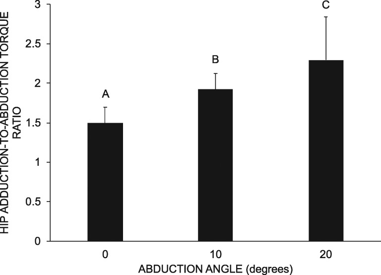
Adductor-to-abductor torque ratio increased from 1.49 (SD = 0.20), to 1.92 (SD = 0.20) and to 2.30 (SD = 0.54) with successively increasing hip abduction angle (p < 0.001). Peak torque was significantly different between all angles (p ≤ 0.016) except between adduction exertions performed at 10 and 20 degrees of abduction (p = 0.895). Small changes in hip angle during the exertion were coincident with exertion direction, which confirmed the isometric nature of the task.
Conclusion
Hip abduction angle has a significant impact on the measured adductor-to-abductor torque ratio. The ratio increased due to a combination of increased adductor torque and decreased abductor torque as the hip abduction angle increased.
Level of Evidence
2b
Keywords: Athletes, isometric dynamometry, groin pain, hip injuries, hip strength
INTRODUCTION
Hip adductor strains are significant injuries at both the minor and professional levels of ice hockey, representing up to 10% of all injuries and 43% of all muscle strains.1-3 Strength imbalances between agonist and antagonist muscle groups have been associated with a variety of sport-related injuries including muscle strains.4-6 Of particular interest is the link drawn between agonist and antagonist strength imbalances and adductor strains in elite hockey players.7 Specifically, the average adduction-to-abduction strength ratio (measured using a handheld dynamometer) of players that sustained an adductor strain was 0.78, and the average ratio for players who did not become injured was 0.95. Since the original study, several other researchers have reported hip strength8-11 and torque ratios12-14 for injured and uninjured elite athletes participating in a variety of sports and non-athletes with femoroacetabular impingement; however, protocol inconsistencies hinder the potential for comparing hip adductor-to-abductor strength/torque ratios across studies. Particular inconsistencies include participant positioning (e.g. supine-lying, side-lying), hip posture (e.g. abduction/adduction angle), participant compensations/restraints (e.g. using assessment table for support), and task (e.g. isometric, isokinetic).
Hip abduction posture is particularly important for isometric dynamometry in the frontal plane. Hip abductor muscle lengths decrease while the adductor muscle lengths increase as the hip is abducted.15 Changes in hip abductor and adductor muscle length with increasing hip abduction angle are likely to impact the position on the force-length relationship at which the muscles operate. Previous work has demonstrated that peak hip abduction force/torque decreases and peak hip adduction force/torque increases with increasing hip abduction angle.9,16 Hypothetically this means that the adduction-to-abduction torque ratio would also increase with increasing hip abduction angle; however, the impact of changing the hip abduction angle on the adduction-to-abduction torque ratio has not been directly investigated.
The primary goal of this investigation was to determine the influence of hip joint abduction angle on measured ratios of hip adduction to abduction torque in experienced, recreational, male hockey players. It was hypothesized that the ratio would increase with greater hip abduction angles. A secondary objective was to evaluate the accuracy of participant positioning and to monitor the effectiveness of restraints to preserve the isometric nature of the task.
METHODS
Participants
Male participants were recruited from local recreational ice hockey teams. Inclusion criteria stipulated that participants had to currently play recreational ice hockey (minimum once per week)17 and have a minimum level of experience equivalent to or greater than midget AAA/minor competitive. Goaltenders were excluded from participating due to the difference in their functional demands compared to skaters. Additional exclusions were those with a current lower body injury or low back pain, an adductor strain within the prior year, neurological impairments, previous surgery in the lower limb or spine, current involvement in a concussion return-to-play protocol, diagnosed hip pathology, and uncontrolled diabetes. All participants provided written informed consent and the study protocol was approved by the Research Ethics Board at the Canadian Memorial Chiropractic College (REB #1504B01).
Instrumentation
Kinetic
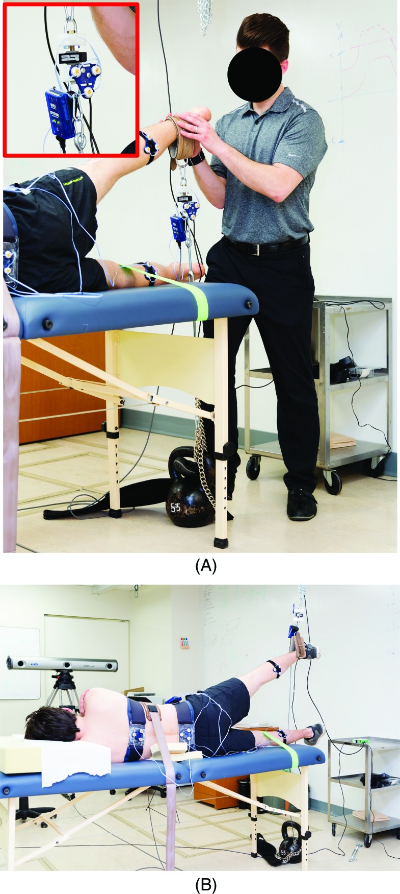
Forces exerted during each task were measured by a uniaxial load cell (MLP-1K, Transducer Techniques, Temecula, CA, USA) that was fixed to a chain. Stated measurement error for the load cell was 0.05% of the full-scale (1000 pounds).18 The chain was secured to the ceiling for adduction trials (Figure 1A) or to an immovable weight on the floor for abduction trials (Figure 1B). Analog data were digitally sampled at a rate of 1000 Hz using a ± 10V range on a 16-bit analog-to-digital conversion board (ODAU III, Northern Digital Inc., Waterloo, ON, Canada).
Figure 1.
Patient positioning for an abduction (A) and adduction (B) trial.
Kinematic
Three-dimensional kinematic data were recorded from the shank bilaterally, pelvis, and thorax with two optoelectronic cameras (Optotrak Certus, Northern Digital Inc., Waterloo, ON, Canada). The Optotrak Certus cameras are capable of measuring the position of an infrared-light emitting diode (IRED) with an accuracy of 0.1 mm and resolution of 0.01 mm.19 Separate rigid bodies holding three IREDs were strapped to each of the participant's shanks at the widest point of the gastrocnemius, to the pelvis at the level of the anterior superior iliac spines (ASISs), and around the thorax at the approximate level of the sixth thoracic vertebra (T6). A fifth rigid body was attached to the uniaxial load cell to continuously monitor its position and orientation. An investigator digitized additional anatomical landmarks while the participant stood in an upright and anatomically neutral posture. Specific bilateral landmarks were the ASISs, iliac crests, greater trochanters, medial and lateral aspects of the knee joints, tibial tuberosities, medial and lateral malleoli, and acromion processes. Unilateral landmarks were the suprasternal notch, xiphoid process, and the spinous processes of the twelfth thoracic (T12) and fifth lumbar (L5) vertebrae. Two marked points were also digitized on either side of the load cell and referenced to the load cell's rigid body. Three-dimensional coordinates for all digitized locations were continuously monitored throughout data collection by assuming a fixed geometrical relationship between the position and orientation of the segment's rigid body and the digitized location. All kinematic data from the rigid bodies and digitized landmarks were synchronized with the kinetic data and recorded at a rate of 100 Hz.
Protocol
Upon arriving at the lab, participants were asked to complete an 11-item inventory to determine their leg dominance,20 and their Q-angle was measured.21 Participants followed a three-minute standardized warm-up consisting of squats, lunges and side-to-side resistance band walks to challenge the muscles targeted during the procedure.22,23 Following the warm-up, participants were outfitted with the kinematic instrumentation. As a baseline measure, kinematic data were obtained during an upright standing trial prior to beginning the maximal exertion protocol. The participant was instructed to look directly ahead of them while standing with their arms at their side, and feet pointed forward and approximately shoulder width apart.
Participants were then positioned into side-lying on a massage table with their dominant limb on the up side for all hip abduction and adduction strength trials (Figure 1). This position has been shown to be more reliable compared to supine and standing.24 All exertions were performed with the dominant limb. A strap was placed around the participants’ thorax to control its motion and minimize artifacts due to differences in trunk/pelvis orientation between the ascribed hip abduction angles. The participant's non-dominant lower leg was also strapped to the table in addition to manual stabilization of the pelvis provided by an examiner. Foam cushions were used to mitigate lateral bending of the lumbar spine. Investigators positioned the participant and cued them to maintain their body in the frontal plane during all exertions. Participants were also instructed to have their arms crossed to avoid utilizing the upper body and core musculature to generate additional force through muscle irradiation.25,26 A strap was placed around the ankle of the participant's dominant lower limb and connected to the chain with the load cell.
Participants acclimated themselves to the instrumentation and isometric task by performing several practice trials at submaximal effort. The task required participants to isometrically exert either an upward (hip abduction) or downward (hip adduction) force with a straight leg in 0 degrees of hip flexion/extension and internal/external rotation.27 After the participant had indicated familiarity with the task, they performed maximal isometric adduction and abduction exertions in the side-lying position at 0, 10, and 20 degrees of ascribed hip abduction. These angles have previously been studied24,28 and represent angles utilized by ice hockey players. Ascribed hip abduction angles were determined using a goniometer with the stationary arm of the goniometer aligned between both ASISs and the moving arm extended along the long axis of the femur.7 The 0 degree position was defined as a 90 degree angle between the stationary and moving arms.29 Each exertion was three seconds in duration with a one-second ramp up to their maximum and a two-second hold at the maximum. Investigators provided verbal encouragement to participants to achieve maximal performance during each exertion. A minimum of two minutes rest was given between each exertion to minimize the potential for fatigue development.30 Participants were instructed to notify the examiners of any pain during the procedure, as determined by a verbal numeric pain scale.
Each participant performed three abduction trials at each abduction angle and three adduction trials at each abduction angle for a total of 18 maximum voluntary isometric contractions (MVICs). The orders of the ascribed hip abduction angles, and direction of exertion (i.e. abduction or adduction) were administered in a block-randomized manner.
Data Processing and Biomechanical Analysis
Load cell voltages and kinematic data were initially imported to Visual3D (C-Motion Inc., Germantown, MD, USA). Three-dimensional coordinates for the digitized locations from the upright standing trial were used to construct anatomical frames of reference for the shanks, femurs, pelvis, and trunk. Femoral kinematics were determined using the digitized locations for the knee joint and the greater trochanter.31 Hip joint centers were defined as a quarter of the intertrochanteric distance from the digitized positions for each greater trochanter. Hip joint angle for the dominant lower limb was determined from the relative orientations of the femur and pelvis. The elevation angle of the participant's dominant femur with respect to the lab's horizontal plane was also calculated.
All kinematic data from the isometric exertions were digitally filtered using a dual pass of a second order Butterworth filter with a cutoff frequency of 6 Hz. Load cell voltages were digitally filtered with a dual pass of a second order Butterworth filter at a cutoff frequency of 20Hz before calibration to units of force (Newtons). The direction for the exerted force was determined by mathematically connecting the two digitized points on the load cell. The anatomical point of force application was derived by intersecting the force vector with the shank of the participant's dominant lower limb. Hip adduction and abduction torques were determined using the following equation (1):
| (1) |
In this equation, T = hip torque in the frontal plane; r = moment arm connecting the hip center to the point of force application near the ankle on the participant's dominant lower limb; F = force magnitude; = unit vector representing the force's direction; = unit vector representing the direction of the hip's anterior axis. Peak hip torque was expressed relative to baseline for each exertion.
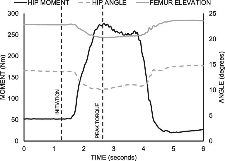
Hip abduction and femoral elevation angles were obtained at two instances, baseline and peak torque, for each exertion (Figure 2). Movements of the hip joint and femur during exertion were determined as the relative changes in hip joint abduction and femoral elevation angles from baseline to peak torque. Hip abduction angles and femoral elevation angles at baseline and peak torque, as well as movements of the hip joint and femur for each ascribed hip abduction angle were averaged across the three abduction and adduction trials for subsequent statistical analysis. Averages of the baseline adjusted peak hip torques across the three trials in abduction and adduction at each of the ascribed hip abduction angles were used to derive the adduction to abduction torque ratio. These values were also used as dependent measures in subsequent statistical analyses.
Figure 2.
Sample data of a single adduction exertion at 20 degrees of hip abduction. Hip adduction moment, hip abduction angle, and femur elevation angle are illustrated. The two vertical dashed lines denote the identified points in time for the exertion initiation (baseline) and peak torque.
Figure 3.
• • •
Statistical Analysis
All statistical analyses were performed with SPSS software (SPSS Corporation, Chicago, IL, USA). Descriptive measures (averages and standard deviations) were determined for the hip positions at baseline and peak torque, as well as the change in hip position between baseline and peak torque. A one-way repeated measures analysis of variance (ANOVA) was conducted to identify the effect of ascribed hip abduction angle on the hip adductor-to-abductor torque ratio. A two-way repeated measures ANOVA was performed to determine the effect of exertion direction and ascribed hip angle on the absolute value of the hip torque in the frontal plane. Three additional two-way repeated measures ANOVAs (one for each of the ascribed hip abduction angles) were performed to determine the effects of exertion direction and instance (initiation or peak torque) on the measured hip joint abduction and femoral elevation angles. Pairwise post-hoc comparisons with Holm's adjustments were used to determine differences for dependent measures with a statistically significant main and/or interaction effects. A total of nine paired comparisons were performed as post-hoc analyses for a statistically significant interaction between exertion direction and the ascribed hip angle. Three paired comparisons were performed for a statistically significant main effect of ascribed hip angle on the hip adductor-to-abductor torque ratio. Four paired comparisons were performed for statistically significant interactions between the exertion direction and instance (initiation or peak torque) for kinematic data at each of the three ascribed hip angles. The level of statistical significance was set to 0.05 for all analyses.
RESULTS
Participants
Data were obtained from a total of 12 participants with one participant's data being excluded due to absence of a suitable baseline prior to each exertion. Demographics for all participants are summarized in Table 1.
Table 1.
Participant demographics (N = 12 males).
| Participant Characteristics | Mean (SD) |
|---|---|
| Age (years) | 25.4 (2.2) |
| Height (cm) | 178.6 (5.2) |
| Weight (kg) | 83.0 (7.4) |
| Dominant limb | 11/12 (R/L) |
| Q-angle (degrees) | 12.1 (1.6) |
| Hockey experience (years) | 20.3 (3.5) |
Kinetic Analysis
All kinetic data are summarized in Table 2. The hip adductor-to-abductor torque ratio increased with increasing hip abduction angle (p ≤ 0.019). A statistically significant interaction was observed between the ascribed hip angle and the direction of exertion (p < 0.001). Adduction torque was greater than abduction torque for all three hip abduction angles, which was reflected by all ratios being greater than 1 (p < 0.001). Abduction torque decreased with increasing hip abduction angle (p ≤ 0.013), and adduction torque was lowest for the 0 degree of hip abduction trials (p ≤ 0.016). There was no difference between adduction torques for the exertions at 10 degrees and 20 degrees of hip abduction (p = 0.895).
Table 2.
Group averages for hip adductor and abductor torques (Nm) and the adductor-to-abductor torque ratio for the three ascribed hip abduction angles. Standard deviations are presented in parentheses. Italicized values are the adjusted p-values for post hoc paired comparisons of means corresponding to statistically significant interactions between exertion direction and the ascribed hip angle (for torques) or main effects of the ascribed hip angle (for torque ratios). Shaded cells represent potential paired comparisons that were not performed.
| ADDUCTOR TORQUE | ABDUCTOR TORQUE | ADDUCTOR-TO-ABDUCTOR RATIO | ||||||||||
|---|---|---|---|---|---|---|---|---|---|---|---|---|
| HIP ANGLE | 0 | 10 | 20 | 0 | 10 | 20 | 0 | 10 | 20 | |||
| MEAN | 228 | 247 | 246 | 151 | 130 | 111 | 1.49 | 1.92 | 2.3 | |||
| (SD) | (49) | (48) | (48) | (22) | (21) | (25) | (0.20) | (0.20) | (0.54) | |||
| ADDUCTOR TORQUE | 0 | 228 | (49) | 0.019 | 0.016 | <0.001 | ||||||
| 10 | 247 | (48) | 0.895 | <0.001 | ||||||||
| 20 | 246 | (48) | <0.001 | |||||||||
| ABDUCTOR TORQUE | 0 | 151 | (22) | 0.004 | 0.001 | |||||||
| 10 | 130 | (21) | 0.013 | |||||||||
| 20 | 111 | (25) | ||||||||||
| ADDUCTOR-TO-ABDUCTOR RATIO | 0 | 1.49 | (0.20) | <0.001 | <0.001 | |||||||
| 10 | 1.92 | (0.20) | 0.019 | |||||||||
| 20 | 2.3 | (0.54) | ||||||||||
The following are examples for reading the table: For an ascribed hip abduction angle of 10 degrees, the average adductor torque was 247 Nm (standard deviation of 48 Nm) and the average abductor torque was 130 Nm (standard deviation of 21 Nm. These average torques were statistically different from each other (p < 0.001). The average adductor torque at an ascribed hip angle of 20 degrees was 246 Nm (standard deviation of 48 Nm), which was not statistically different from the average adductor torque at an ascribed hip angle of 10 degrees (p = 0.895). The average adductor-to-abductor torque ratio at an ascribed hip angle of 0 degrees was 1.49 (standard deviation of 0.20), which was statistically smaller than the average ratio of 1.92 (standard deviation of 0.20) at an ascribed hip angle of 10 degrees (p < 0.001).
Kinematics Analysis
Hip angles and femoral elevation angles at initiation and peak torque during each of the six combinations of ascribed hip angle and exertion direction are presented in Tables 3 and 4. Statistically significant interactions between exertion direction and instance were observed for hip angles and femoral elevation angles at each of the three ascribed hip angles (p ≤ 0.037).
Table 3.
Hip abduction/adduction angles (degrees) at the start and at peak exertion for each direction of exertion and ascribed hip angle. Abduction angles are represented by negative values. Standard deviation of the means is represented in parentheses. Italicized values are the adjusted p-values for post hoc paired comparisons of means corresponding to statistically significant interactions between exertion direction and time (initial, peak). Shaded cells represent potential paired comparisons that were not performed.
| HIP ANGLE | EXERTION | ABDUCTOR | |||||
|---|---|---|---|---|---|---|---|
| 0 | ADDUCTOR | INITIAL | PEAK | ||||
| MEAN (SD) | -3.1 | -4.2 | 0.235 | ||||
| (4.5) | (4.0) | ||||||
| INITIAL | 1.6 | (4.7) | 0.007 | ||||
| PEAK | 2.8 | (4.4) | 0.001 | ||||
| 0.135 | |||||||
| 10 | INITIAL | PEAK | |||||
| MEAN (SD) | -12.5 | -13.2 | 0.326 | ||||
| (2.2) | (2.9) | ||||||
| INITIAL | -8.8 | (3.6) | 0.002 | ||||
| PEAK | -6.3 | (3.2) | <0.001 | ||||
| 0.010 | |||||||
| 20 | INITIAL | PEAK | |||||
| MEAN (SD) | -18.2 | -20.0 | 0.111 | ||||
| (3.4) | (3.4) | ||||||
| INITIAL | -16.2 | (3.2) | 0.100 | ||||
| PEAK | -12.3 | (3.4) | <0.001 | ||||
| <0.001 | |||||||
The following is an example for reading the table: For an ascribed hip abduction angle of 10 degrees, there was a difference in the hip abduction angle at the start of the exertion (p = 0.002) and at peak exertion (p < 0.001). A statistically significant reduction in the abduction angle occurred between the start and peak of adduction exertions (p = 0.010) and no such change during abduction exertions (p = 0.326).
Table 4.
Femoral elevation angles (degrees) at the start and at peak exertion for each direction of exertion and ascribed hip angle. Standard deviation of the means is represented in parentheses. Italicized values are the adjusted p-values for post hoc paired comparisons of means corresponding to statistically significant interactions between exertion direction and time (initial, peak). Shaded cells represent potential paired comparisons that were not performed.
| HIP ANGLE | EXERTION | ABDUCTOR | |||||
|---|---|---|---|---|---|---|---|
| 0 | ADDUCTOR | INITIAL | PEAK | ||||
| 9.0 | 12.1 | <0.001 | |||||
| (4.2) | (3.8) | ||||||
| INITIAL | 7.6 | (2.4) | 0.208 | ||||
| PEAK | 6.2 | (2.7) | 0.002 | ||||
| 0.001 | |||||||
| 10 | INITIAL | PEAK | |||||
| 20.5 | 23.4 | 0.001 | |||||
| (2.8) | (2.5) | ||||||
| INITIAL | 19.9 | (3.4) | 0.402 | ||||
| PEAK | 17.9 | (3.2) | <0.001 | ||||
| 0.002 | |||||||
| 20 | INITIAL | PEAK | |||||
| 28.0 | 31.6 | <0.001 | |||||
| (2.6) | (2.1) | ||||||
| INITIAL | 27.1 | (3.2) | 0.361 | ||||
| PEAK | 24.8 | (3.4) | <0.001 | ||||
| <0.001 | |||||||
The following is an example for reading the table: For an ascribed hip abduction angle of 10 degrees, there was no difference in the femoral elevation angle at the start of the exertion (p = 0.402) and a statistically significant difference at peak exertion (p < 0.001). A statistically significant reduction in the femoral elevation angle occurred between the start and peak of adduction exertions (p = 0.002) and a statistically significant increase during abduction exertions (p = 0.001).
Hip
The average discrepancy between the ascribed hip angle and hip angle at initiation was 2.3 degrees. At initiation, the hip angle was, on average, 4.2 degrees greater for abduction exertions than adduction exertions at 0 degrees and 10 degrees of ascribed hip abduction (p ≤ 0.007). Hip angle at peak torque was also significantly different between abduction and adduction exertions for all three ascribed hip abduction angles (average difference = 7.2 degrees, p ≤ 0.002). Significant changes in hip angle from initiation to peak torque were also observed for adduction exertions at 10 degrees (average change = 2.5 degrees) and 20 degrees (average change = 3.9 degrees) of ascribed hip abduction (p ≤ 0.010). There was no significant change in hip angle during abduction exertions at any of the ascribed hip angles (p ≥ 0.111).
Femur
Femoral elevation at peak torque was greater for abduction exertions than adduction exertions at all three ascribed hip angles (p ≤ 0.002). Conversely, there were no differences in femoral elevation between abduction and adduction exertions at initiation for any of the ascribed hip angles (p ≥ 0.208). Femoral elevation angle changed significantly from initiation to peak torque for all six combinations of ascribed hip angle and exertion direction (p ≤ 0.001).
DISCUSSION
Previous studies have investigated the change in hip abduction and adduction force with different hip abduction angles.9 The current study was the first to directly demonstrate that hip abduction angle can significantly influence the hip adduction-to-abduction torque ratio. Furthermore, this investigation was the first to the authors’ knowledge to evaluate patient positioning and monitor the kinematics of the lower limb, pelvis, and thorax during maximal isometric hip abduction and adduction exertions. This information is particularly useful considering that the hip adductor-to-abductor torque ratio has been used to determine potential injury risk.
Manual muscle testing is widely used by many health practitioners during pre-season testing to guide training or rehabilitation.32-35 Agonist-antagonist strength ratios from these tests are often used to inform injury risk, as demonstrated in hockey players,7,12 soccer players,4 and Gaelic football players.33 The ratios reported in the current investigation were higher than those reported in previous studies that used hockey and soccer players;7,36,37 however direct comparisons should not be made due to differences in the testing parameters. Tyler and colleagues7 performed adduction trials with the athlete in a side-lying position and the hip adducted, which may reduce the force-producing capacity of the adductor muscles. Furthermore, these authors performed abduction trials with the hip abducted “above horizontal”. This discrepancy in hip posture for the adduction and abduction trials possibly provided a mechanical advantage to the hip abductors compared to the testing position of the adductors. Hip adduction-to-abduction ratios in the current investigation were determined for the same ascribed hip abduction angle. This decision was made due to the fact that co-contraction at a given angle is an important aspect of normal joint motion.38 An acute muscle injury, such as a hip adductor strain, occurs at a given joint angle, most often in the eccentric phase of the hockey stride as the hip moves into an abducted position.39 Therefore, testing two opposing muscle groups at the same joint angle is likely more representative of the interaction between these muscle groups when an injury occurs. Although the hockey stride involves a combination of hip abduction, extension, and external rotation, the intention for this study was to evaluate the abduction-adduction component as a risk factor for injury. Therefore, hip extension or external rotation strength were not tested in the current investigation.
An optoelectronic motion-capture system provided data to allow the authors to monitor three-dimensional orientations for the lower limbs, pelvis, thorax, and the direction of the exerted force during all trials in this investigation. The kinematic information allowed the authors to derive the hip torque exerted in the frontal plane during each trial. This analysis accounts for differences that might occur in the direction of the exerted force and the point of application on the lower limb.
Measuring kinematics of the lower limbs, pelvis, and thorax also provided an opportunity to determine the accuracy of the ascribed hip abduction angles at both initiation and peak torque during exertions, as traditional goniometric assessments of the hip tend to overestimate hip ROM.40 Measured hip angles at the initiation of exertions accurately matched the ascribed hip angles, but were different between abduction and adduction exertions. Conversely, femoral elevation angles at the initiation of exertions were greater than their expected elevation (e.g. a femoral elevation angle of 10 degrees would be expected for exertions with an ascribed hip abduction angle of 10 degrees). Furthermore, there were no statistically significant differences in the femoral elevation angle at the initiation of the abduction and adduction exertions. These conflicting findings indicate that the observed differences in hip posture at the initiation of abduction and adduction exertions was likely the result of pelvic positioning at the initiation of the exertions. Monitoring the kinematics throughout the test also allowed for an investigation into the extent to which the isometric nature of the task was maintained. Small (average changes of 2.5 degrees and 3.9 degrees), yet statistically significant, changes in hip posture were observed for adduction exertions performed with 10 and 20 degrees of ascribed hip abduction. The small changes in hip posture indicate that the isometric nature of the task was adequately maintained by the experimental setup. Overall, any movement of the hip joint was consistent with the direction of exertion (i.e. hip abduction decreased during adduction exertions and increased during abduction exertions).
There are several limitations to this study. First, only male hockey players were utilized and therefore this work cannot be extrapolated to female hockey players. Studies have shown reduced abductor torque in female youth athletes compared to male youth athletes.41 It is possible that due to anatomical differences of the pelvis and Q-angle in females that the adductor-to-abductor ratio may differ in this population. The small sample size and cross-sectional design also prevents the use of this data for normative or injury risk factor purposes. Other limitations pertain to the experimental setup and protocol. The decision to evaluate abduction and adduction torque in a lateral recumbent position was consistent with the position used by Tyler and colleagues.7 Previous work has demonstrated that the lateral recumbent position is the most reliable method for evaluating isometric hip abduction strength.24 Furthermore, a rigid mechanical restraint was used to ensure that the exertions were isometric.24 This may reduce the clinical validity of findings presented in the current investigation; however, previous work has recommended the use of an externally fixed dynamometer (i.e. rigid mechanical restraint) on the basis that intertester bias exists when using a handheld dynamometer.42,43 Finally, this study did not test the hip in an adducted position, which is commonly used clinically, because a decision was made to test strength in ranges-of-motion representative of ice hockey.44
CONCLUSION
The results of this study demonstrated that the hip abduction angle has a significant impact on the adductor-to-abductor strength ratio, therefore the ability of this ratio to determine injury risk could be dependent upon the angle at which the hip muscles are tested. The adductor-to-abductor strength ratio is a reported risk factor for adductor strain; however, previous work has provided insufficient details regarding hip positioning and joint angles as well as a rationale for the chosen testing parameters. The value of using this ratio to infer injury risk may be limited by the data collection methods and clinicians should use caution when interpreting the ratio when the testing parameters are not standardized. Using one angle to test both adduction and abduction is likely to be more representative of the agonist/antagonist relationship between opposing muscles. While the chosen angle can vary, it should fall within the functional range of the task or the position in which an injury most often occurs. In addition, this study demonstrated that femoral and hip posture can change during the exertion, which changes the intended hip position of this test. Because of this finding, we recommend that specific measures are taken to stabilize the pelvis and femur during isometric testing of the hip, as many studies do not adequately address this compensatory motion.27,45
REFERENCES
- 1.Emery CA Meeuwisse WH. Injury rates, risk factors, and mechanisms of injury in minor hockey. Am J Sports Med. 2006;34(12): 1960–1969. [DOI] [PubMed] [Google Scholar]
- 2.Lorentzon R Wedren H Pietila T. Incidence, nature, and causes of ice hockey injuries. A three-year prospective study of a Swedish elite ice hockey team. J Am Coll Sports Med. 1988;16(4): 392–396. [DOI] [PubMed] [Google Scholar]
- 3.Molsa J Airaksinen O Nasman O et al. Ice hockey injuries in Finland. A prospective epidemiologic study. J Am Coll Sports Med. 1997;25(4): 495–499. [DOI] [PubMed] [Google Scholar]
- 4.Belhaj K Meftah S Mahir L, et al. Isokinetic imbalance of adductor–abductor hip muscles in professional soccer players with chronic adductor-related groin pain. Eur J Sport Sci. 2016;16(8): 1226–1231. [DOI] [PubMed] [Google Scholar]
- 5.Knapik JJ Bauman CL Jones BH, et al. Preseason strength and flexibility imbalances associated with athletic injuries in female collegiate athletes. Am J Sports Med. 1991;19(1): 76–81. [DOI] [PubMed] [Google Scholar]
- 6.Magalhaes E Silva A Sacramento S, et al. Isometric strength ratios of the hip musculature in females with patellofemoral pain: A comparison to pain-free controls. J Strength Cond Res. 2013;27(8): 2165–2170. [DOI] [PubMed] [Google Scholar]
- 7.Tyler TF Nicholas SJ Campbell RJ, et al. The association of hip strength and flexibility with the incidence of adductor muscle strains in professional ice hockey players. Am J Sports Med. 2001;29(2): 124–128. [DOI] [PubMed] [Google Scholar]
- 8.Diamond LE Wrigley TV Hinman RS, et al. Isometric and isokinetic hip strength and agonist/antagonist ratios in symptomatic femoroacetabular impingement. J Sci Med Sport. 2016;19(9): 696–701. [DOI] [PubMed] [Google Scholar]
- 9.Kulig K Andrews JG Hay JG. Human strength curves. Exerc Sports Sci Rev. 1984;12: 417–466. [PubMed] [Google Scholar]
- 10.Prendergast N Hopper D Finucane M, et al. Hip adduction and abduction strength profiles in elite, sub-elite and amateur Australian footballers. J Sci Med Sport. 2016;19(9): 766–770. [DOI] [PubMed] [Google Scholar]
- 11.Tyler TF Nicholas SJ Campbell RJ, et al. The effectiveness of a preseason exercise program to prevent adductor muscle strains in professional ice hockey players. Am J Sports Med. 2002;30(5): 680–683. [DOI] [PubMed] [Google Scholar]
- 12.Kea J Kramer J Forwell L, et al. Hip abduction-adduction strength and one-leg hop tests: test-retest reliability and relationship to function in elite ice hockey players. J Orthop Sports Phys Ther. 2001;31(8): 446–455. [DOI] [PubMed] [Google Scholar]
- 13.O’Connor D. Groin injuries in professional rugby league players: a prospective study. J Sports Sci. 2004;22(7): 629–636. [DOI] [PubMed] [Google Scholar]
- 14.Thorborg K Serner A Petersen J, et al. Hip adduction and abduction strength profiles in elite soccer players: implications for clinical evaluation of hip adductor muscle recovery after injury. Am J Sports Med. 2011;39(1): 121–126. [DOI] [PubMed] [Google Scholar]
- 15.Florence PK Patricia GP Rodgers M, et al. Muscles: Testing and Function, with Posture and Pain. 5th ed. Philadelphia, PA: Lippincott Williams & Wilkins; 2005. [Google Scholar]
- 16.Kindel C Challis J. Joint moment-angle properties of the hip abductors and hip extensors. Physiother Theory Pract. 2017;33(7): 568–576. [DOI] [PubMed] [Google Scholar]
- 17.Struyf F Nijs J Meeus M, et al. Does scapular positioning predict shoulder pain in recreational overhead athletes? Int J Sports Med. 2014;35(1): 75–82. [DOI] [PubMed] [Google Scholar]
- 18.Transducer techniques. Mini low profile load cell universal/tension or compression. https://www.transducertechniques.com/mlp-load-cell.aspx. Accessed March 29, 2019.
- 19.Northern Digital Inc. Optotrak Certus Technical Specifications. https://www.ndigital.com/msci/products/optotrak-certus/#optotrak-certus-technical-specifications. Accessed March 29, 2019.
- 20.Chapman JP Chapman LJ Allen JJ. The measurement of foot preference. Neuropsychologia. 1987;25(3): 579–584. [DOI] [PubMed] [Google Scholar]
- 21.Smith TO Hunt NJ Donell ST. The reliability and validity of the Q-angle: a systematic review. Knee Surg Sports Traumatol Arthrosc. 2008;16(12): 1068–1079. [DOI] [PubMed] [Google Scholar]
- 22.Berry JW Lee TS Foley HD, et al. Resisted side stepping: the effect of posture on hip abductor muscle activation. J Orthop Sports Phys Ther. 2015;45(9): 675–682. [DOI] [PMC free article] [PubMed] [Google Scholar]
- 23.Gooyers CE Beach TA Frost DM, et al. The influence of resistance bands on frontal plane knee mechanics during body-weight squat and vertical jump movements. Sports Biomech. 2012;11(3): 438–439. [DOI] [PubMed] [Google Scholar]
- 24.Widler KS Glatthorn JF Bizzini M et al. Assessment of hip abductor muscle strength. A validity and reliability study. J Bone Joint Surg. 2009;91(11): 2666–2672. [DOI] [PubMed] [Google Scholar]
- 25.Gontijo LB Pereira PD Neves CDC, et al. Evaluation of strength and irradiated movement pattern resulting from trunk motions of the proprioceptive neuromuscular facilitation. Rehabil Res Pract. 2012;2012: 281937. [DOI] [PMC free article] [PubMed] [Google Scholar]
- 26.Gupta S Hamdani N Sachdev H. Effect of irradiation by proprioceptive neuromuscular facilitation on lower limb extensor muscle force in adults. J Yoga Phys Ther. 2015;5(2): 1–7. [Google Scholar]
- 27.Lee JH Cynn HS Kwon OY, et al. Different hip rotations influence hip abductor muscles activity during isometric side-lying hip abduction in subjects with gluteus medius weakness. J Electromyogr Kinesiol. 2014;24(2): 318–324. [DOI] [PubMed] [Google Scholar]
- 28.Brunner R Maffiuletti NA Casartelli NC, et al. Prevalence and functional consequences of femoroacetabular impingement in young male ice hockey players. Am J Sports Med. 2015;44(1): 46–53. [DOI] [PubMed] [Google Scholar]
- 29.Kang SY Choung SD Jeon HS. Modifying the hip abduction angle during bridging exercise can facilitate gluteus maximus activity. Man Ther. 2016;22: 211–215. [DOI] [PubMed] [Google Scholar]
- 30.Rafn BS Tang L Nielsen MP, et al. Hip strength testing of soccer players with long-standing hip and groin pain: what are the clinical implications of pain during testing? Clin J Sport Med. 2016;26(3): 210–215. [DOI] [PubMed] [Google Scholar]
- 31.Schulz BW Kimmel WL. Can hip and knee kinematics be improved by eliminating thigh makers? Clin Biomech. 2010;25(7): 687–692. [DOI] [PMC free article] [PubMed] [Google Scholar]
- 32.Chamorro C Armijo-Olivo S De La Fuente C, et al. Absolute reliability and concurrent validity of hand held dynamometry and isokinetic dynamometry in the hip, knee and ankle joint: systematic review and meta-analysis. Open Med. 2017;12: 359–375. [DOI] [PMC free article] [PubMed] [Google Scholar]
- 33.Delahunt E Fitzpatrick H Blake C. Pre-season adductor squeeze test and HAGOS function sport and recreation subscale scores predict groin injury in Gaelic football players. Phys Ther Sport. 2017;23: 1–6. [DOI] [PubMed] [Google Scholar]
- 34.Jackson SM Cheng MS Smith AR, et al. Intrarater reliability of hand held dynamometry in measuring lower extremity isometric strength using a portable stabilization device. Musculoskelet Sci Pract. 2017;27: 137–141. [DOI] [PubMed] [Google Scholar]
- 35.Haroy J Thorborg K Serner A, et al. Including the Copenhagen Adduction Exercise in the FIFA 11 + provides missing eccentric hip adduction strength effect in male soccer players: A randomized controlled trial. Am J Sports Med. 2017;45(13): 3052-3059. [DOI] [PubMed] [Google Scholar]
- 36.Mosler AB Crossley KM Thorborg K, et al. Hip strength and range of motion: Normal values from a professional football league. J Sci Med Sport. 2017;20(4): 339–343. [DOI] [PubMed] [Google Scholar]
- 37.Wilcox CRJ Osgood CT White HSF, et al. Investigating strength and range of motion of the hip complex in ice hockey athletes. J Sport Rehabil. 2015;24(3): 300–306. [DOI] [PubMed] [Google Scholar]
- 38.Catani F Hodge A Mann RW, et al. The role of muscular co-contraction of the hip during movement. Chir Organi Mov. 1995;80(2): 227–236. [PubMed] [Google Scholar]
- 39.Sim FH Chao EY. Injury potential in modern ice hockey. Am J Sports Med. 1978;6(6): 378–384. [DOI] [PubMed] [Google Scholar]
- 40.Nussbaumer S Leunig M Glatthorn JF, et al. Validity and test-retest reliability of manual goniometers for measuring passive hip range of motion in femoroacetabular impingement patients. BMC Musculoskelet Disord. 2010;11: 194. [DOI] [PMC free article] [PubMed] [Google Scholar]
- 41.Bittencourt NF Santos TR Goncalves GG, et al. Reference values of hip abductor torque among youth athletes: Influence of age, sex and sports. Phys Ther Sport. 2016;21: 1–6. [DOI] [PubMed] [Google Scholar]
- 42.Thorborg K Bandholm T Schick M, et al. Hip strength assessment using handheld dynamometry is subject to intertester bias when testers are of different sex and strength. Scand J Med Sci Sports. 2013;23(4): 487–493. [DOI] [PubMed] [Google Scholar]
- 43.Ryan S Kempton T Pacecca E, et al. Measurement properties of an adductor strength-assessment system in professional Australian footballers. Int J Sports Physiol Perform. 2019;14(2): 256-259. [DOI] [PubMed] [Google Scholar]
- 44.Hellyer MR Alexander MJ Glazebrook CM, et al. Differences in lower body kinematics during forward treadmill skating between two different hockey skate designs. Int J Kinesiol Sports Sci. 2016;4(1): 1–16. [Google Scholar]
- 45.Bazett-Jones DM Tylinksi T Krstic J, et al. Peak hip muscle torque measurement are influenced by sagittal plane hip position. Int J Kinesiol Sports Sci. 2017;12(4): 535–542. [PMC free article] [PubMed] [Google Scholar]


