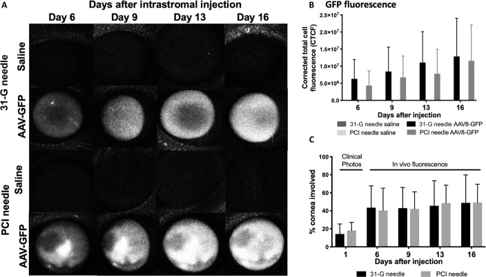
FIGURE 4.
In vivo expression of GFP. A, Using a SLO, GFP expression was detected on days 6, 9, 13, and 16 after intrastromal injection of 25 μL of saline (balanced salt solution) or AAV8-GFP using either a 31-G or a PCI needle. Fluorescence was not visible in the saline-dosed eyes; however, corneal expression was noted in increasing density in the right eyes (representative images, n = 6/injection). B, Mean fluorescence CTCF using the 31-G or PCI needle in the right corneas increased at each time point after injection but was not significantly different from each other at any day. C, Area of the cornea with GFP expression in vivo. The area of GFP fluorescence was higher at 6, 9, 13, and 16 days compared with the visible injection site immediately after injection (measured on clinical photographs), suggesting that diffusion of the virus beyond the injection site occurred. There was no significant difference with type of injection.

