SUMMARY:
The round window serves to decompress acoustic energy that enters the cochlea via stapes movement against the oval window. Any inward motion of the oval window via stapes vibration leads to outward motion of the round window. Occlusion of the round window is a cause of conductive hearing loss because it increases the resistance to sound energy and consequently dampens energy propagation. Because the round window niche is not adequately evaluated by otoscopy and may be incompletely exposed during an operation, otologic surgeons may not always correctly identify associated pathology. Thus, radiologists play an essential role in the identification and classification of diseases affecting the round window. The purpose of this review is to highlight the developmental, acquired, neoplastic, and iatrogenic range of pathologies that can be encountered in round window dysfunction.
The round window (RW) serves as a boundary between the basal turn of the cochlea anteriorly and the round window niche posteriorly.1 It, along with the oval window, is 1 of 2 natural openings between the inner and middle ear. The round window is often overshadowed by the “first window” (the oval window) and pathologic “third windows” (eg, superior semicircular canal dehiscence). Nevertheless, numerous developmental, acquired, neoplastic, and iatrogenic processes can affect the round window membrane and niche. Any of these can cause conductive hearing loss because occlusion of the round window prevents propagation of acoustic energy along the cochlear axis.2 In operative interventions in which a primary goal of surgery is to improve conductive hearing loss or access the round window region for cochlear implantation, accurate preoperative identification of round window abnormalities is essential to first determine whether surgery is a worthwhile endeavor and subsequently to guide the surgical strategy. In this article, the various pathologic entities and surgical considerations of the round window that can be encountered on imaging are reviewed.
Physiology and Functionality
The inner ear “windows” refer to openings in the otic capsule that connect the fluid in the inner ear to either the middle ear or intracranial space.3 The 2 primary natural openings are the oval and round windows.3 Other windows include the cochlear and vestibular aqueducts and tiny foramina that transmit vessels and/or nerves to the inner ear and adjacent structures (eg, the petromastoid canal and singular canal).
Functionally, the role of the oval and round windows is related to sound transmission: Vibratory acoustic energy enters through the oval window, is transmitted through the cochlea, and exits into the middle ear cavity via the round window.3 The fluid in the cochlea through which sound is transmitted is functionally incompressible due to the surrounding osseous structures.4 Movement of the cochlear fluid is thereby dependent on the mobility of the round and oval window membranes: Inward displacement of the oval window membrane via the stapes by ossicular vibration is matched by outward round window membrane displacement.4
Developmental Anomalies
Normal Anatomy.
The round window is located along the posterior aspect of the cochlear promontory and measures 1.5–2.1 mm horizontally, 1.9 mm vertically, and 0.65 mm in thickness (Fig 1).1,5 The round window membrane is thicker along its edges and thinner in the middle and is made up of 3 layers: 2 epithelial layers facing the inner and middle ear, respectively, and connective tissue in the core.6 Contrary to its name, the shape of the round window is typically skewed, ovoid, and nonplanar according to a recent study.7 The round window niche is primarily defined by the relatively thin overhanging bone that naturally extends from the promontory. This overhanging bone may obscure complete direct visualization of the round window membrane during routine middle ear surgery and cochlear implantation (Fig 2).8 In addition, most ears have a thin layer of mucosa covering the round window membrane, often called a “pseudomembrane,” that blocks direct visualization of the window if not removed.
FIG 1.
Normal round window anatomy. Axial (A), coronal (B), Stenvers (C), and Pöschl (D) images centered on the round window membrane (straight white arrows), situated between the basal turn of the cochlea (black arrows) and the round window niche of the middle ear (curved white arrows). The oval window is closely adjacent (dashed arrow).
FIG 2.
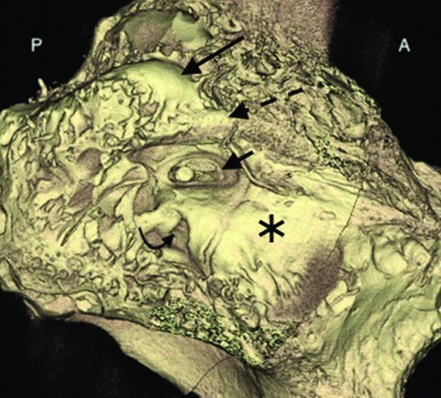
Surface-rendered image of a microCT (GE Healthcare, Milwaukee, Wisconsin) of the temporal bone shows the relationship of the round window niche (curved arrow) to the adjacent anatomic structures. Also seen are the lateral semicircular canal (long straight arrow), facial nerve canal (dashed arrow), stapes and oval window (short arrow), and cochlear promontory (asterisk). A indicates anterior; P, posterior.
Round Window Stenosis and Atresia.
Round window absence is a rare abnormality that may be seen in conjunction with various syndromes, including incomplete partition anomalies, mandibulofacial dysostosis, and Coloboma of the eye, Heart defects, Atresia of the choanae, Retardation of growth and/or development, Genital and/or urinary abnormalities, and Ear abnormalities and deafness (CHARGE) syndrome, in addition to cases of aural atresia (Fig 3).9-11 In very rare cases, it may also occur without an associated syndrome (Fig 4).12,13 Some authors have posited that such nonsyndromic cases may represent an inherited autosomal dominant genetic disorder with variable penetrance.10 Even in nonsyndromic cases, few reports exist of round window atresia as an isolated finding; most of these patients have associated middle ear or pinna abnormalities.10
FIG 3.
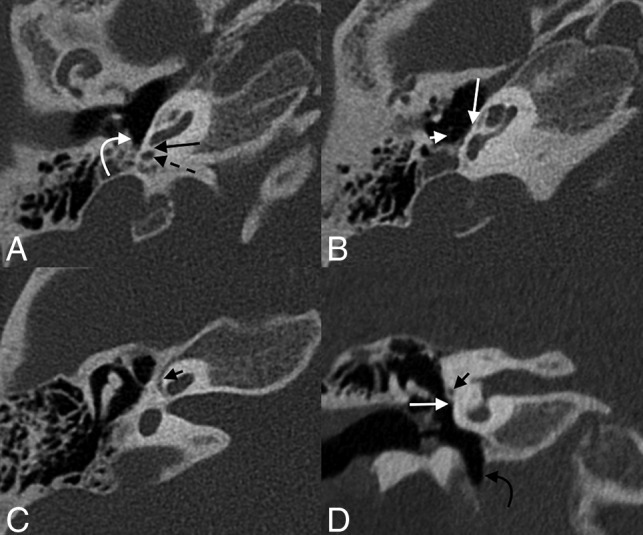
A 5-year-old girl with round window atresia in the setting of CHARGE syndrome. Axial (A–C) and coronal (D) images of the right temporal bone demonstrate complete absence of the round window (long black arrow, A); the round window niche is hypoplastic and surrounded by bone (dashed arrow, A), adjacent to a preserved sinus tympani (curved white arrow, A). Multiple other anomalies are the following: The stapes is dysmorphic with absence of the neck, crura, and footplate (short white arrow, B); the oval window is absent (long white arrow, B and D); the facial nerve canal is diminutive (short black arrow, C) and inferiorly displaced (short black arrow, D); and the hypotympanum is enlarged (curved black arrow, D). The semicircular canals are absent.
FIG 4.
Nonsyndromic round window atresia. Axial (A), coronal (B), and Pöschl (C) views of the right temporal bone of a 10-year-old girl demonstrate an ossification at the expected location of the round window membrane (arrows). The patient had no known syndromic association.
Patients with round window atresia typically have a mixed but predominantly conductive hearing loss, with an associated typical air-bone gap of 30–40 dB.14 Attempts to surgically recreate the round window do not always produce substantial gains in hearing.14 The reasons remain uncertain because few such surgical case reports exist. However, the inconsistent results may be related to the presence of associated anomalies such as congenital stapes fixation, otosclerosis, or re-ossification following an operation.
Unfortunately, because surgeons often have incomplete exposure of the round window niche during routine otologic procedures and because of its rarity, round window atresia can easily be overlooked intraoperatively.14 It also is frequently missed on imaging, and many patients with round window atresia are not diagnosed until middle ear exploration.10 Close attention must therefore be paid to the morphology of the round window and round window niche on imaging performed for conductive or mixed hearing loss.
Stenosis of the round window membrane and/or recess is more common than complete round window absence. Like round window atresia, stenosis of the round window can have syndromic associations and contribute to hearing loss.15 The lower limit of normal size for the round window niche is generally cited as being 1.5 mm.1 Nevertheless, imaging characteristics of stenotic recesses can be quite variable, ranging from mild to severe (Fig 5).
FIG 5.
Round window stenosis. A 52-year-old woman with profound bilateral hearing loss. Axial (A), coronal (B), and Pöschl (C) images of the right temporal bone demonstrate advanced stenosis of the round window (arrows).
Acquired Abnormalities
Acquired abnormalities that affect the round window include a range of traumatic, inflammatory, and iatrogenic processes. Otitis media associated with mucosal thickening and effusion may obscure the round window or adjacent niche (Fig 6). Less commonly, barotrauma or increased pressure may cause the round window membrane to rupture, leading to perilymph fistula with sensorineural hearing loss and vertigo.17-19 Temporal bone fractures through the round window may disrupt the membrane. Several primary osseous processes such as Paget disease, fibrous dysplasia, otosyphilis, and osteogenesis imperfecta can affect the temporal bone, including the annular ring of the round window.15,20,21 Below, several of the most common causes of acquired abnormalities of the round window are discussed, with imaging correlates.
FIG 6.
Round window niche occlusion. A 48-year-old woman who presented with fullness in the right ear and conductive hearing loss following a bout of flulike symptoms. Axial (A), Pöschl (B), and coronal (C) images of the right temporal bone demonstrate a tiny focus of debris adjacent to the right round window membrane (straight arrow) within the round window niche (curved arrow), possibly sequelae of her recent inflammatory illness.
Labyrinthitis Ossificans.
Labyrinthitis ossificans (LO) refers to ossification within the membranous labyrinth, most frequently occurring within the scala tympani.22 Typically, LO is secondary to inflammatory changes from infection such as suppurative otitis media, labyrinthitis, or meningitis, though trauma and otosclerosis have also been indicated as inciting processes.23,24 In cases of LO associated with bacterial meningitis, the basal turn of the cochlea is often preferentially affected because infection may spread from the subarachnoid space through the cochlear aqueduct to the proximal scala tympani (though spread may also occur through the modiolus).25 In some cases, LO may involve the round window, involvement thought to occur when the inciting event is otitis media or meningitis that spreads through the round window along the scala tympani (Fig 7).26 The etiology of LO cannot be predicted on the basis of mineralization patterns seen on CT.27 LO is associated with the development of sensorineural hearing loss and may render cochlear implantation more challenging and outcomes less rewarding.28,29 Extensive LO may preclude the placement of a cochlear implant. Expected CT findings of LO involving the round window include thickening/high attenuation along the membrane, likely with coexistent ossific material in the basal turn of the cochlea or elsewhere in the membranous labyrinth. In the early stages of labyrinthitis, fibrosis may be evident only on heavily weighted T2 imaging, where low signal is seen within or partially replacing normally high-signal perilymph.
FIG 7.
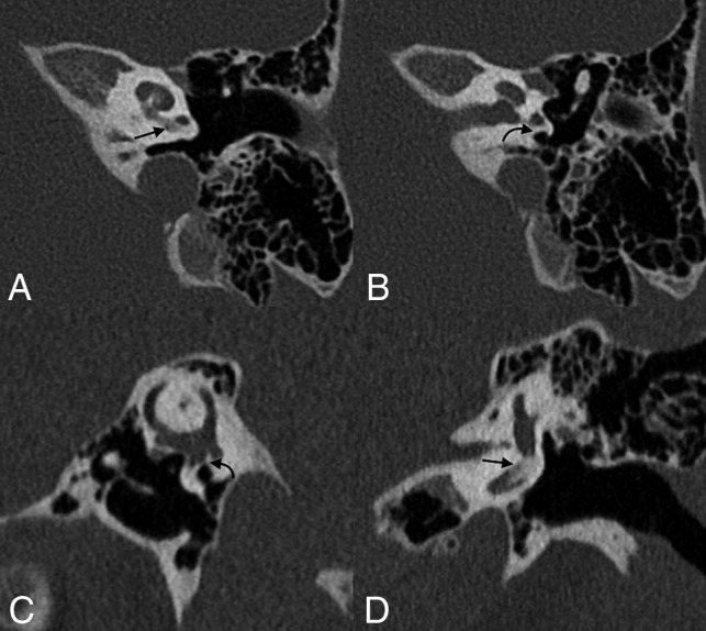
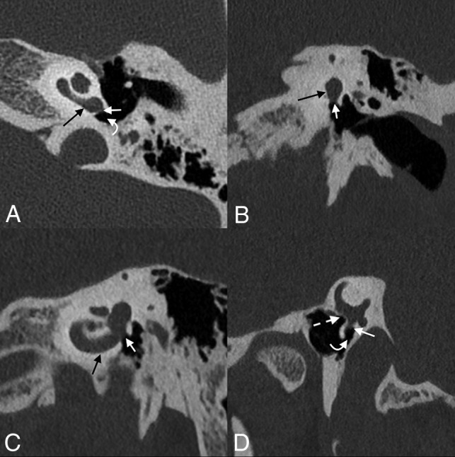
Labyrinthitis ossificans with round window involvement. Axial (A and B), Pöschl (C), and coronal (D) images of the left temporal bone of a 5-year-old with a history of prior tympanoplasty tube placements. Labyrinthitis ossificans is seen in the basal turn of the cochlea (straight arrows, A and D) with mineralization of the round window (curved arrows, B and C).
Otosclerosis.
Otosclerosis is an acquired condition in which spongiotic bone replaces mature endochondral bone of the otic capsule.30 Typically, otosclerosis involves the oval window and stapes footplate in the region of the fissula ante fenestram.30,31 Fenestral otosclerosis may also secondarily involve the round window in approximately 3%–13% of cases.32,33 Isolated round window otosclerosis has been reported, though the prevalence is rare (approximately 0.3% of cases).32 Nevertheless, secondary involvement or isolated involvement of the round window by otosclerosis is an important radiologic observation because it portends a poorer chance of surgical success.30 Specifically, round window involvement diminishes the movement of perilymphatic fluid following stapedectomy and prothesis placement, the surgery typically used in cases of otosclerosis.34,35
The appearance of otosclerosis on imaging depends on the disease phase. In the active (otospongiotic) stage, the affected bone surrounding the round window will appear lucent and demineralized (Fig 8).36,37 Later on, these areas are replaced by sclerotic bone during the nonactive (otosclerotic) stage.37 As this replacement occurs, the round window membrane may become thickened or irregular. Heaped-up osseous plaques may narrow the round window or adjacent niche.34
FIG 8.
Otosclerosis with round window involvement. Axial (A), Pöschl (B), and coronal (C) images of the right temporal bone demonstrate bony changes compatible with otosclerosis adjacent to the round window, with marked narrowing of the round window niche (arrows).
Mansour et al32 categorized otosclerosis of the round window on the basis of the extent of involvement of the membrane and adjacent structures. Under this classification scheme, RW-I represented hypodensities about the round window edge, RW-II had partial thickening of the membrane, RW-III showed global membrane thickening with a persistent air-filled recess, RW-IV had obliteration of the recess, and RW-V demonstrated overgrowth of otosclerotic foci. Predictably, higher grades of round window involvement were found to be associated with more severe hearing loss, likely related to increased impedance within the scala tympani.32
Jugular Bulb Dehiscence and Diverticula.
The positional anatomy of the jugular bulb is variable throughout a person’s life.38 Jugular bulb abnormalities consequently develop with time and are typically acquired by the fourth decade of life.38 High-riding jugular bulbs are found in approximately 8% of patients, both on pathologic specimens and CT images.38 Dehiscent jugular bulbs and jugular bulb diverticula are more rare, occurring at a rate of 2.6% and 1.2%, respectively.39,40
High-riding jugular bulbs, dehiscent bulbs, and jugular diverticula are often asymptomatic.41 However, they may also present with pulsatile tinnitus or, less commonly, conductive hearing loss, likely related to encroachment by the bulb on the round window, ossicular chain, or tympanic membrane (Fig 9).41,42 The incidence in which the round window membrane is specifically involved is rare; 1 histologic analysis of temporal bones identified 2 such cases in 1579 specimens (0.1%).43
FIG 9.
Jugular bulb anomalies. Coronal (A), Pöschl (B), and axial (C) images demonstrate a high-riding jugular bulb (asterisk) that extends into the round window niche (straight black arrows). The bony margins overlying the jugular bulb within the middle window are markedly thinned, compatible with dehiscence (white arrows).
Imaging findings vary on the basis of the type of jugular bulb abnormality. High-riding bulbs typically occur as an isolated finding, in which the dome of the jugular bulb is within 2 mm of the internal auditory canal (IAC) floor (though definitions vary).44,45 A high-riding bulb may extend further superolaterally, erode the sigmoid plate, and protrude into the middle ear cavity; this dehiscence is best seen on CT as thinning and/or absence of bone between the bulb and middle ear structures.44 Finally, diverticula will appear as distinct outpouchings from the bulb. Any jugular bulb anomaly seen on CT should be closely examined for the presence of abutment of the bulb on the round window membrane, niche, or other middle ear structures.
Neoplastic Processes.
Several neoplastic processes may affect the round window membrane or round window niche. Some, such as metastases and primary osseous tumors, are centered in the bone; secondary round window involvement by these lesions depends on their size and location. Notably, most primary osteodystrophies and osseous neoplasms spare the otic capsule, given divergent embryology and composition. Other tumors have an anatomic proclivity to involve the round window membrane. For example, intralabyrinthine schwannomas may extend through the round window membrane into the niche (Fig 10).46,47 CT images of such patients may demonstrate a soft-tissue mass within the niche; visualization of the entire lesion often requires MR imaging.
FIG 10.
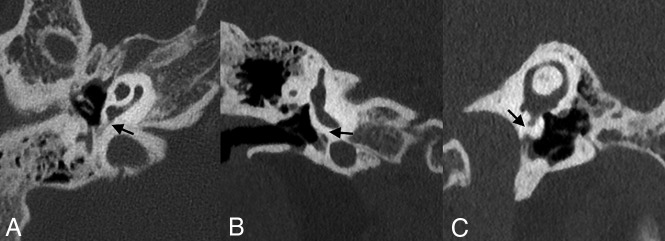
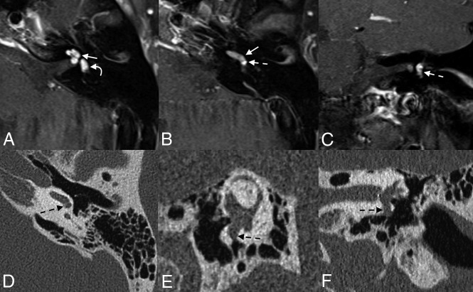
Intralabyrinthine schwannoma with involvement of the round window. Axial (A and B) and coronal (C) contrast-enhanced T1WI shows an avidly enhancing mass in the left cochlea (straight arrows) and vestibule (curved arrow), compatible with a schwannoma. The mass extends through the round window membrane and into the round window niche (dashed arrows), better demonstrated on the follow-up axial (D), Pöschl (E), and coronal (F) CT images.
Jugulotympanic paragangliomas—comprising glomus jugulare and glomus tympanicum tumors—may also involve the round window.48,49 Glomus tympanicum tumors arise from the tympanic nerve (Jacobson nerve) and grow within and along the cochlear promontory; if large enough, they may extend into and occlude the round window niche.48 Glomus jugulare tumors begin within the jugulare foramen. Although histologically benign, glomus jugulare tumors are locally destructive and can erode into adjacent middle ear structures such as the round window niche (Fig 11).
FIG 11.
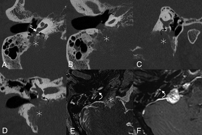
Glomus jugulare tumor with round window obstruction. Axial (A and B), Pöschl (C), and coronal (D) CT images of the right temporal bone show a permeative destructive mass centered in the right jugular foramen (asterisk), which extends into the right middle ear and fills the round window niche (curved arrows); opacification of the niche extends up to the round window membrane (straight arrow). Axial T2 sampling perfection with application-optimized contrasts by using different flip angle evolutions (SPACE sequence; Siemens, Erlangen, Germany) (E) and T1WI with gadolinium (F) also show the mass (asterisk) and round window niche occlusion (curved arrows).
Surgical Considerations
Surgical Access and Round Window Visualization.
Several conditions may require surgical dissection at the round window niche. Most commonly, the niche is accessed during cochlear implantation. Most cases are performed using a standard cortical mastoidectomy with facial recess. The bony overhang of the round window niche is then drilled to allow direct round window membrane visualization during electrode insertion. Conversely, when surgical access of the round window or niche is performed to treat infectious or neoplastic middle ear processes, the region is typically visualized via a transcanal approach. In addition, there are uncommon situations in which round window occlusion is performed to treat traumatic perilymphatic fistulas or superior canal dehiscence syndrome. In these cases, the middle ear is typically accessed via a transcanal approach, the round window is visualized with an operating microscope or endoscope, and fascia with or without cartilage is packed in the round window niche.
Recently, endoscopes have gained popularity in otologic surgery. Endoscopy allows superior visualization around corners, particularly during removal of a cholesteatoma in the sinus tympani and anterior epitympanum. Thus, endoscopes are sometimes used to augment microscopic techniques in complex cases. However, for visualization and access of the round window, the use of an endoscope does not carry a substantial advantage over microscope visualization in most cases50 because the round window niche is readily seen when performing either transcanal or transmastoid-facial recess surgery via a direct line of sight.
Postoperative Changes in Third-Window Lesions.
Third-window lesions refer to any range of pathology that creates an abnormal connection between the inner ear and either the middle ear or the intracranial cavity.51 Acoustic energy is lost through these windows, often causing a so-called “pseudoconductive” hearing loss, which manifests as increased bone and decreased air conduction.3,52 There are many such connections: vestibular aqueduct enlargement, semicircular canal dehiscence, and any other type of osseous thinning between the inner ear and adjacent vascular or nervous channels.52 Patients can present with Tullio phenomenon (vertigo symptoms induced by loud noises) or Hennebert sign (similar symptoms induced by increases in pressure within the ear canal) due to deflection of the superior canal cupula by endolymphatic fluid escaping the osseous defect.53-55
For cases of superior semicircular canal dehiscence, surgical plugging of the pathologic osseous defect can be completed, either via a middle cranial fossa craniotomy or transmastoid approach.55,56 Alternatively, the round window can be targeted; surgeons may reinforce the round window with overlying tissue (eg, fascia, cartilage, fat) or occlude the round window niche (Fig 12).55,57-60 Currently, most authors favor the former approach over the latter; although round window occlusion is considered low-risk, this strategy may induce conductive hearing loss. Furthermore, the theoretic physiologic justification for this approach is lacking because occlusion of the round window should theoretically create preferential shunting toward the pathologic third window.20,61,62
FIG 12.
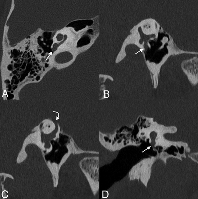
Postoperative changes of round window niche occlusion for superior semicircular canal dehiscence. Axial (A), Pöschl (B and C), and coronal (D) images of the right temporal bone demonstrate a soft-tissue opacity corresponding to a mixed temporalis fascia and irradiated rib cartilage against the round window and within the adjacent niche (straight arrow). There is frank dehiscence of the superior semicircular canal (curved arrow).
Cochlear Implant.
Cochlear implants may be inserted through a cochleostomy adjacent to the round window or directly through the round window membrane.63,64 Most surgeons surveyed in 2007 preferred the former approach, though these decisions may be based on historical presumptions; early multichannel leads were thought to traumatize the cochlea if placed through the round window.65,66 A more recent survey found that today most surgeons prefer round window membrane electrode insertions, given the natural and less traumatic access provided to the scala tympani—the preferred location of electrode placement.67 A recent study found no difference in the number of audiometric outcomes or postoperative complications among groups undergoing electrode placement via either approach; however, most cochlear implant surgeons now prefer the round window approach when trying to preserve any residual natural acoustic hearing.66,67
Regardless of the cochlear entry point, complications do occur. The electrode can kink, flip over at its tip, or migrate.68,69 Postoperative imaging can also demonstrate various degrees of electrode displacement, including within the semicircular canal, internal carotid artery, internal auditory canal, and vestibule.69,70 Across time, electrodes may also migrate from their initial position (Fig 13). Postoperative images should be evaluated in the context of the surgical approach (ie, via the round window or adjacent cochleostomy) and should include an assessment of electrode position, integrity, and change since prior examinations.
FIG 13.
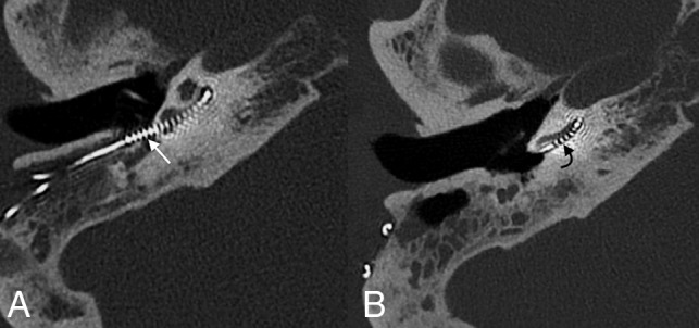
A 64-year-old woman who had poor progress of hearing following the placement of a cochlear implant. Axial CT image at the time of presentation (A) shows that the electrode array is retracted from its expected location, with multiple electrode leads located outside of the cochlea (straight white arrow). Follow-up imaging after surgical revision (B) shows normal positioning of the implant, with the first electrode located approximately 4 mm past the round window (curved black arrow).
CONCLUSIONS
Numerous acquired and developmental processes may affect the round window, presenting with varying clinical symptoms. Such cases can be challenging to radiologists who are unfamiliar with the local anatomy and pathologies. Nevertheless, because the round window and round window niche are often anatomically obscured during an operation, imaging plays a uniquely important role. Thus, radiologists should scrutinize the round window and familiarize themselves with the anomalies and disease processes that may be encountered in this important anatomic region.
ABBREVIATIONS:
- LO
labyrinthitis ossificans
- RW
round window
Footnotes
Internal departmental funding was used without commercial sponsorship or support.
References
- 1.Veillon F, Riehm S, Emachescu B, et al. Imaging of the windows of the temporal bone. Semin Ultrasound CT MR 2001;22:271–80 10.1016/S0887-2171(01)90011-3 [DOI] [PubMed] [Google Scholar]
- 2.Curtin HD. Imaging of conductive hearing loss with a normal tympanic membrane. AJR Am J Roentgenol 2016;206:49–56 10.2214/AJR.15.15060 [DOI] [PubMed] [Google Scholar]
- 3.Merchant SN, Rosowski JJ. Conductive hearing loss caused by third-window lesions of the inner ear. Otol Neurotol 2008;29:282–89 10.1097/MAO.0b013e318161ab24 [DOI] [PMC free article] [PubMed] [Google Scholar]
- 4.Stenfelt S, Goode RL. Bone-conducted sound: physiological and clinical aspects. Otol Neurotol 2005;26:1245–46 10.1097/01.mao.0000187236.10842.d5 [DOI] [PubMed] [Google Scholar]
- 5.Paparella MM, Oda M, Hiraide F, et al. Pathology of sensorineural hearing loss in otitis media. Ann Otol Rhinol Laryngol 1972;81:632–47 10.1177/000348947208100503 [DOI] [PubMed] [Google Scholar]
- 6.Goycoolea MV, Lundman L. Round window membrane: structure function and permeability—a review. Microsc Res Tech 1997;36:201–11 [DOI] [PubMed] [Google Scholar]
- 7.Atturo F, Barbara M, Rask-Andersen H. Is the human round window really round? An anatomic study with surgical implications. Otol Neurotol 2014;35:1354–60 10.1097/MAO.0000000000000332 [DOI] [PubMed] [Google Scholar]
- 8.Roland PS, Wright CG, Isaacson B. Cochlear implant electrode insertion: the round window revisited. Laryngoscope 2007;117:1397–1402 10.1097/MLG.0b013e318064e891 [DOI] [PubMed] [Google Scholar]
- 9.Vesseur AC, Verbist BM, Westerlaan HE, et al. CT findings of the temporal bone in CHARGE syndrome: aspects of importance in cochlear implant surgery. Eur Arch Otorhinolaryngol 2016;273:4225–40 10.1007/s00405-016-4141-z [DOI] [PMC free article] [PubMed] [Google Scholar]
- 10.Borrmann A, Arnold W. Non-syndromal round window atresia: an autosomal dominant genetic disorder with variable penetrance? Eur Arch Otorhinolaryngol 2007;264:1103–08 10.1007/s00405-007-0305-1 [DOI] [PubMed] [Google Scholar]
- 11.Morimoto AK, Wiggins RH, Hudgins PA, et al. Absent semicircular canals in CHARGE syndrome: radiologic spectrum of findings. AJNR Am J Neuroradiol 2006;27:1663–71 [PMC free article] [PubMed] [Google Scholar]
- 12.Martin C, Tringali S, Bertholon P, et al. Isolated congenital round window absence. Ann Otol Rhinol Laryngol 2002;111:799–801 10.1177/000348940211100907 [DOI] [PubMed] [Google Scholar]
- 13.Clifford AR, Fagan PA, Doust BD. Isolated congenital round window absence. J Laryngol Otol 1990;104:980–81 10.1017/S0022215100114549 [DOI] [PubMed] [Google Scholar]
- 14.Linder TE, Ma F, Huber A. Round window atresia and its effect on sound transmission. Otol Neurotol 2003;24:259–63 10.1097/00129492-200303000-00021 [DOI] [PubMed] [Google Scholar]
- 15.Alkadhi H, Rissmann D, Kollias SS. Osteogenesis imperfecta of the temporal bone: CT and MR imaging in Van der Hoeve-de Kleyn syndrome. AJNR Am J Neuroradiol 2004;25:1106–09 [PMC free article] [PubMed] [Google Scholar]
- 16.Lemmerling M, Foer Bd. Temporal Bone Imaging. Berlin: Springer-Verlag; 2014 [Google Scholar]
- 17.Choo YB. Microscopic characteristics of round window problems in otology. Laryngoscope 1984;94:1–9 10.1002/lary.5540940101 [DOI] [PubMed] [Google Scholar]
- 18.Lyons GD, Dodson ML, Casey DA, et al. Round window rupture secondary to acoustic trauma. South Med J 1978;71:71–73 10.1097/00007611-197801000-00020 [DOI] [PubMed] [Google Scholar]
- 19.Al Felasi M, Pierre G, Mondain M, et al. Perilymphatic fistula of the round window. Eur Ann Otorhinolaryngol Head Neck Dis 2011;128:139–41 10.1016/j.anorl.2010.12.004 [DOI] [PubMed] [Google Scholar]
- 20.Andreu-Arasa VC, Sung EK, Fujita A, et al. Otosclerosis and dysplasias of the temporal bone. Neuroimaging Clin N Am 2019;29:29–47 10.1016/j.nic.2018.09.004 [DOI] [PubMed] [Google Scholar]
- 21.Ogungbemi A, Dudau C, Morley S, et al. Computed tomography features of luetic osteitis (otosyphilis) of the temporal bone. J Laryngol Otol 2014;128:185–88 10.1017/S0022215113003575 [DOI] [PubMed] [Google Scholar]
- 22.deSouza C, Paparella MM, Schachern P, et al. Pathology of labyrinthine ossification. J Laryngol Otol 1991;105:621–24 10.1017/S0022215100116846 [DOI] [PubMed] [Google Scholar]
- 23.Kaya S, Paparella MM, Cureoglu S. Pathologic findings of the cochlea in labyrinthitis ossificans associated with the round window membrane. Otolaryngol Head Neck Surg 2016;155:635–40 10.1177/0194599816651245 [DOI] [PubMed] [Google Scholar]
- 24.d’Archambeau O, Parizel PM, Koekelkoren E, et al. CT diagnosis and differential diagnosis of otodystrophic lesions of the temporal bone. Eur J Radiol 1990;11:22–30 10.1016/0720-048X(90)90098-V [DOI] [PubMed] [Google Scholar]
- 25.Du Y, Wu X, Li L. Mechanisms of bacterial meningitis-related deafness. Drug Discov Today Dis Mech 2006;3:115–18 10.1016/j.ddmec.2006.02.002 [DOI] [Google Scholar]
- 26.Suga F, Lindsay JR. Labyrinthitis ossificans due to chronic otitis media. Ann Otol Rhinol Laryngol 1975;84:37–44 10.1177/000348947508400106 [DOI] [PubMed] [Google Scholar]
- 27.Buch K, Baylosis B, Fujita A, et al. Etiology-specific mineralization patterns in patients with labyrinthitis ossificans. AJNR Am J Neuroradiol 2019;40:551–57 10.3174/ajnr.A5985 [DOI] [PMC free article] [PubMed] [Google Scholar]
- 28.Swartz JD, Mandell DM, Faerber EN, et al. Labyrinthine ossification: etiologies and CT findings. Radiology 1985;157:395–98 10.1148/radiology.157.2.3931172 [DOI] [PubMed] [Google Scholar]
- 29.Green JD, Marion MS, Hinojosa R. Labyrinthitis ossificans: histopathologic consideration for cochlear implantation. Otolaryngol Head Neck Surg 1991;104:320–26 10.1177/019459989110400306 [DOI] [PubMed] [Google Scholar]
- 30.Thomson S, Madani G. The windows of the inner ear. Clin Radiol 2014;69:e146–52 10.1016/j.crad.2013.10.020 [DOI] [PubMed] [Google Scholar]
- 31.Wichova H, Alvi S, Boatright C, et al. High-resolution computed tomography of the inner ear: effect of otosclerosis on cochlear aqueduct dimensions. Ann Otol Rhinol Laryngol 2019;128:749–54 10.1177/0003489419842579 [DOI] [PubMed] [Google Scholar]
- 32.Mansour S, Nicolas K, Ahmad HH. Round window otosclerosis: radiologic classification and clinical correlations. Otol Neurotol Off Neurotol 2011;32:384–92 10.1097/MAO.0b013e3182096e80 [DOI] [PubMed] [Google Scholar]
- 33.Lagleyre S, Sorrentino T, Calmels M-N, et al. Reliability of high-resolution CT scan in diagnosis of otosclerosis. Otol Neurotol 2009;30:1152–59 10.1097/MAO.0b013e3181c2a084 [DOI] [PubMed] [Google Scholar]
- 34.Purohit B, Hermans R, Op de Beeck K. Imaging in otosclerosis: a pictorial review. Insights Imaging 2014;5:245–52 10.1007/s13244-014-0313-9 [DOI] [PMC free article] [PubMed] [Google Scholar]
- 35.Bae YJ, Shim YJ, Choi BS, et al. “Third window” and “single window” effects impede surgical success: analysis of retrofenestral otosclerosis involving the internal auditory canal or round window. J Clin Med 2019;8 10.3390/jcm8081182 [DOI] [PMC free article] [PubMed] [Google Scholar]
- 36.Batson L, Rizzolo D. Otosclerosis: an update on diagnosis and treatment. JAAPA 2017;30:17–22 10.1097/01.JAA.0000511784.21936.1b [DOI] [PubMed] [Google Scholar]
- 37.Wolfovitz A, Luntz M. Impact of imaging in management of otosclerosis. Otolaryngol Clin North Am 2018;51:343–55 10.1016/j.otc.2017.11.005 [DOI] [PubMed] [Google Scholar]
- 38.Friedmann DR, Eubig J, Winata LS, et al. Prevalence of jugular bulb abnormalities and resultant inner ear dehiscence: a histopathologic and radiologic study. Otolaryngol Head Neck Surg 2012;147:750–56 10.1177/0194599812448615 [DOI] [PubMed] [Google Scholar]
- 39.Woo CK, Wie CE, Park SH, et al. Radiologic analysis of high jugular bulb by computed tomography. Otol Neurotol 2012;33:1283–87 10.1097/MAO.0b013e318259b6e7 [DOI] [PubMed] [Google Scholar]
- 40.Bilgen C, Kirazli T, Ogut F, et al. Jugular bulb diverticula: clinical and radiologic aspects. Otolaryngol Head Neck Surg 2003;128:382–86 10.1067/mhn.2003.32 [DOI] [PubMed] [Google Scholar]
- 41.Koo YH, Lee JY, Lee JD, et al. Dehiscent high-riding jugular bulb presenting as conductive hearing loss: a case report. Medicine (Baltimore) 2018;97:e11067 10.1097/MD.0000000000011067 [DOI] [PMC free article] [PubMed] [Google Scholar]
- 42.Weiss RL, Zahtz G, Goldofsky E, et al. High jugular bulb and conductive hearing loss. Laryngoscope 1997;107:321–27 10.1097/00005537-199703000-00008 [DOI] [PubMed] [Google Scholar]
- 43.Kuhn MA, Friedmann DR, Winata LS, et al. Large jugular bulb abnormalities involving the middle ear. Otol Neurotol 2012;33:1201–06 10.1097/MAO.0b013e31825d97fa [DOI] [PubMed] [Google Scholar]
- 44.Friedmann DR, Eubig J, Winata LS, et al. A clinical and histopathologic study of jugular bulb abnormalities. Arch Otolaryngol Head Neck Surg 2012;138:66–71 10.1001/archoto.2011.231 [DOI] [PubMed] [Google Scholar]
- 45.Rauch SD, Xu WZ, Nadol JB. High jugular bulb: implications for posterior fossa neurotologic and cranial base surgery. Ann Otol Rhinol Laryngol 1993;102:100–07 10.1177/000348949310200204 [DOI] [PubMed] [Google Scholar]
- 46.Mafee MF, Lachenauer CS, Kumar A, et al. CT and MR imaging of intralabyrinthine schwannoma: report of two cases and review of the literature. Radiology 1990;174:395–400 10.1148/radiology.174.2.2404313 [DOI] [PubMed] [Google Scholar]
- 47.Noguchi Y, Takahashi M, Momiyama N, et al. Intralabyrinthine schwannoma extending into the round window niche and internal auditory canal [in Japanese]. Nippon Jibiinkoka Gakkai Kaiho 2012;115:687–692 10.3950/jibiinkoka.115.687 [DOI] [PubMed] [Google Scholar]
- 48.Carlson ML, Sweeney AD, Pelosi S, et al. Glomus tympanicum: a review of 115 cases over 4 decades. Otolaryngol Head Neck Surg 2015;152:136–42 10.1177/0194599814555849 [DOI] [PubMed] [Google Scholar]
- 49.Singh VK, Badhwar S, D’Souza J, et al. Glomus tympanicum. Med J Armed Forces India 2004;60:200–03 10.1016/S0377-1237(04)80125-4 [DOI] [PMC free article] [PubMed] [Google Scholar]
- 50.Fujita T, Shin JE, Cunnane M, et al. Surgical anatomy of the human round window region: implication for cochlear endoscopy through the external auditory canal. Otol Neurotol Off Neurotol 2016;37:1189–94 10.1097/MAO.0000000000001074 [DOI] [PubMed] [Google Scholar]
- 51.Ho ML. Third window lesions. Neuroimaging Clin N Am 2019;29:57–92 10.1016/j.nic.2018.09.005 [DOI] [PubMed] [Google Scholar]
- 52.Ho ML, Moonis G, Halpin CF, et al. Spectrum of third window abnormalities: semicircular canal dehiscence and beyond. AJNR Am J Neuroradiol 2017;38:2–9 10.3174/ajnr.A4922 [DOI] [PMC free article] [PubMed] [Google Scholar]
- 53.Kaski D, Davies R, Luxon L, et al. The Tullio phenomenon: a neurologically neglected presentation. J Neurol 2012;259:4–21 10.1007/s00415-011-6130-x [DOI] [PubMed] [Google Scholar]
- 54.Yu A, Teich DL, Moonis G, et al. Superior semicircular canal dehiscence in East Asian women with osteoporosis. BMC Ear Nose Throat Disord 2012;12:8 10.1186/1472-6815-12-8 [DOI] [PMC free article] [PubMed] [Google Scholar]
- 55.Silverstein H, Van Ess MJ. Complete round window niche occlusion for superior semicircular canal dehiscence syndrome: a minimally invasive approach. Ear Nose Throat J 2009;88:1042–56 10.1177/014556130908800808 [DOI] [PubMed] [Google Scholar]
- 56.Spasic M, Trang A, Chung LK, et al. Clinical characteristics of posterior and lateral semicircular canal dehiscence. J Neurol Surg B Skull Base 2015;76:421–25 10.1055/s-0035-1551667 [DOI] [PMC free article] [PubMed] [Google Scholar]
- 57.Nikkar-Esfahani A, Whelan D, Banerjee A. Occlusion of the round window: a novel way to treat hyperacusis symptoms in superior semicircular canal dehiscence syndrome. J Laryngol Otol 2013;127:705–07 10.1017/S0022215113001096 [DOI] [PubMed] [Google Scholar]
- 58.Chemtob RA, Noij KS, Qureshi AA, et al. Superior canal dehiscence surgery outcomes following failed round window surgery. Otol Neurotol 2019;40:535–42 10.1097/MAO.0000000000002185 [DOI] [PubMed] [Google Scholar]
- 59.Suzuki M, Okamoto T, Ushio M, et al. Two cases of Tullio phenomenon in which oval and round window reinforcement surgery was effective. Auris Nasus Larynx 2019;46:636–40 10.1016/j.anl.2018.10.022 [DOI] [PubMed] [Google Scholar]
- 60.Wegner I, Eldaebes M, Landry TG, et al. Effect of round window reinforcement on hearing: a temporal bone study with clinical implications for surgical reinforcement of the round window. Otol Neurotol 2016;37:598–601 10.1097/MAO.0000000000001023 [DOI] [PubMed] [Google Scholar]
- 61.Silverstein H, Kartush JM, Parnes LS, et al. Round window reinforcement for superior semicircular canal dehiscence: a retrospective multi-center case series. Am J Otolaryngol 2014;35:286–93 10.1016/j.amjoto.2014.02.016 [DOI] [PubMed] [Google Scholar]
- 62.Palma Diaz M, Cisneros Lesser JC, Vega Alarcón A. Superior semicircular canal dehiscence syndrome: diagnosis and surgical management. Int Arch Otorhinolaryngol 2017;21:195–98 10.1055/s-0037-1599785 [DOI] [PMC free article] [PubMed] [Google Scholar]
- 63.Deep NL, Dowling EM, Jethanamest D, et al. Cochlear implantation: an overview. J Neurol Surg Part B Skull Base 2019;80:169–77 10.1055/s-0038-1669411 [DOI] [PMC free article] [PubMed] [Google Scholar]
- 64.Paprocki A, Biskup B, Kozłowska K, et al. The topographical anatomy of the round window and related structures for the purpose of cochlear implant surgery. Folia Morphol (Warsz) 2004;63:309–12 [PubMed] [Google Scholar]
- 65.Adunka OF, Buchman CA. Scala tympani cochleostomy, I: results of a survey. Laryngoscope 2007;117:2187–94 10.1097/MLG.0b013e3181453a6c [DOI] [PubMed] [Google Scholar]
- 66.Gudis DA, Montes M, Bigelow DC, et al. The round window: is it the “cochleostomy” of choice? Experience in 130 consecutive cochlear implants. Otol 2012;33:1497–1501 10.1097/MAO.0b013e31826a52c7 [DOI] [PubMed] [Google Scholar]
- 67.Carlson ML, O’Connell BP, Lohse CM, et al. Survey of the American Neurotology Society on Cochlear Implantation, Part 2: surgical and device-related practice patterns. Otol Neurotol 2018;39:e20–27 10.1097/MAO.0000000000001631] [DOI] [PubMed] [Google Scholar]
- 68.Sabban D, Parodi M, Blanchard M, et al. Intra-cochlear electrode tip fold-over. Cochlear Implants Int 2018;19:225–29 10.1080/14670100.2018.1427823 [DOI] [PubMed] [Google Scholar]
- 69.Vogl TJ, Tawfik A, Emam A, et al. Pre-, intra- and post-operative imaging of cochlear implants. Rofo 2015;187:980–89 10.1055/s-0035-1553413 [DOI] [PubMed] [Google Scholar]
- 70.Jain R, Mukherji SK. Cochlear implant failure: imaging evaluation of the electrode course. Clin Radiol 2003;58:288–93 10.1016/S0009-9260(02)00523-8 [DOI] [PubMed] [Google Scholar]


