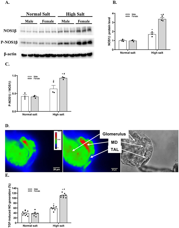
Figure 1. High salt diet induces greater increases of macula densa NOS1β expression and activity in female mice than male mice.
(A) The immunoblots of NOS1, P-NOS1 and the loading control of β-actin. The renal cortical levels of NOS1β (B) and P-NOS1β/NOS1β (C) in male and female C57BL/6 mice with normal salt diet or high salt diet. n=3; *P<0.01 versus normal salt; #P<0.01 versus male mice. (D) The NO generation in the macula densa was measured in isolated perfused JGA with DAF-2 DA. The bright field image exhibited the anatomic structure of the perfused JGA. The florescent image of DAF-2 DA loaded JGA showed the NO generation in the macula densa. (E) The TGF-induced NO generation by the macula densa in male and female C57BL/6 mice with normal salt diet or high salt diet. n=7-9; *P<0.01 versus normal salt; #P<0.05 versus male mice. Statistical difference was calculated by two-way ANOVA followed by Sidak multiple comparisons test.

