Abstract
Reinforcer devaluation is a task often used to model flexible goal-directed behavior. Here, we inactivated basolateral amygdala (BLA), orbitofrontal cortex (OFC), mediodorsal thalamus (MD.) (Exp. 1) and prelimbic cortex (PL) (Exp. 3) in rats during multiple-response/multiple-reinforcer operant training with levers available to earn reinforcers during cued trials. After two training days with each lever-food relationship, a reinforcer was devalued through selective satiety and devaluation was assessed in a choice test with the brain areas non-inactivated. The control (PBS) and OFC and PL inactivation groups exhibited a devaluation effect, but the BLA or MD groups did not. Since the OFC is proposed to be required in devaluation tasks when a discrete cue signals an outcome and PL is proposed to be required when responses based on lever spatial-location guide behavior, we ran new rats through a cue-switching experiment (Exp. 2) to determine the strategy rats use to complete our task (attending to the discrete light cue or spatial lever location). Both groups (cue switched and cue normal) showed a devaluation effect based on the lever spatial location, suggesting that rats rely on the spatial lever location to guide behavior. Future studies will determine whether OFC and PL can compensate for each other to show intact devaluation when the functioning of one of them is impaired.
Keywords: devaluation, decision-making, orbitofrontal cortex, prelimbic cortex, basolateral amygdala, mediodorsal thalamus
1. Introduction
The ability to flexibly alter behavior in the face of changing goals is critical to organismic success. One way to model flexible goal-directed decision-making is through a reinforcer devaluation task. In this task, an individual learns that a response or a cue predicts a reward and the value of the reward is then reduced using motivational (selective satiety) or associative (conditioned taste aversion) methods. Devaluation is then assessed by presenting the response option or cue that previously led to the devalued outcome and measuring responding, which is typically reduced in normal task performance. Notably, proper goal-directed action (in which responses decrease as the value of the reward is decreased) is impaired in people with schizophrenia [1] and obsessive-compulsive disorder [2, 3]. There is also mixed evidence for [4, 5] and against [6, 7] an impairment in individuals dependent on addictive drugs.
In nonhuman primates, basolateral amygdala (BLA) [8–10], orbitofrontal cortex (OFC) [8, 11–14] and mediodorsal thalamus (MD) [15, 16] are required for devaluation tasks. BLA lesions/inactivations also impair goal-directed action in most devaluation tasks in rodents [17–22]. However, in rodents, results for the other two brain areas differ. Lesions/inactivations of MD or lateral OFC impair [19, 23–30] or have no effect on goal-directed action [29–31]. In light of these discrepancies, we investigated the role of these 3 brain areas in a multiple-reinforcer/multiple-response cued operant devaluation task. Our task had discrete trials in which each lever was available and accompanied by a cuelight above the lever (with inter-trial intervals [ITIs] where the lever-light compounds were not available). This task differs from the common free-operant versions of the task, in which levers are continuously available without any obvious ITIs that would indicate reinforcer availability. In free operant tasks, the lever spatial location indicates the available reinforcer, and goal-directed action is usually attributed to response-outcome (R-O) associations. In our task, the lever spatial location also indicated the identity of the available reinforcer, although the limited access to the lever (with ITIs between trials) and distinct cues above each lever may lead to a greater role of stimulus-outcome (S-O) associations in goal-directed action. For all three brain areas, evidence suggests that, if the brain areas was required for a particular task version, inactivation during the initial Pavlovian/operant training would impair later devaluation [19, 21, 28, 30]. Therefore, we inactivated only during initial training to determine a role in learning the necessary associations. We targeted lateral OFC (and refer to this region as “OFC”), as most rodent devaluation research has targeted this area [19, 23–26, 32, 33], although medial OFC may play a different role in devaluation than lateral OFC [34, 35].
In Exp. 1, we inactivated OFC, BLA, or MD throughout operant training and then assessed devaluation without inactivation. We found that BLA and MD inactivation during training impaired subsequent devaluation, but the controls and OFC inactivation group showed intact devaluation, suggesting that BLA and MD, but not OFC, are necessary for learning the information later needed for goal-directed action in our devaluation task.
Since each cuelight is consistently associated with one of the levers, rats could use either the cuelight identity (S-O associations) or the lever spatial location (R-O associations) to guide behavior. To assess the strategy/association used, we ran a second experiment with no inactivations in which the lever-cuelight combinations were different between operant training and devaluation testing (ex: flashing light above the right lever during training and above the left lever during testing). We found that the rats reduced responding based on the lever’s fixed spatial location rather than the cuelight location (ex: decreased responding for the lever spatial location associated with the sated food, regardless of the cuelight above it), suggesting that rats primarily employ an R-O strategy to guide goal-directed action in our task.
Prelimbic cortex (PL) is required for goal-directed action in free operant versions of the devaluation task, [36–38]. As such, PL may be required to learn R-O associations that allow for later goal-directed action [39, 40], with the fixed lever-location defining “responses”. Since the results of Exp. 2 suggest that R-O associations (based on lever-location) primarily guide behavior in our task, we investigated whether PL was necessary for our task. In Exp. 3, we inactivated PL throughout training, but not during the devaluation test. However, we found that PL inactivation during training did not impair later goal-direction action.
2. Methods
2.1. General methods
2.1.1. Subjects
Naïve Long-Evans rats (n = 220 from Charles River Laboratories (Kingston, NY and Raleigh, NC) were used for the experiments. Males and females were 150-225g and 225-275g, respectively, upon arrival in the facility. All animals were individually housed and maintained on a 12-hour-12 hour reverse light-dark cycle with lights off at 07:30 am in a temperature and humidity-controlled room. For the experiments with surgical manipulations (Experiments 1 and 3), the rats were given time to acclimate to the facility and grow to a minimum weight of 250 g (for females) or 295 g (for males), before they received intracranial surgery for cannula implantation. The rats were then allowed to recover for at least 5 days after surgery and reach minimum weights (250 g for females or 295 g for males) before beginning food restriction. In Experiment 2, there were no surgical manipulations, but the rats were allowed to grow to a minimum of 295 g before they began food restriction.
For food restriction in all experiments, initial free-feeding body weights were recorded and the rats were food-restricted to 85% of their free-feeding weight and subsequently allowed to grow 1 g/day for males and 0.25 g/day for females. The difference in feeding procedures between males and females is intended to maintain similar rates of lever-pressing across both sexes, based on pilot data within our lab. Water was available ad libitum throughout the experiment.
Behavioral training took place once rats were stabilized on the food restriction conditions. Training took place during the dark cycle and rats were weighed and fed after the daily sessions. All procedures and animal care were in accordance with the Kansas State University Institutional Animal Care and Use Committee guidelines, the National Institutes of Health Guidelines for the Care and Use of Laboratory Animals, and United States federal law.
2.1.2. Apparatus
Experiments were conducted in 12 standard operant chambers (Med Associates, St Albans, VT). Each chamber was equipped with a pellet dispenser that delivered either a 45-mg grain pellet (catalog #1811156; TestDiet, Richmond, IN, USA), a 45-mg precision pellet (catalog #1811155; TestDiet, Richmond, IN) or a 45-mg chocolate-flavored sucrose pellet (product #F07256; BioServ, Frenchtown, NJ). The identity of the pellet dispensed was dependent on the phase of the task. The chambers had two retractable levers on either side of the food cup at approximately one-third of the total height of the chamber, with a white cuelight located above each lever. A red houselight was mounted on the top–center of the back wall. A Dell Optiplex computer (Dell Inc., Round Rock, TX) was equipped with Med-PC for Windows (Med Associates, St Albans, VT), which controlled the equipment and recorded lever-presses.
2.1.3. Surgical procedures
Rats underwent surgery for Experiments 1 and 3. Animals were anesthetized with 3-5% isoflurane in an induction chamber and then placed in a stereotaxic device (Kopf Instruments, Tujunga, CA) and maintained on 1-3% isoflurane for the duration of the surgery. Once the skull was exposed, bregma was used to determine guide cannula placement, for which holes were burred with a drill. Stainless steel guide cannula (PlasticsOne, Roanoke, VA) were implanted bilaterally to target specific brain areas according to coordinates based on a rat brain atlas by Paxinos and Watson (2009). For BLA, two single 23G cannulae were bilaterally implanted −2.9 anterior-posterior (AP), ±5.3 medial-lateral (ML; angled 4 degrees), and −7.6 dorsal-ventral (DV) mm relative to bregma. For OFC, two single 23G cannulae were bilaterally implanted +3.2 AP, ±4.2 ML (angled 10 degrees), and −4.3 DV relative to bregma. For MD, a single bilateral 22G cannula was implanted −2.9 AP, ±0.8 ML, −4.5 DV relative to bregma. For PL, a single bilateral 22G guide cannula was implanted +3.0 AP, ±0.6 ML, −2.6 DV relative to bregma. Cannulae were fixed in place using 4 bone anchor screws and dental acrylic. The injection channels of the guide cannulae were kept clear and contaminants were kept out using dummy cannulae and dustcaps. Animals were given 2mg/kg meloxicam (VetOne, Boise, ID) subcutaneously upon completion of surgery and also 24-h later during the post-op health check.
2.1.4. Intracranial infusions
Figure 1 shows the experimental timeline. In Experiments 1 and 3, animals received bilateral infusions of either sterile 1X phosphate buffered saline (PBS) or a muscimol/baclofen (M/B) cocktail prior to each cue training session. The cocktail consisted of 0.125μg of muscimol (Bachem, Torrance, CA) and 0.125μg baclofen (Sigma-Aldrich, St. Louis, MO) in 0.3μl sterile PBS in each hemisphere. Injection cannulae (30G for OFC and BLA infusions, 28G for MD and PL infusions, PlasticsOne, Roanoke, VA) extending 1mm beyond the guide cannulae were placed inside the guide cannula for the intracranial microinfusions. A volume of 0.3 μl was infused at a rate of 0.3 μl/min using a microinfusion pump (model 101, KD Scientific, Hollison, MA) and 10μl Hamilton syringes (model 1701 LT, Hamilton Company, Reno, NV). Upon completion of the infusions, the injector cannulae were left in place for 1 minute to allow for the diffusion of the infused solutions. Injector cannulae were then removed and replaced with dummy cannulae and dustcaps. Subjects began their cued-trial training session 5 minutes after injector cannulae removal.
Figure 1:
Experimental timeline. * = Only Exps. 1 and 3. ** = Intracranial infusions occurred prior to the training sessions in Exps. 1 and 3.
2.1.5. Behavioral training and testing
Animals received 3 once-daily 40-min magazine training sessions, with each session delivering one of the three different types of pellet every 125 seconds (precision, grain, or chocolate). Animals then underwent 2 lever-press training sessions during a single day (with morning and afternoon sessions 2-4 hours apart) to earn grain pellets on a fixed-interval-1 (FI1) reinforcement schedule (lever-presses could earn a pellet each second). Each lever was trained in a separate session. These sessions ended when rats received 50 pellets or after one hour had elapsed, whichever came first. Rats that failed to earn 50 pellets during one of these sessions received up to 2 additional FI1 training sessions. All rats earned 50 pellets within a session by the end of the FI1 sessions. Grain pellets were used throughout FI1 training. We chose to use a different reinforcer than those used in the later cued-trial training (precision and chocolate pellets) because we did not want the rats to experience pairings of lever-presses with the cued-trial reinforcers prior to the start of infusions (in Exps. 1 and 3), as this could allow R-O associations to form before the target brain areas were inactivated.
Next rats received 4 once-daily cued-trial operant training sessions, two sessions for each cue-lever-reinforcer combination. Throughout training, the left-lever earned precision pellets and was always associated with a steady white cuelight. The right lever earned chocolate pellets and was always associated with a flashing (2 Hz) white cuelight. All rats received the steady light-left lever-precision pellet combination during the first and fourth sessions. On the second and third sessions, all rats received the flashing light-right lever-chocolate pellet combination. Regardless of the cue-lever-reinforcer relationship present, every cued-trial training session was 40-min long with 40 trials. All trials lasted 40 sec and rats could earn two pellets per trial, one for pressing at a randomized time within the first half of the trial and one for pressing at a randomized time within the second half of the trial, which represents a modified variable-interval-20 schedule. Thus, animals could earn up to 80 pellets during a session. The levers were retracted and the cuelights extinguished during the inter-trial interval (ITI), which ranged from 13-29 seconds (average 20 sec). This pattern of trials, with 40-sec cues and the levers retracted for an average of 20 sec during each minute, was used for both operant training sessions and the non-reinforced devaluation test sessions. We used a relatively short ITI in order to allow for multiple trials in a short period of time after the end of the satiation period in the upcoming devaluation choice tests, so that later trials would not after a longer delay when satiation might be weaker. Although longer ITIs are often used in tasks where responses are possible during the ITI (in order to extinguish the context and increase stimulus control by the discriminative stimuli), the retracted levers during the ITI prevented any such extinction and longer ITIs would have less of a benefit. In Exps. 1 and 3, the cued-trial operant training sessions were always preceded by intracranial infusions, as described above.
After initial cued-trial training was completed, a choice test was administered. For the choice tests, animals first received a 1-h satiation session in which they had access to 30 g of either precision or chocolate pellets (counterbalanced across group and sex) in the operant chambers. The satiation period began with 20g of pellets in a ceramic bowl and an additional 10g of pellets were added 45 min into the session. After the satiation session was complete, animals received a 15-min break in their home cage during which the remaining pellets were weighed to determine consumption. Animals were subsequently placed back into the operant chambers for a 12-min choice test consisting of 12 40-sec trials, conducted in extinction (no food pellets for pressing either lever). In Exps. 1 and 3 and for the Cue Normal group in Exp. 2, both levers were extended simultaneously with the corresponding cuelight illuminated above them (the same lever-light compound as in training). For the Cue Switched group in Exp. 2, the identity of the cuelight above the levers was switched such that the cuelight above the left lever was flashing and the cuelight above the right lever was steadily illuminated (the opposite of the configurations during training). After the first choice test was administered, all animals underwent 2 retraining sessions over 2 days, with 1 session for each of the two lever-cuelight-pellet compounds. For all groups (including the Cue Switched group in Exp. 2), the lever-light compounds in these retraining sessions were the same as those during the initial cued-trial operant training. Animals in Exps. 1 and 3 received intracranial infusions prior to these retraining sessions. Once the retraining sessions were completed, animals underwent a second choice test. For this second choice test, the rats were sated with the opposite pellet type as in the first satiation session. The second choice test was otherwise identical to the first. We have found that rats in our experiments sometimes prefer precision pellets to chocolate pellets, so performing two choice tests with each of the pellets sated in one of the two tests allows this preference to average out in the devaluation assessment.
Finally, at least one day after the final choice test, animals in Exps. 1 and 3 began consumption tests. Each consumption test included a 60-min satiation period for one of the two pellet types (counterbalanced across group and sex), identical to the satiation periods that preceded the choice tests. After the satiation session was complete, animals received a 15-min break in their home cage during which the remaining pellets were weighed to determine initial pellet consumption. Animals were subsequently placed back into the operant chambers for the consumption test. During the consumption test, the animals had access to two ceramic bowls. One bowl contained 10g of one type of pellet and the other bowl contained 10g of the other pellet type. The consumption test lasted 10 min. Upon completion of the consumption test, animals were returned to their home cages and the remaining pellets were sorted by type and weighed to determine consumption. Animals received at least one day off after the first consumption test before completing a second consumption test identical to the first, except that animals were sated on the opposite pellet type as in the first consumption test. These consumption tests were run on separate days after all devaluation choice testing was completed (rather than immediately after the choice tests) to allow for similar satiation levels during the choice and consumption tests. A consumption test run after the choice test would likely begin about 42 min after the end of satiation, with 15 min after satiation to set up the choice test, 12 min for the choice test, and 15 min to set up the consumption test after the choice test. The level of satiation might be reduced after this 42-min period, and would likely not match the level of satiety during the choice test that began almost 30 min prior. However, while rats consumed pellets until they were sated (all rats had some pellets left over at the end of each satiation period before the choice and consumption tests) and the choice and consumption tests began at the same interval after the end of the satiation period, we cannot be certain that the levels of satiety were equal across the choice and consumption tests.
2.1.6. Histological analysis
Animals were deeply anesthetized using 1-2mL of Fatal Plus (Vortech Parmaceuticals, LTD, Dearborn, MI). Once animals stopped breathing, they were perfused transcardially with 100mL 1X PBS (pH: 7.2-7.4) followed by 400mL 4% paraformaldehyde (PFA, pH 7.2-7.4). Brains were post-fixed in 4% PFA for 2-24 hours and then placed in 30% sucrose in 1X PBS until they sank, at which point they were flash-frozen in dry ice and stored in a −80°C freezer. The tissue was sliced into 40um sections using a cryostat (model HM 550, Thermo Scientific, Waltham, MA) and mounted onto microscope slides, at which point they were Nissl stained using thionin. Slides were coverslipped immediately after staining using Permount (Fisher Scientific, Waltham, MA) and placement was assessed using a light microscope (model BX41, Olympus) and SPOT 5.1 Advanced Software. Only the data of animals whose injection cannula were located within the boundaries of the targeted brain region (according to the rat brain atlas by Paxinos and Watson, 2009) were included in analyses.
2.1.7. Statistical analysis
Data were analyzed by Statistica 13.3 software (Palo Alto, CA). The factors used in the statistical analyses are described in the Results section for each analysis and significant effects (p<0.05) in the different ANOVAs were followed by post-hoc Tukey’s HSD tests. Importantly, test data was collapsed across both test sessions of the same type, i.e., data from both choice tests was analyzed together and data from both consumption tests was analyzed together. This addressed any potential issues with side or pellet preference.
2.2. Individual experiments
2.2.1. Experiment 1
Rats (n=128) underwent surgery to place cannulae targeting OFC, MD, or BLA. We performed OFC cannulation surgery on 22 females and 24 males, MD cannulation surgery on 18 females and 19 males, and BLA cannulation surgeries on 23 females and 22 males. The animals were trained and tested according to the procedure outlined in the general methods. All rats were trained with pre-training infusions and had the same lever-light configurations during training and testing. For intracranial infusions, 27 rats (14 female and 13 male) received M/B injections into BLA, 26 rats (12 female and 14 male) received M/B injections into MD, 28 rats (13 female and 15 male) received M/B injections into OFC, and 30 rats (15 female and 15 male) received PBS injections into BLA, MD or OFC. Of the PBS-injected rats, 10 rats (5 female and 5 male) received injections into BLA, 9 rats (5 female and 4 male) received injections into MD, and 11 rats (5 female and 6 male) received injections into OFC. Seventeen rats underwent surgery, but experienced health problems before the first intracranial injection day and did not receive infusions.
2.2.2. Experiment 2
Male rats (n=24) underwent behavioral training as described in the general methods. There were two groups: one that had incongruent light cues presented during testing (the Cue Switched group, n=12) and one that had congruent light cues presented during testing (the Cue Normal Group, n=12). There were no infusions before any of the training sessions. All rats had identical training in all phases other than the choice test phase, when the two groups had either the same (Cue Normal group) or different (Cue Switched group) lever-light configurations during training and testing.
2.2.3. Experiment 3
Rats (n=68) underwent surgery to place cannulae targeting PL. The animals were trained and tested according to the procedure outlined in the general methods. We performed PL cannulation surgery on 36 females and 32 males. All rats were trained with pre-training infusions and had the same lever-light configurations during training and testing. For pre-training infusions, 33 rats (17 female and 16 male) received M/B injections into PL and 33 rats (17 female and 16 male) received PBS injections into PL. Two rats underwent surgery, but experienced health problems before the first intracranial injection day and did not receive infusions.
3. Results
3.1. Experiment 1
3.1.1. Histological and behavioral exclusions.
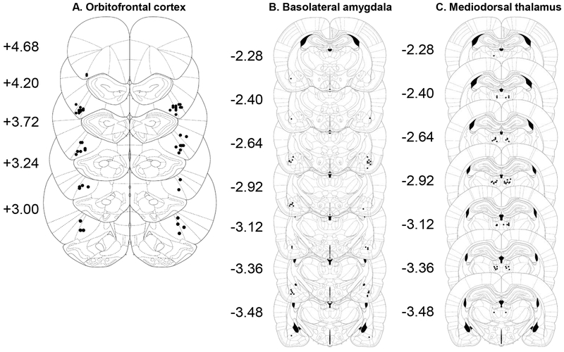
Figure 2 shows the placement of infusion sites in the OFC, BLA, and MD. For OFC (Figure 2A), our criterion for inclusion of data in the analysis was that the injector tip must be within or on the border of lateral orbitofrontal cortex (comprised of the ventral orbital, lateral orbital, or agranular insular cortices [41]) in both hemispheres. Although placement in ventral orbital would be acceptable, no injector tip in an included animal was located within ventral orbital cortex. For BLA (Figure 2B), our criterion for inclusion of data in the analysis was that the injector tip must be within the basolateral complex (comprised of the lateral, basal/basolateral and accessory basal/basomedial amygdaloid nuclei [42, 43]) in both hemispheres. However, while some injector tips were on the lateral-basolateral border, no injector tip in an included animal was located entirely within lateral nucleus. For MD (Figure 2C), our criterion for inclusion of data in the analysis was that the injector tip must be within the medial, central or lateral mediodorsal thalamic nuclei. Based on these criteria, 11 rats (4 Female-BLA, 1 Female-PBS, 2 Male-MD, 2 Male-OFC, 2 Male-PBS) were excluded. Seventeen rats (4 Female-BLA, 2 Female-MD, 1 Female-OFC, 6 Male-BLA, 1 Male-MD, 3 Male-OFC, 1 Male-PBS) were also excluded from training and test analyses because they earned <6 rewards in at least one operant training or retraining session. These rats received additional training sessions (with additional intracranial infusions) on the same lever-light-pellet combination used in any session with low responding to avoid the possibility that they had insufficient experience with the experimental contingencies to learn the expected outcome (regardless of brain area activity). However, due to concerns about uneven training with the two levers, operant over-training, and unwanted effects of extra intracranial infusions, we excluded their data from the analyses. The final group sizes in Experiment 1 were as follows: Female OFC group, n=11; Male OFC group, n=8; Female BLA group, n=5; Male BLA group, n=7; Female MD group, n=10; Male MD group, n=9; Female PBS group, n=14; Male PBS group, n=12.
Figure 2:
A. Placement of included injector tips in OFC. B. Placement of included injector tips in BLA. C. Placement of included injector tips in MD. Numbers represent anterior-posterior coordinates in relation to bregma from Paxinos & Watson, 2009, reproduced with permission. Black circles represent injector tips.
3.1.2. Training.
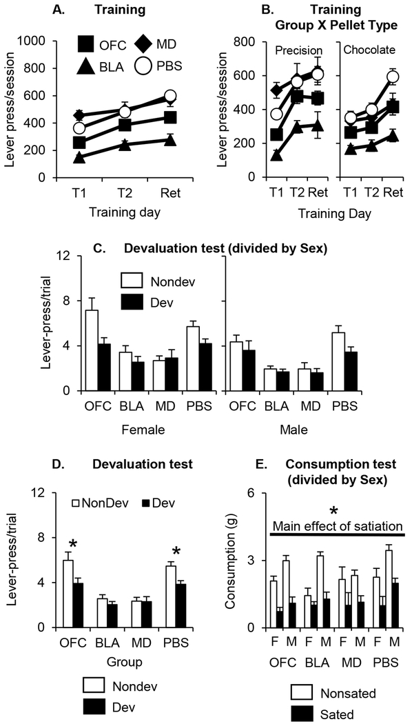
Inactivation of BLA, but not MD or OFC, decreased lever-pressing for food rewards during training (Figure 3A). A mixed factor ANOVA with the between-subjects factors of Group (OFC, BLA, MD, PBS) and Sex (Female, Male) and the within-subjects factors of Pellet Type (Precision, Chocolate) and Training Day (First, Second, and Retrain days of training) found a significant main effect of Group (F(3, 68) = 8.2, p<0.01) and a significant interaction of Group X Pellet Type (F(3, 68) = 6.6, p<0.01). In addition, there were significant effects of Pellet Type, Training Day, and a Pellet Type X Training Day interaction (all p<0.05; see Table 1). The Group X Pellet Type interaction can be observed in Figure 3B. A post hoc analysis of the Group X Pellet Type interaction revealed that the BLA group made significantly fewer lever-presses for both pellet types than the PBS group (all p<0.05) and the MD group made more responses for precision pellets than chocolate pellets (p<0.05), but no other comparisons were significant. There were no effects or interactions of Sex, and no other effects or interactions were significant (all p>0.05; Table 1).
Figure 3:
A. Lever presses/session (mean ± SEM) in each infusion group during the first and second training cued-trial training days and retraining days for each pellet in Exp. 1 (averaged across sex and pellet type). B. Lever presses/session (mean ± SEM) in each infusion group during the first and second training cued-trial training days and retraining days for each pellet type in Exp. 1 (averaged across sex and separated by pellet type). Left panel represents responding for precision pellets. Right panel represents responding for chocolate-flavored sucrose pellets. For A and B: Black squares represent rats that received OFC inactivations. Black diamonds represent rats that received MD inactivations. Black Triangles represent rats that received BLA inactivations. White circles represent rats that received PBS infusions. T1 and T2 represent the first and second day of cued trial training for each pellet type and Ret represents the retraining day for each pellet type (averaged across the first, second and retraining day for each pellet in A and presented separately for each pellet on in B). C. Lever presses/trial (mean ± SEM) during the devaluation test in Exp. 1 (averaged across trial and separated by sex). Left panel represents female groups. Right panel represents male groups. D. Lever presses/trial (mean ± SEM) during the devaluation test in Exp. 1 (averaged across sex and trial). For C and D: White bars represent responding on the nondevalued lever. Black bars represent responding on the devalued lever. Nondev = nondevalued lever. Dev = devalued lever. E. Consumption in grams (mean ± SEM) of the sated and nonsated pellet types during the consumption test in Exp. 1 (separated by sex). White bars = nonsated pellet type. Black bars = sated pellet type. F = female. M = male. * = p<0.05
Table 1:
Results of ANOVA for main effects and interactions for the training data in Experiment 1.
| Main effect: Group | Main effect: Sex | Main effect: Pellet Type | Main effect: Training Day | Interaction: Group*Sex |
|---|---|---|---|---|
| F(3, 68) = 8.2* | F(1, 68) = 0.1 | F(1, 68) = 46.4* | F(2, 136) = 28.3* | F(3, 68) = 0.1 |
| Interaction: Group*Pellet Type | Interaction: Group*Training Day | Interaction: Sex*Pellet Type | Interaction: Sex*Training Day | Interaction: Pellet Type*Training Day |
| F(3, 68) = 6.6* | F(6, 136) = 1.0 | F(1, 68) = 3.6 | F(2, 136) = 1.6 | F(2, 136) = 7.9* |
| Interaction: Group*Sex* Pellet Type | Interaction: Group*Sex* Training Day | Interaction: Group*Pellet Type*Training Day | Interaction: Sex*Pellet Type*Training Day | Interaction: Group*Sex* Pellet Type* Training Day |
| F(3, 68) = 0.3 | F(6, 136) = 0.7 | F(6, 136) = 1.0 | F(2, 136) = 1.6 | F(6, 136) = 0.7 |
Values in bold with an asterisk represent significant effects (p<.05).
3.1.3. Devaluation test.
The statistical results revealed three patterns. First, the PBS and OFC groups made more responses on the nondevalued lever compared to the devalued lever, indicative of a devaluation effect, but the BLA and MD groups did not (Figure 3C; collapsed across Sex in Figure 3D). Second, female rats responded on the levers more than the males but there were no interactions of Sex with the devaluation effect or the effects of inactivation. Third, all rats decreased responding on the levers across the 6 trials but responding decreased faster for the nondevalued lever compared to the devalued lever. A mixed factor ANOVA with the between-subjects factors of Group (OFC, BLA, MD, PBS) and Sex (Female, Male) and the within-subjects factors of Lever (the levers associated with the Devalued and Nondevalued foods) and Trial (Trials 1-6) found significant main effects of Sex (F(1, 68) = 8.4, p<0.01), Group (F(3, 68) = 13.5, p<0.01), Lever (F(1, 68) = 16.0, p<0.01), and Trial (F(5, 340) = 19.9, p<0.01) and significant interactions of Group X Lever (F(3, 68) = 3.0, p<0.05) and Lever X Trial (F(5, 340) = 3.4, p<0.01). A post-hoc analysis of the Group X Lever interaction showed that the PBS and OFC groups pressed the nondevalued lever significantly more than the devalued lever (both p<0.05), but the BLA and MD groups responded on the two levers equally. The main effect of Sex showed that females pressed more than males but this did not interact with any other factor (all p>0.05; Table 2). A post-hoc analysis of the Lever X Trial interaction showed that the peak responding on the non-devalued lever was on trial 2, and responding on the non-devalued lever significantly decreased from trial 2 to trials 4-6 and responding on the devalued lever only decreased from trial 2 to trial 6.
Table 2:
Results of ANOVA for main effects and interactions for the devaluation test data in Experiment 1.
| Main effect: Group | Main effect: Lever | Main effect: Sex | Main effect: Trial | Interaction: Group*Lever |
|---|---|---|---|---|
| F(3, 68) = 13.5* | F(1, 68) = 16.0* | F(1, 68) = 8.4* | F(5, 340) = 16.9* | F(3, 68) = 3.0* |
| Interaction: Group*Sex | Interaction: Group*Trial | Interaction: Sex*Lever | Interaction: Sex*Trial | Interaction: Lever*Trial |
| F(3, 68) = 0.4 | F(15, 340) = 1.0 | F(1, 68) = 1.0 | F(5, 340) = 0.9 | F(5, 340) = 3.4* |
| Interaction: Group*Sex* Lever | Interaction: Group*Sex*Trial | Interaction: Group*Lever* Trial | Interaction: Lever*Trial*Sex | Interaction: Group*Lever* Sex*Trial |
| F(3, 68) = 1.7 | F(15, 340) = 1.0 | F(15, 340) = 0.7 | F(5, 340) = 0.8 | F(15, 340) = 0.5 |
Values in bold with an asterisk represent significant effects (p<.05).
There was no difference in the effects of PBS injections into the different brain areas. All rats in the PBS group made more responses on the nondevalued lever compared to the devalued lever and this effect did not interact with the brain region infused with PBS. A mixed factor ANOVA with the between-subjects factors Brain Region (OFC, BLA, MD) and Sex (Female, Male) and the within-subjects factor Lever (the levers associated with the Devalued and Nondevalued foods) found a significant main effect of Lever (F(1, 20) = 20.6, p<0.01). No other main effects or interactions were significant (all p>0.05).
3.1.4. Consumption test.
The rats ate significantly more of the pellet type they were not sated on (i.e. the nondevalued pellet type) regardless of whether they had OFC, BLA, or MD inactivated during training or not (Figure 3E). In addition, male rats ate greater amounts of food than female rats. A mixed factor ANOVA with the between-subjects factors of Group (OFC, BLA, MD, PBS) and Sex (Female, Male) and the within-subjects factor of Pellet Satiation (the pellets that were sated or nonsated) found significant main effects of Sex (F(1, 68) = 20.5, p<0.01) and Pellet Satiation (F(1, 68) = 86.9, p<0.01) and a significant Sex X Pellet Satiation interaction (F(1, 68) = 4.2, p<0.05). A post hoc analysis of the Sex X Pellet Satiation interaction revealed that the difference in consumption between the sated and non-sated food was significant in both the males and in the females. Thus, although the interaction is driven by a larger numerical decrease in consumption to the sated pellet in males, satiation led to a significant decrease in consumption in both sexes. There was no Group X Pellet Satiation interaction (F(3, 68) = 0.8, p>0.05), suggesting the groups were able to devalue the sated pellet properly when presented with both pellet types. There were no other significant main effects or interactions (all p>0.05; Table 3).
Table 3:
Results of ANOVA for main effects and interactions for the consumption test data in Experiment 1 and Experiment 3.
| Main effect: Group | Main effect: Sex | Main effect: Pellet Satiation | Interaction: Group*Sex | Interaction: Group*Pellet Satiation | Interaction: Sex*Pellet Satiation | Interaction: Group*Sex*Pellet Satiation | |
|---|---|---|---|---|---|---|---|
| Exp. 1 | F(3, 68) = 2.1 | F(1, 68) = 20.5* | F(1, 68) = 86.9* | F(3, 68) = 1.0 | F(3, 68) = 0.8 | F(1, 68) = 4.2* | F(3, 68) = 1.1 |
| Exp. 3 | F(1, 44) = 0.0 | F(1, 44) = 4.4* | F(1, 44) = 40.7* | F(1, 44) = 0.2 | F(1, 44) = 0.7 | F(1, 44) = 0.9 | F(1, 44) = 0.8 |
Values in bold with an asterisk represent significant effects (p<.05).
3.2. Experiment 2
3.2.1. Training.
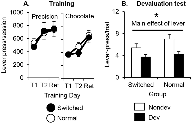
The Normal and Switched groups responded at equal rates during training for both pellet types (Figure 4A). A mixed factor ANOVA with the between-subjects factor of Group (Normal, Switched) and the within-subjects factors of Pellet Type (Precision, Chocolate) and Training Day (First, Second, and Retrain days of training) found significant main effects of Pellet Type (F(1, 22) = 22.4, p<0.01) and Training Day (F(2, 44) = 28.1, p<0.01) and a significant interaction of Pellet Type X Training Day (F(2, 44) = 7.9, p<0.01). There was no main effect of Group or interactions with Group (all p>0.05), indicating the groups did not differ in baseline lever-pressing.
Figure 4:
A. Lever presses/session (mean ± SEM) during the first and second training cued-trial training days and retraining days for each pellet in Exp. 2 (divided by pellet type). Left panel represents responding for precision pellets. Right panel represents responding for chocolate-flavored sucrose pellets. Black circles represent rats in the Cue Switched group. White circles represent rats in the Cue Normal group. B. Lever presses/trial (mean ± SEM) during the devaluation test session in Exp 2 (averaged across trial). White bars represent responding on the nondevalued lever. Black bars represent responding on the devalued lever. Nondev = nondevalued lever. Dev = devalued lever. * = p<0.05
3.2.2. Devaluation test.
The rats made more responses on the nondevalued lever compared to the devalued lever, but there was no effect of switching the training cue location on devaluation performance (Figure 4B). For the analysis, we defined the Devalued and Nondevalued levers in terms of the fixed lever-location rather than the location of each cuelight. A mixed-factor ANOVA with the between-subjects factor of Group (Normal, Switched) and the within-subjects factors of Lever (the fixed lever-location associated with the Devalued and Nondevalued foods) and Trial (Trials 1-6) found a significant main effect of Lever (F(1, 22) = 19.8, p<0.01) and Trial (F(5, 110) = 18.0, p<0.01) and a marginal Lever X Trial interaction (F(5, 110) = 2.3, p=.052. The main effect of Lever showed that rats pressed the nondevalued lever more than the devalued lever. Post-hoc tests on the main effect of Trial showed that rats significantly decreased responding across the two levels on Trials 4-6 compared to Trial 1. There was no main effect of Group (F(1, 22) = 1.7, p>0.05) or Group X Lever interaction (F(1, 22) = 1.3, p>0.05), suggesting the cue mismatch between training and test did not affect devaluation.
3.3. Experiment 3
3.3.1. Histology.
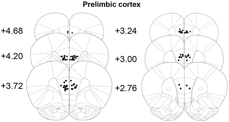
For PL (Figure 5), our criterion for inclusion of data in the analysis was that the injector tip must be within the prelimbic cortex posterior to +4.68 AP in both hemispheres. Based on these criteria, 11 rats (3 Female-PL, 1 Female-PBS, 5 Male-PL, 2 Male-PBS) were excluded. The final group sizes in Experiment 3 were as follows: Female PL group, n=14; Male PL group, n=10; Female PBS group, n=12; Male PBS group, n=13.
Figure 5:
Placement of included injector tips in PL. Numbers represent anterior-posterior coordinates in relation to bregma from Paxinos & Watson, 2009, reproduced with permission. Black circles represent injector tips.
3.3.2. Training.
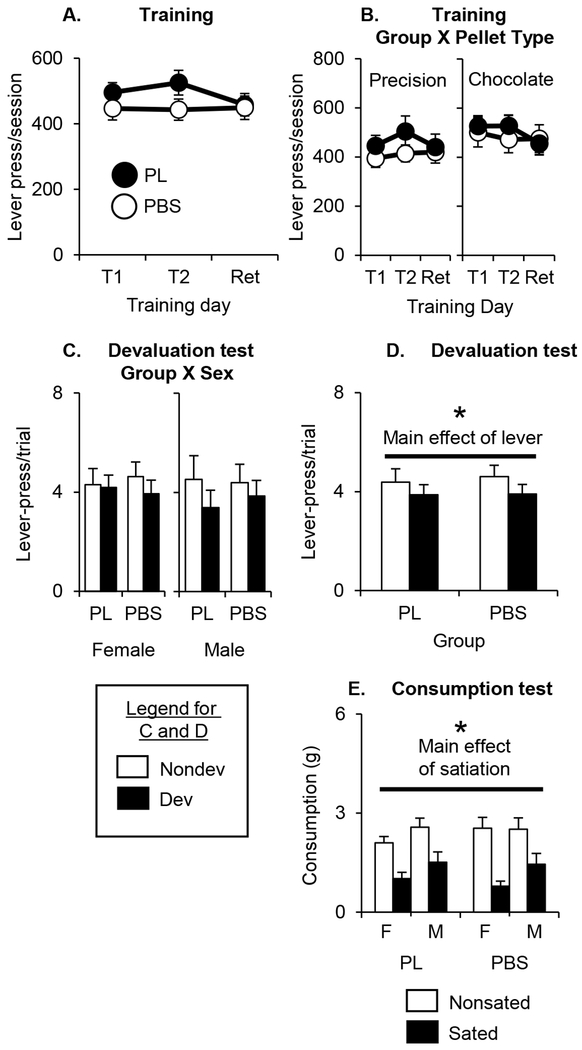
The PBS and PL groups responded at equal rates during training for both pellet types without any sex differences in behavior (Figure 6A, to parallel our figures for Exp. 1 we present responding broken down by pellet type in Figure 6B). A mixed factor ANOVA with the between-subjects factors of Group (PL, PBS) and Sex (Female, Male) and the within-subjects factors of Pellet Type (Precision, Chocolate) and Training Day (First, Second, and Retrain days) found no significant main effects or interactions with Group or Sex (all p>0.05). There were also no main effects or interactions with Pellet Type or Training Day (all p>0.05; Table 4).
Figure 6:
A. Lever presses/session (mean ± SEM) during the first and second training cued-trial training days and retraining days for each pellet in Exp. 3 (averaged across sex and pellet type). B. Lever presses/session (mean ± SEM) during the first and second training cued-trial training days and retraining days for each pellet in Exp. 3 (averaged across sex and separated by pellet type). Left panel represents responding for precision pellets. Right panel represents responding for chocolate-flavored sucrose pellets. For A and B: Black circles represent rats that received PL inactivations. White circles represent rats that received PBS infusions. T1 and T2 represent the first and second day of cued trial training for each pellet type and Ret represents the retraining day for each pellet type (averaged across the first, second and retraining day for each pellet in A and presented separately for each pellet on in B). C. Lever presses/trial (mean ± SEM) during the devaluation test in Exp. 3 (averaged across trial and separated by sex). Left panel represents female responses. Right panel represents male responses. D. Lever presses/trial (mean ± SEM) during the devaluation test session in Exp. 3 (averaged across sex and trial). For C and D: White bars represent responding on the nondevalued lever. Black bars represent responding on the devalued lever. Nondev = nondevalued lever. Dev = devalued lever. E. Consumption in grams (mean ± SEM) of the sated and nonsated pellet types during the consumption test in Exp. 3 (averaged across sex). White bars = nonsated pellet type. Black bars = sated pellet type. F = female. M = male. * = p<0.05.
Table 4:
Results of ANOVA for main effects and interactions for the training data in Experiment 3.
| Main effect: Group | Main effect: Sex | Main effect: Pellet Type | Main effect: Training Day | Interaction: Group*Sex |
|---|---|---|---|---|
| F(1, 44) = 1.2 | F(1, 44) = 1.9 | F(1, 44) = 2.3 | F(2, 88) = 1.4 | F(1, 44) = 1.4 |
| Interaction: Group*Pellet Type | Interaction: Group*Training Day | Interaction: Sex*Pellet Type | Interaction: Sex*Training Day | Interaction: Pellet Type*Training Day |
| F(1, 44) = 0.8 | F(2, 88) = 1.6 | F(1, 44) = 0.5 | F(2, 88) = 0.4 | F(2, 136) = 1.9 |
| Interaction: Group*Sex* Pellet Type | Interaction: Group*Sex* Training Day | Interaction: Group*Pellet Type*Training Day | Interaction: Sex*Pellet Type*Training Day | Interaction: Group*Sex*Pellet Type*Training Day |
| F(1, 44) = 3.4 | F(2, 88) = 0.9 | F(2, 88) = 0.2 | F(2, 88) = 2.9 | F(2, 88) = 0.6 |
The absence of values in bold with an asterisk represent a lack of significant effects (all p>.05).
3.3.3. Devaluation test.
The rats made more responses on the nondevalued lever than the devalued lever, but there was no effect of previous PL inactivation and no sex differences in performance (Figure 6C; collapsed across Sex in Figure 6D). The rats also extinguished responding across the six trials. A mixed factor ANOVA with the between-subjects factors of Group (PL, PBS) and Sex (Female, Male) and the within-subjects factors of Lever (the levers associated with the Devalued and Nondevalued foods) and Trial (Trials 1-6) found significant main effects of Lever (F(1, 44) = 5.6, p<0.05) and Trial (F(5, 220) = 14.9, p<0.01). The main effect of Lever showed that all rats decreased responding for the devalued lever. Post-hoc tests on the main effect of Trial showed that rats significantly decreased responding across the two levels on Trials 4-6 compared to Trial 1. There were no main effects or interactions with Group or Sex (all p>0.05; Table 5).
Table 5:
Results of ANOVA for main effects and interactions for the devaluation test data in Experiment 3.
| Main effect: Group | Main effect: Lever | Main effect: Sex | Main effect: Trial | Interaction: Group*Lever |
|---|---|---|---|---|
| F(1, 44) = 0.1 | F(1, 44) = 5.6* | F(1, 44) = 0.1 | F(5, 220) = 14.9 | F(1, 44) = 0.0 |
| Interaction: Group*Sex | Interaction: Group*Trial | Interaction: Sex*Lever | Interaction: Sex*Trial | Interaction: Lever*Trial |
| F(1, 44) = 0.0 | F(5, 220) = 0.5 | F(1, 44) = 0.9 | F(5, 220) = 0.3 | F(5, 220) = 0.8 |
| Interaction: Group*Sex* Lever | Interaction: Group*Sex*Trial | Interaction: Group*Lever* Trial | Interaction: Lever*Trial*Sex | Interaction: Group*Lever* Sex*Trial |
| F(1, 44) = 0.8 | F(5, 220) = 0.2 | F(5, 220) = 0.1 | F(5, 220) = 1.5 | F(5, 220) = 0.8 |
Values in bold with an asterisk represent significant effects (p<.05).
3.3.4. Consumption test.
The rats ate significantly more of the pellet type they were not sated on (i.e. the nondevalued pellet type) regardless of whether they had PL inactivated during training or not (Figure 6E). Male rats ate greater amounts of food than female rats. A mixed factor ANOVA with the between-subjects factors of Group (PL, PBS) and Sex (Female, Male) and the within-subjects factor of Pellet Satiation (the pellets that were sated or nonsated) found significant main effects of Sex (F(1, 44) = 4.4, p<0.05) and Pellet Satiation (F(1, 44) = 40.7, p<0.01). There was no Group X Pellet Satiation interaction (F(1, 44) = 0.7, p>0.05), suggesting the groups were able to devalue the sated pellet properly when presented with both pellet types. There were no other main effects or interactions (all p>0.05; Table 3).
4. Discussion
In the current experiments, we showed that BLA and MD, but not OFC or PL, was necessary at the time of operant training in order for rats to exhibit normal goal-directed action in a devaluation test in our multi-response/multi-reinforcer task with cued trials. We also showed that, despite the fact that each reinforcer had a unique cuelight stimulus associated with it (steady cuelight for precision, flashing cuelight for chocolate), intact rats primarily used the lever-location rather than cuelight identity to guide their goal-directed action if there was a mismatch between the training and testing lever location+cuelight compounds. Below, we discuss the parameters of our task and how these may have affected the strategies available for rats to maintain goal-directed action, the effects of MD and BLA inactivation on devaluation, and the null-effects found for OFC and PL inactivation.
4.1. Response-outcome associations primarily guide goal-directed action in our task if no neural manipulation occurs
In our task, outcome-specific behavior could be guided by either the lever-location (R-O associations) or cuelight (S-O associations). In Exp. 2, we determined whether neurologically intact rats normally rely on the lever-location, the cuelight, or a combination of the two to guide behavior. During the test in the Cue Switched group, the cuelight associated with precision pellets during training was presented above the lever-location associated with chocolate pellets. If rats were primarily guiding their behavior based on lever-location, then they should decrease responding on the lever-location associated with the sated outcome (even though this lever is below the cuelight associated with the non-sated outcome in training). We found a pattern largely consistent with rats guiding their behavior based on the lever-location (R-O associations) rather than the cuelight (S-O associations), although the smaller devaluation magnitude in the Cue Switched group than in the Cue Normal group (if calculated based on the lever location) suggests that the cuelight is not entirely ignored.
4.2. Inactivation of BLA and MD limited to operant acquisition phase impairs devaluation
We found evidence that inactivation of BLA or MD did impair later devaluation. While there was indirect evidence from previous comparisons of pre- vs. post-training permanent lesions or NMDA receptor antagonism that this would be the case, our results more directly tested the effects of decreasing general neuronal function exclusively during operant training.
BLA is necessary for goal-directed action in most devaluation tasks [8–10, 17–22], and there is evidence that NMDA receptor blockade limited to the operant training phase can impair later devaluation in a free-operant task [22]. One research study in rats comparing the effects of pre- vs. post-training permanent lesions suggested that impairment in function during the initial acquisition phase (for Pavlovian conditioning) is sufficient to impair later devaluation if only one reinforcer is used in initial training [19]. However, a pair of experiments using the same Pavlovian conditioning procedure with two reinforcers in rats found that post-training/pre-value manipulation (e.g.: taste aversion or selective satiation) lesions impair devaluation (see also [10, 20, 21, 44] for examples of operant tasks with two reinforcers where post-training BLA lesions or temporary inactivation during selective satiety impaired devaluation). It was unclear whether these procedural changes that made BLA required after the initial operant/Pavlovian training could have possibly caused BLA to no longer be required for outcome-specific encoding during the initial training phase in cued-trial tasks (rather than free operant tasks). However, our data clearly show that BLA is still required at the time of initial operant training to form outcome-specific representation that can guide later goal-directed action, even if cued-trial operant training involves multiple reinforcers.
MD is needed for goal-directed action in devaluation tasks where multiple responses to earn reinforcers have been trained [15, 16, 28, 29] (but see [31]), but not where only one response to earn reinforcers has been trained [29]. However, there is a discrepancy between the rodent and macaque literature in the time-course of this involvement. For MD, an experiment in macaques found devaluation impairments (in a discrete trial task) when this brain area was inactivated prior to selective satiety or prior to the devaluation test [16]. However, a comparison of the effects of pre- vs. post-training permanent lesions in rats suggested that impairment in function during the initial acquisition phase (for free operant conditioning) is sufficient to impair later devaluation [21, 28] (although see [31] for an example of MD chemogenetic inactivation during operant training or satiation/testing having no effect on devaluation in mice). Thus, task parameters, the method of decreasing MD function, and/or species differences may affect the role of MD in different phases of devaluation task. Our results suggested that MD activity during operant training is needed for goal-directed action in our devaluation task in rats. Our results cannot resolve the different results found in macaques and mice. However, our results provide converging evidence with prior pre- vs post-training lesion studies [21, 28] that MD plays a role in encoding stimulus-specific information during initial training in operant tasks in rats, while extending this to a different experimental procedure (cued-trial operant devaluation) and method of decreasing function (inactivation rather than permanent lesions).
4.3. Inactivation of OFC and PL during operant training did not impair later devaluation
The lack of effect of OFC inactivation on later devaluation was a surprise. When responses to a particular spatial location always earn the same reinforcer without discrete cue presentations (“free operant” tasks), goal-directed action in rodents tends to not be dependent on OFC [25, 45] (but see [27, 30]) or connections between BLA and OFC [32]. This suggests that OFC might not be required for learning or using response-outcome (R-O) associations to guide behavior, and could be limited to tasks measuring Pavlovian goal-tracking responses or operant tasks where the location of the correct response alternates and cues indicate the correct response (presumably requiring S-O associations) [11, 13, 14, 19, 23–26, 32, 46]. However, there are several examples where OFC lesions or BLA-OFC disconnections in rats [33] or macaques [8, 47] impair goal-directed action in operant devaluation tasks where responses to a particular spatial location are always associated with the same reinforcer, but cues indicate when reinforced responses are possible. As such, our experiment was not a critical test of the hypothesis that OFC is important for cue-based action learning, but it seemed possible that OFC might be required for goal-directed action in our cued-trial task (with a flashing or steady cuelight as the cues).
Once we found no effect of OFC inactivation in Exp. 1 and that rats are primarily guiding their behavior based on the lever-location in Exp. 2, we then expected that PL inactivation during training would impair later devaluation in our task. Prelimbic cortex (PL) is required for goal-directed action in free operant versions of the devaluation task, where lever spatial location indicates the available reinforcer without discrete cues presentations [36–40] (see also [8] for partially supportive results where cues indicate the time responses for reinforcement are possible). However, we found that PL inactivation also did not impair devaluation. While it is possible that one or both brain regions play a role in later phases of the task (such as during satiation or testing), results in free operant tasks suggest that PL plays a selective role in acquiring the necessary outcome-specific information needed for later devaluation [36–40] and OFC inactivation limited to operant training impairs later devaluation [30].
Our null results should be interpreted with caution for several reasons, particularly in light of many previous demonstrations that OFC and/or PL play a key role in devaluation. First, although the devaluation effect was the same magnitude or larger in the PL group than the PBS group in Exp. 3, our small devaluation effect (even in the PBS-infused group in Exp. 3) could represent a floor effect that would make it difficult to observe devaluation impairments. Second, failure to reject the null hypothesis does not strictly indicate that the null hypothesis is supported, so the non-significant Group X Lever interactions in Exp. 1 and 3 do not firmly establish a lack of effect of OFC or PL inactivation. Thus, procedural differences between our task and those used previously (combined with statistical issues with supporting the null hypothesis and a potential floor effect) mean that our results do not invalidate previous demonstrations that these brain areas are needed for other versions of the devaluation task. However, below, we discuss one possible explanation for our null effects.
In our task, information provided by the lever-location or the cuelights could support goal-directed action and rats may have compensated for impairment in the function of one of the brain areas responsible for one strategy by using the alternative strategy. Although our results in Exp. 2 suggest that rats normally guide their behavior based on R-O associations, behavior in the task could be guided by S-O associations if R-O associations were unable to be formed (see, for example, the smaller magnitude devaluation effect in the Cue Switched group in Exp. 2, although the Group X Lever interaction was non-significant). In this case, the lever-location (for R-O associations) could be overshadowing the cuelights (for S-O associations), similar to how auditory or visual cues can overshadow one another [48, 49]. The cuelight stimulus and the lever-location might each be capable of forming outcome-specific associations with the food reinforcer, but the lever-location might be a more salient cue that overshadows the cuelight if both indicators of the expected outcome are present. In that case, S-O associations (potentially encoded by OFC) may normally play a secondary role in the task. However, inactivating PL during operant training may prevent both the formation of R-O associations and overshadowing of the cuelight, with S-O associations that allow for outcome-specific goal-directed action in the devaluation task learned instead. This would represent a form of behavioral compensation in which a different strategy (often using different neural substrates) is used to maintain or recover function after a neurological disruption (see [50–54] for examples of behavioral compensation in other domains). Our future research will determine whether the PL-inactivated rats maintain outcome-specific behavior in our devaluation task through behavioral compensation. If so, our task could represent a sensitive procedure for future research on behavioral compensation in outcome-specific associative processing.
It is notable that this manner of compensation (and a null effect of OFC inactivation) might be expected to have occurred in two previous reports where chemogenetic inactivation of OFC impaired devaluation in free operant tasks in rodents. To the best of our knowledge, these represent the only experiments examining chemogenetic inactivation of OFC in a free operant task, so it is possible that this represents a distinction in which chemogenetic OFC inactivation impairs free operant devaluation [27, 30] while OFC lesions have no effect [25, 45]. However, another experiment found that chemogenetic inactivation of BLAOFC projection neurons had no effect on free operant devaluation [32], so this explanation would require the assumption that chemogenetic inactivation focused on OFC neurons (regardless of their afferents) has a different effect than chemogenetic inactivation of BLAOFC projections. An alternative explanation is that each of the two experiments in which OFC chemogenetic inactivation impaired free-operant devaluation had unusual procedures in which a lever in the same position in a chamber had multiple consequences. Gremel and Costa trained in two different contexts, in which a lever in the same position in the two chambers was reinforced under different schedules [27]. Likewise, Parkes and colleagues found no effect of OFC inactivation if each lever consistently earned the same reinforcer throughout training, but that OFC inactivation impaired devaluation if the reinforcer earned by each lever was switched during training [30]. As such, there may be a role for OFC in cases that include ambiguity in the consequences of actions, such as cases where a response (defined by a spatial location) has multiple R-O associations and the rodent needs to determine which is currently relevant. However, this explanation does not fit perfectly with our data, as we performed our initial FI1 operant training with a different reinforcer than that used in cued-trial operant training. More research will need to be done to determine the situations in which OFC is required for operant devaluation with or without discrete or contextual cues to guide behavior.
4.4. Conclusion
In conclusion, we found that inactivation of BLA and MD, but not PL or OFC, during operant training impaired later devaluation (with the brain areas non-inactivated during test) in a cued trial multi-response/multi-reinforcer devaluation task. As our task had redundant information from the cuelights presented above each lever and the lever-location that both indicated the outcome, we investigated which indicator rats use to guide behavior by pitting the lever-location information against the cuelight information. We found that rats without any inactivations primarily guide their behavior based on the lever-location. While previous research findings from pre- vs. post-training lesion or NMDA receptor antagonism studies suggested that BLA and MD would be required during training for later goal-directed action, our experiment added to this previous evidence to show that temporary inactivation of general neuronal activity limited to the initial operant training phase impairs later goal-directed action. The lack of an effect of inactivating OFC and PL was surprising, but procedural differences between our task and those used previously mean that our results do not invalidate previous demonstrations that these brain areas are needed for other versions of the devaluation task. It is possible that OFC and/or PL have no role in encoding the outcome-specific information needed for later goal-directed action in our task (with a possible role at later phases of the task). However, we suggest that these results may indicate that there are two strategies/types of associations that can guide outcome-specific goal-directed action in our task, with OFC and PL each being required for one of the two strategies/associations, and rats exhibit behavioral compensation to use the available strategy/association when one of the two brain areas is inactivated. If so, our task could be used to examine behavioral compensation in associative encoding for information needed for flexible decision-making based on one’s goals. Future research will be needed to determine whether this behavioral compensation occurs in our task.
Acknowledgements
We would like to thank Samantha Fox, Skyler Gilbert, and Charday Long for technical assistance. All procedures and animal care were in accordance with the Kansas State University Institutional Animal Care and Use Committee guidelines, the National Institutes of Health Guidelines for the Care and Use of Laboratory Animals, and United States federal law. There are no conflicts of interest arising from this work.
Funding
This project was supported by funds from Kansas State University, grant GM113109 from the National Institute of General Medical Science of the National Institutes of Health (C.L.P.).
References
- [1].Morris RW, Quail S, Griffiths KR, Green MJ, Balleine BW, Corticostriatal control of goal-directed action is impaired in schizophrenia, Biol Psychiatry 77(2) (2015) 187–95. [DOI] [PubMed] [Google Scholar]
- [2].Gillan CM, Papmeyer M, Morein-Zamir S, Sahakian BJ, Fineberg NA, Robbins TW, de Wit S, Disruption in the balance between goal-directed behavior and habit learning in obsessive-compulsive disorder, Am J Psychiatry 168(7) (2011) 718–26. [DOI] [PMC free article] [PubMed] [Google Scholar]
- [3].Gottwald J, de Wit S, Apergis-Schoute AM, Morein-Zamir S, Kaser M, Cormack F, Sule A, Limmer W, Morris AC, Robbins TW, Sahakian BJ, Impaired cognitive plasticity and goal-directed control in adolescent obsessive-compulsive disorder, Psychol Med 48(11) (2018) 1900–1908. [DOI] [PMC free article] [PubMed] [Google Scholar]
- [4].Ersche KD, Gillan CM, Jones PS, Williams GB, Ward LH, Luijten M, de Wit S, Sahakian BJ, Bullmore ET, Robbins TW, Carrots and sticks fail to change behavior in cocaine addiction, Science 352(6292) (2016) 1468–71. [DOI] [PMC free article] [PubMed] [Google Scholar]
- [5].Sjoerds Z, de Wit S, van den Brink W, Robbins TW, Beekman AT, Penninx BW, Veltman DJ, Behavioral and neuroimaging evidence for overreliance on habit learning in alcohol-dependent patients, Transl Psychiatry 3 (2013) e337. [DOI] [PMC free article] [PubMed] [Google Scholar]
- [6].Luijten M, Gillan CM, De Wit S, Franken IHA, Robbins TW, Ersche KD, Goal-Directed and Habitual Control in Smokers, Nicotine Tob Res (2019). [DOI] [PMC free article] [PubMed] [Google Scholar]
- [7].Hogarth L, Lam-Cassettari C, Pacitti H, Currah T, Mahlberg J, Hartley L, Moustafa A, Intact goal-directed control in treatment-seeking drug users indexed by outcome-devaluation and Pavlovian to instrumental transfer: critique of habit theory, Eur J Neurosci (2018). [DOI] [PubMed] [Google Scholar]
- [8].Rhodes SE, Murray EA, Differential effects of amygdala, orbital prefrontal cortex, and prelimbic cortex lesions on goal-directed behavior in rhesus macaques, J Neurosci 33(8) (2013) 3380–9. [DOI] [PMC free article] [PubMed] [Google Scholar]
- [9].Malkova L, Gaffan D, Murray EA, Excitotoxic lesions of the amygdala fail to produce impairment in visual learning for auditory secondary reinforcement but interfere with reinforcer devaluation effects in rhesus monkeys, J Neurosci 17(15) (1997) 6011–20. [DOI] [PMC free article] [PubMed] [Google Scholar]
- [10].Wellman LL, Gale K, Malkova L, GABAA-mediated inhibition of basolateral amygdala blocks reward devaluation in macaques, J Neurosci 25(18) (2005) 4577–86. [DOI] [PMC free article] [PubMed] [Google Scholar]
- [11].Izquierdo A, Suda RK, Murray EA, Bilateral orbital prefrontal cortex lesions in rhesus monkeys disrupt choices guided by both reward value and reward contingency, J Neurosci 24(34) (2004) 7540–8. [DOI] [PMC free article] [PubMed] [Google Scholar]
- [12].Murray EA, Moylan EJ, Saleem KS, Basile BM, Turchi J, Specialized areas for value updating and goal selection in the primate orbitofrontal cortex, Elife 4 (2015). [DOI] [PMC free article] [PubMed] [Google Scholar]
- [13].West EA, DesJardin JT, Gale K, Malkova L, Transient inactivation of orbitofrontal cortex blocks reinforcer devaluation in macaques, J Neurosci 31(42) (2011) 15128–35. [DOI] [PMC free article] [PubMed] [Google Scholar]
- [14].Rudebeck PH, Murray EA, Dissociable effects of subtotal lesions within the macaque orbital prefrontal cortex on reward-guided behavior, J Neurosci 31(29) (2011) 10569–78. [DOI] [PMC free article] [PubMed] [Google Scholar]
- [15].Mitchell AS, Browning PG, Baxter MG, Neurotoxic lesions of the medial mediodorsal nucleus of the thalamus disrupt reinforcer devaluation effects in rhesus monkeys, J Neurosci 27(42) (2007) 11289–95. [DOI] [PMC free article] [PubMed] [Google Scholar]
- [16].Wicker E, Turchi J, Malkova L, Forcelli PA, Mediodorsal thalamus is required for discrete phases of goal-directed behavior in macaques, Elife 7 (2018). [DOI] [PMC free article] [PubMed] [Google Scholar]
- [17].Balleine BW, Killcross AS, Dickinson A, The effect of lesions of the basolateral amygdala on instrumental conditioning, J Neurosci 23(2) (2003) 666–75. [DOI] [PMC free article] [PubMed] [Google Scholar]
- [18].Hatfield T, Han JS, Conley M, Gallagher M, Holland P, Neurotoxic lesions of basolateral, but not central, amygdala interfere with Pavlovian second-order conditioning and reinforcer devaluation effects, J Neurosci 16(16) (1996) 5256–65. [DOI] [PMC free article] [PubMed] [Google Scholar]
- [19].Pickens CL, Saddoris MP, Setlow B, Gallagher M, Holland PC, Schoenbaum G, Different roles for orbitofrontal cortex and basolateral amygdala in a reinforcer devaluation task, J Neurosci 23(35) (2003) 11078–84. [DOI] [PMC free article] [PubMed] [Google Scholar]
- [20].Johnson AW, Gallagher M, Holland PC, The basolateral amygdala is critical to the expression of pavlovian and instrumental outcome-specific reinforcer devaluation effects, J Neurosci 29(3) (2009) 696–704. [DOI] [PMC free article] [PubMed] [Google Scholar]
- [21].Ostlund SB, Balleine BW, Differential involvement of the basolateral amygdala and mediodorsal thalamus in instrumental action selection, J Neurosci 28(17) (2008) 4398–405. [DOI] [PMC free article] [PubMed] [Google Scholar]
- [22].Parkes SL, Ferreira G, Coutureau E, Acquisition of specific response-outcome associations requires NMDA receptor activation in the basolateral amygdala but not in the insular cortex, Neurobiol Learn Mem 128 (2016) 40–5. [DOI] [PubMed] [Google Scholar]
- [23].Gallagher M, McMahan RW, Schoenbaum G, Orbitofrontal cortex and representation of incentive value in associative learning, J Neurosci 19(15) (1999) 6610–4. [DOI] [PMC free article] [PubMed] [Google Scholar]
- [24].Pickens CL, Saddoris MP, Gallagher M, Holland PC, Orbitofrontal lesions impair use of cue-outcome associations in a devaluation task, Behav Neurosci 119(1) (2005) 317–22. [DOI] [PMC free article] [PubMed] [Google Scholar]
- [25].Panayi MC, Killcross S, Functional heterogeneity within the rodent lateral orbitofrontal cortex dissociates outcome devaluation and reversal learning deficits, Elife 7 (2018). [DOI] [PMC free article] [PubMed] [Google Scholar]
- [26].West EA, Forcelli PA, McCue DL, Malkova L, Differential effects of serotonin-specific and excitotoxic lesions of OFC on conditioned reinforcer devaluation and extinction in rats, Behav Brain Res 246 (2013) 10–4. [DOI] [PMC free article] [PubMed] [Google Scholar]
- [27].Gremel CM, Costa RM, Orbitofrontal and striatal circuits dynamically encode the shift between goal-directed and habitual actions, Nat Commun 4 (2013) 2264. [DOI] [PMC free article] [PubMed] [Google Scholar]
- [28].Corbit LH, Muir JL, Balleine BW, Lesions of mediodorsal thalamus and anterior thalamic nuclei produce dissociable effects on instrumental conditioning in rats, Eur J Neurosci 18(5) (2003) 1286–94. [DOI] [PubMed] [Google Scholar]
- [29].Pickens CL, A limited role for mediodorsal thalamus in devaluation tasks, Behav Neurosci 122(3) (2008) 659–76. [DOI] [PMC free article] [PubMed] [Google Scholar]
- [30].Parkes SL, Ravassard PM, Cerpa JC, Wolff M, Ferreira G, Coutureau E, Insular and Ventrolateral Orbitofrontal Cortices Differentially Contribute to Goal-Directed Behavior in Rodents, Cereb Cortex 28(7) (2018) 2313–2325. [DOI] [PubMed] [Google Scholar]
- [31].Parnaudeau S, Taylor K, Bolkan SS, Ward RD, Balsam PD, Kellendonk C, Mediodorsal thalamus hypofunction impairs flexible goal-directed behavior, Biol Psychiatry 77(5) (2015) 445–53. [DOI] [PMC free article] [PubMed] [Google Scholar]
- [32].Lichtenberg NT, Pennington ZT, Holley SM, Greenfield VY, Cepeda C, Levine MS, Wassum KM, Basolateral Amygdala to Orbitofrontal Cortex Projections Enable Cue-Triggered Reward Expectations, J Neurosci 37(35) (2017) 8374–8384. [DOI] [PMC free article] [PubMed] [Google Scholar]
- [33].Zeeb FD, Winstanley CA, Functional disconnection of the orbitofrontal cortex and basolateral amygdala impairs acquisition of a rat gambling task and disrupts animals’ ability to alter decision-making behavior after reinforcer devaluation, J Neurosci 33(15) (2013) 6434–43. [DOI] [PMC free article] [PubMed] [Google Scholar]
- [34].Bradfield LA, Dezfouli A, van Holstein M, Chieng B, Balleine BW, Medial Orbitofrontal Cortex Mediates Outcome Retrieval in Partially Observable Task Situations, Neuron 88(6) (2015) 1268–1280. [DOI] [PubMed] [Google Scholar]
- [35].Bradfield LA, Hart G, Balleine BW, Inferring action-dependent outcome representations depends on anterior but not posterior medial orbitofrontal cortex, Neurobiol Learn Mem 155 (2018) 463–473. [DOI] [PubMed] [Google Scholar]
- [36].Balleine BW, Dickinson A, Goal-directed instrumental action: contingency and incentive learning and their cortical substrates, Neuropharmacology 37(4–5) (1998) 407–19. [DOI] [PubMed] [Google Scholar]
- [37].Corbit LH, Balleine BW, The role of prelimbic cortex in instrumental conditioning, Behav Brain Res 146(1–2) (2003) 145–57. [DOI] [PubMed] [Google Scholar]
- [38].Killcross S, Coutureau E, Coordination of actions and habits in the medial prefrontal cortex of rats, Cereb Cortex 13(4) (2003) 400–8. [DOI] [PubMed] [Google Scholar]
- [39].Ostlund SB, Balleine BW, Lesions of medial prefrontal cortex disrupt the acquisition but not the expression of goal-directed learning, J Neurosci 25(34) (2005) 7763–70. [DOI] [PMC free article] [PubMed] [Google Scholar]
- [40].Tran-Tu-Yen DA, Marchand AR, Pape JR, Di Scala G, Coutureau E, Transient role of the rat prelimbic cortex in goal-directed behaviour, Eur J Neurosci 30(3) (2009) 464–71. [DOI] [PubMed] [Google Scholar]
- [41].Schoenbaum G, Roesch MR, Stalnaker TA, Orbitofrontal cortex, decision-making and drug addiction, Trends Neurosci 29(2) (2006) 116–24. [DOI] [PMC free article] [PubMed] [Google Scholar]
- [42].Janak PH, Tye KM, From circuits to behaviour in the amygdala, Nature 517(7534) (2015) 284–92. [DOI] [PMC free article] [PubMed] [Google Scholar]
- [43].LeDoux J, The amygdala, Curr Biol 17(20) (2007) R868–74. [DOI] [PubMed] [Google Scholar]
- [44].West EA, Forcelli PA, Murnen AT, McCue DL, Gale K, Malkova L, Transient inactivation of basolateral amygdala during selective satiation disrupts reinforcer devaluation in rats, Behav Neurosci 126(4) (2012) 563–74. [DOI] [PMC free article] [PubMed] [Google Scholar]
- [45].Ostlund SB, Balleine BW, Orbitofrontal cortex mediates outcome encoding in Pavlovian but not instrumental conditioning, J Neurosci 27(18) (2007) 4819–25. [DOI] [PMC free article] [PubMed] [Google Scholar]
- [46].Baxter MG, Parker A, Lindner CC, Izquierdo AD, Murray EA, Control of response selection by reinforcer value requires interaction of amygdala and orbital prefrontal cortex, J Neurosci 20(11) (2000) 4311–9. [DOI] [PMC free article] [PubMed] [Google Scholar]
- [47].Fiuzat EC, Rhodes SE, Murray EA, The Role of Orbitofrontal-Amygdala Interactions in Updating Action-Outcome Valuations in Macaques, J Neurosci 37(9) (2017) 2463–2470. [DOI] [PMC free article] [PubMed] [Google Scholar]
- [48].Mackintosh NJ, An analysis of overshadowing and blocking., Quarterly journal of experimental psychology 23(1) (1971) 118–125. [Google Scholar]
- [49].Pavlov IP, Conditioned reflexes, Oxford University Press, London, 1927. [Google Scholar]
- [50].Jones TA, Motor compensation and its effects on neural reorganization after stroke, Nat Rev Neurosci 18(5) (2017) 267–280. [DOI] [PMC free article] [PubMed] [Google Scholar]
- [51].Olsen A, Brunner JF, Indredavik Evensen KA, Finnanger TG, Vik A, Skandsen T, Landro NI, Haberg AK, Altered Cognitive Control Activations after Moderate-to-Severe Traumatic Brain Injury and Their Relationship to Injury Severity and Everyday-Life Function, Cereb Cortex 25(8) (2015) 2170–80. [DOI] [PMC free article] [PubMed] [Google Scholar]
- [52].Ham TE, Bonnelle V, Hellyer P, Jilka S, Robertson IH, Leech R, Sharp DJ, The neural basis of impaired self-awareness after traumatic brain injury, Brain 137(Pt 2) (2014) 586–97. [DOI] [PMC free article] [PubMed] [Google Scholar]
- [53].Bahnemann M, Hamel J, De Beukelaer S, Ohl S, Kehrer S, Audebert H, Kraft A, Brandt SA, Compensatory eye and head movements of patients with homonymous hemianopia in the naturalistic setting of a driving simulation, J Neurol 262(2) (2015) 316–25. [DOI] [PubMed] [Google Scholar]
- [54].Hagoort P, Wassenaar M, Brown C, Real-time semantic compensation in patients with agrammatic comprehension: electrophysiological evidence for multiple-route plasticity, Proc Natl Acad Sci U S A 100(7) (2003) 4340–5. [DOI] [PMC free article] [PubMed] [Google Scholar]


