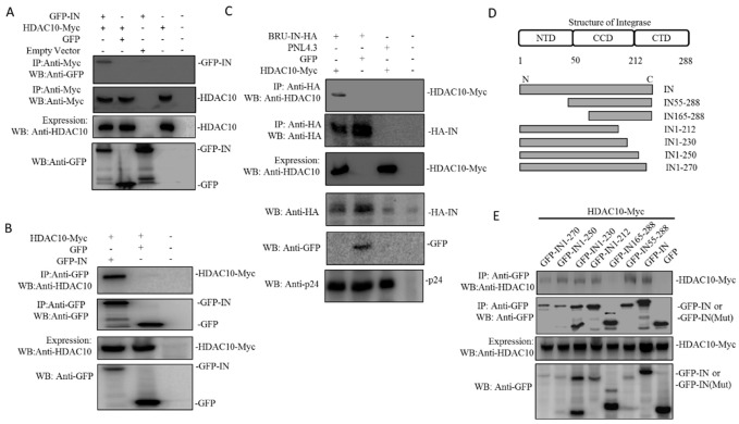
Figure 4.
HDAC10 interacts with HIV-1 IN in co-transfected 293T cells. (A) GFP or GFP-IN was co-expressed with HDAC10-Myc in 293T cells for 48 h and cells were subjected to co-IP analysis by using anti-Myc antibody. Bound GFP-IN was detected by the anti-GFP antibody. (B) Above transfected 293T cells were subjected to co-IP by using rabbit anti-GFP antibody. Bound HDAC10 was detected by the anti-HDAC10 antibody. (C) HIV-1 Bru-IN-HA plasmid was co-expressed with HDAC10-Myc in 293T cells. Meanwhile, HIV-1PNL4.3 plus GFP plasmid was included as a control. Cells were subjected to co-IP analysis by using rabbit anti-HA antibody. Bound HDAC10 was detected by the anti-HDAC10 antibody. (D) The schematic of HIV-1 Integrase deletion mutant tagged with GFP. (E) GFP-IN wild type or its deletion mutant was co-expressed with HDAC10-Myc in 293T cells. Cells were subjected to Co-IP analysis by using anti-GFP antibody. Bound HDAC10 was detected by the anti-HDAC10 antibody. All the above expressions of HDAC10-Myc, GFP-INwt/mut, or IN-HA were detected by WB using anti-HDAC10, anti-GFP, or anti-HA antibody. Panels A–E show the presentative WB images from two independent experiments.

