Abstract
Bone diseases, such as bone cancer, bone infection and osteoporosis, constitute a major issue for modern societies as a consequence of their progressive ageing. Even though these pathologies can be currently treated in the clinic, some of those treatments present drawbacks that may lead to severe complications. For instance, chemotherapy lacks great tumor tissue selectivity, affecting healthy and diseased tissues. In addition, the inappropriate use of antimicrobials is leading to the appearance of drug-resistant bacteria and persistent biofilms, rendering current antibiotics useless. Furthermore, current antiosteoporotic treatments present many side effects as a consequence of their poor bioavailability and the need to use higher doses. In view of the existing evidence, the encapsulation and selective delivery to the diseased tissues of the different therapeutic compounds seem highly convenient. In this sense, silica-based mesoporous nanoparticles offer great loading capacity within their pores, the possibility of modifying the surface to target the particles to the malignant areas and great biocompatibility. This manuscript is intended to be a comprehensive review of the available literature on complex bone diseases treated with silica-based mesoporous nanoparticles—the further development of which and eventual translation into the clinic could bring significant benefits for our future society.
Keywords: mesoporous silica nanoparticles, mesoporous bioactive glasses, bone cancer, bone infection, bone regeneration, osteoporosis, stimuli-responsive drug delivery, targeted drug delivery
1. Introduction
In recent decades, nanotechnology has been applied to a variety of fields, ranging from novel electronic devices to the study of biological processes [1,2,3,4]. In particular, the application of nanotechnology to medicine, the so-called nanomedicine, has attracted the attention of many researchers, and it is expected to revolutionize the pharmaceutical and biotechnological fields in the near future [5,6,7].
The first developments in the field of nanomedicine were reported in the early 1960s, when liposomes were first proposed as carriers [8,9]. Since then, scientists have engineered many different nanocarriers to address effective delivery of therapeutics. Those nanoparticles can be classified as either organic or inorganic. Examples of organic nanocarriers include liposomes, which are amphiphilic lipids that rearrange in water to yield vesicles with an inner aqueous compartment surrounded by lipid bilayers [10]; polymeric nanoparticles produced from polymer chains showing different functionalities [11] or polymeric micelles composed by amphiphilic block copolymers able to rearrange in aqueous media [12]. Examples of inorganic nanocarriers include metal nanoparticles synthesized from noble metals, such as gold or silver [13]; carbon nanoparticles such as carbon nanotubes, fullerenes or mesoporous carbon nanoparticles [14] or silica-based mesoporous nanoparticles, which have been extensively studied owing to their capacity to load large amounts of therapeutic molecules [15]. The main advantages of silica-based mesoporous nanoparticles over other types of particles include the robustness of the silica framework, that allows the use of harsh reaction conditions for their modification, and their excellent textural properties. In fact, conventional polymeric nanoparticles usually present low drug capacity, usually less than 5% of total weight, whereas these silica-based mesoporous nanoparticles offer greater values [16,17]. The main disadvantage over other formulations would be the fact that the translation of these type of particles remains challenging. However, it should be mentioned that silica is “generally recognized as safe” by the US Food and Drug Administration (FDA), and it is often used as excipient in drug formulations and as dietary supplement [18,19]. In this sense, the administration of fenofibrate-loaded ordered mesoporous silica materials in men was found to be safe, and the doses were well tolerated by the patients [20]. In addition, small silica nanoparticles (c-dots, 7 nm) for imaging purposes were approved by the US FDA for a human clinical trial, demonstrating that they were well tolerated by the patients and accumulated in the tumor site [21]. In consequence, silica-based nanoparticles constitute a powerful and promising tool that might be promptly translated into the clinic.
This review will cover the application of silica-based mesoporous nanoparticles for the treatment of complex bone diseases, such as bone cancer, bone infection and osteoporosis. These pathologies are predominantly found in elderly people, who will constitute one-quarter of the European population by 2020 [22]. Then, bone diseases will definitely entail a significant impact on the health care systems and, consequently, bone-targeted nanomedicines, i.e., nanomedicines able to specifically reach bone diseases, could bring significant benefits for our future society.
2. Mesoporous Silica Materials
2.1. The Beginning of a New Era: Ordered Mesoporous Silica Materials
Ordered mesoporous silica materials were first reported in the early 1990s by Mobil Oil Corporation researchers [23] and scientists from Waseda university [24]. These bulk mesoporous materials have attracted great attention because they present (1) tunable and narrow pore size distributions (2–30 nm); (2) adjustable porous structures; (3) high specific surface areas (up to 1500 cm2/g); (4) high pore volumes (ca. 1 cm3/g); (5) high silanol density on the surface that allows further modifications [25,26]. Owing to their exquisite physico-chemical properties, mesoporous silica materials have been broadly applied in a number fields, including heavy metal adsorption [27,28], catalysis [29,30] or energy storage [31,32], among others.
In addition, these materials find broad application within the field of biomaterials, owing to their ability to adsorb molecules within their pores and release them in a sustained fashion. In fact, these materials have been widely studied since Prof. Vallet-Regí and coworkers first reported their suitability as drug delivery systems back in 2001 [33].
In light of their great properties and their potential biomedical application, researchers focused their efforts on translating those excellent features of bulk materials to the nanoscale dimension. As a result, mesoporous silica nanoparticles (MSNs) were developed soon after, opening the gates to multiple biomedical applications, such as controlled drug delivery [34,35], efficient gene transfection [36,37,38], antibacterial treatment [39,40] or bone tissue regeneration [41,42], among others.
2.2. Synthesis and Functionalization of Mesoporous Silica Nanoparticles
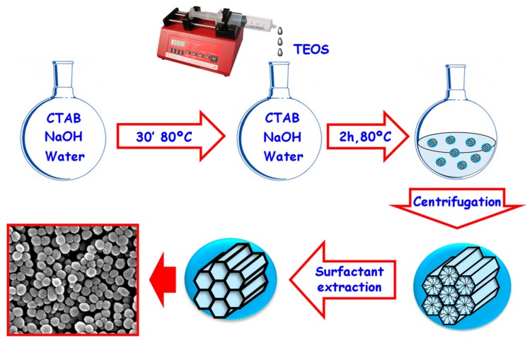
The synthesis of MSNs is based on a modification of the Stöber method, which initially yielded micron-sized monodispersed and non-porous silica spheres [43]. In this sense, the addition of surfactants as structure-directing agents results in silica nanoparticles with excellent physico-chemical properties and showing porosity. This methodology allows obtaining homogenous nanoparticles within the range 50–300 nm [25]. The morphology and dimensions of these surfactant-templated mesoporous silicas can be tailored by controlling the reaction conditions (e.g., pH, temperature, surfactant concentration or silica precursor) [44]. As an example, a synthetic protocol for the synthesis of MCM-41 (Mobil Composition of Matter) MSNs is depicted in Figure 1.
Figure 1.
Synthesis of MCM-41 MSNs using a modification of the Stöber method. The surfactant molecules self-assemble forming rod-like micelles around which the silica precursors polymerize, leading to the formation of a silica backbone with hexagonally ordered mesopores. TEOS: Tetraethyl ortosilicate; CTAB: Cetyltrimethylammonium bromide.
The positively charged polar heads of the surfactant molecules interact with the negatively charged silica precursors, leading to the formation of the silica framework by means of the hydrolysis and condensation of the silica precursor onto the self-assembled rod-like surfactant micelles. Then, the organic template is removed using a solvent extraction method, yielding MSNs with empty pores ready to be filled with therapeutic molecules. This method is usually preferred over calcination, since the latter may cause irreversible aggregation of the particles and cytotoxic byproducts, limiting their potential application [45,46].
One of the most remarkable features of MSNs is their high density of silanol groups on the surface. These chemical groups allow the easy functionalization of the nanoparticles surface, usually using organosilanes bearing different functionalities (amine, carboxylic acid, thiol…), to increase the versatility of the produced nanocarriers. The particular organosilane employed allows tuning the interactions between the payload and the silica matrix, which might be beneficial for particular diseases [47,48]. The functionalization can be accomplished through two different approximations: post-synthesis or co-condensation. The post-synthesis method involves the modification of the surface after the synthesis. This approximation can lead to different groups inside and outside the pores, depending on whether the process is performed before or after removing the template. The co-condensation approach consists in the simultaneous addition of the silica precursor and the functional organosilane during the formation of the particles. This approximation can yield nanoparticles bearing various functional groups homogenously distributed throughout the silica backbone or biodegradable periodic nanoparticles with labile bonds within the silica framework [25].
2.3. Mesoporous Silica Nanoparticles as Smart Drug Delivery Systems
Aside from being biocompatible, any nanoparticle intended to be employed as a drug delivery system should fulfill some basic requirements, such as maximizing the amount of therapeutics loaded, minimizing premature release, reaching the target area and releasing the cargo on-demand only where needed. In this sense, the extraordinary textural properties of MSNs endow them with great loading capacities, being able to load huge amounts of therapeutic molecules within their pores, as demonstrated by Scanning Transmission Electron Microscopy [49]. In addition to serving as a drug reservoir, the silica matrix provides a protective shell for the molecules against potential pH- or enzymatic-mediated drug degradation in the organism.
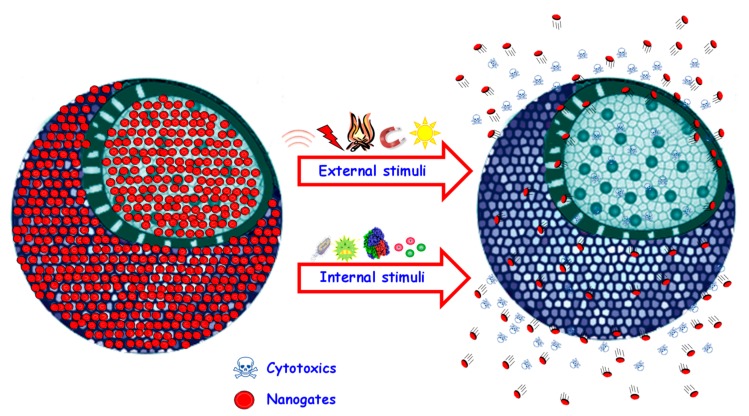
The loading of therapeutic molecules within MSNs can be easily accomplished as consequence of their open porous structure. However, this also means that the cargo molecules might easily diffuse out of the pores before reaching the target area. This premature release can be minimized using the so-called stimuli-responsive gatekeepers, which are structures able to open and close the pore entrances on-demand in response to certain stimuli [16,50,51,52]. In this manner, premature and non-specific drug release would be minimized, and the release would only take place upon application of a convenient stimulus at the diseased area (Figure 2).
Figure 2.
Schematic representation of stimuli-responsive MSNs. In response to the stimulus, the gatekeeper opens the pore entrances, triggering drug release. The origin of the stimulus can be internal (pH, enzymes, redox species, etc.) or external (magnetic fields, light, ultrasounds, etc.).
Stimuli can be applied from inside or outside the organism. The use of internal stimuli is interesting because of the significant variations of various relevant biomarkers that can be found in some diseases. For instance, the pH of some subcellular organelles and that of the tumoral matrix are more acidic compared to the physiological value [53], and analogous behavior is observed in bacterial infections [54]. These pH variations have been employed to trigger the release from pH-responsive smart MSNs [55,56,57,58]. In addition, some enzymes, which have been observed to be overexpressed in osteoporotic [59] or tumoral scenarios [60,61], have the ability to cleave very specific peptidic sequences. In this sense, it is possible to use those peptides to close the pore entrances of MSNs and trigger drug release only in those situations, where the enzymes are overexpressed [62,63,64]. Another relevant example of an internal stimulus is the overexpression of redox species in the cytoplasm of tumoral cells compared to the extracellular fluids [65,66], which has been employed to initiate drug release from different redox-responsive MSNs [67,68,69].
External stimuli, which should be innocuous to the organism, have also attracted great attention. Their main advantage is that they would allow the application of the stimulus directly by the clinician, thereby providing a much higher control of the release kinetics. For instance, the generation of heat through the application of alternating magnetic fields has been employed trigger drug release from MSNs and generate hyperthermia-mediated cell death [70,71,72]. The use of light (ultraviolet, visible, near-infrared) has also attracted the attention of many researchers, and constitutes a non-invasive method to trigger the release from MSNs [73,74,75]. Another relevant example of a non-invasive and innocuous stimulus is ultrasounds, which have been successfully employed to externally trigger the payload release from MSNs [76,77,78].
2.4. Biodistribution and Biodegradation of Mesoporous Silica Nanoparticles
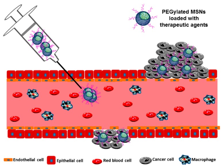
The most common routes of administration of the above-mentioned smart nanoparticles are intravenous, subcutaneous or localized injections in the target area. In particular, the intravenous administration leads to the rapid delivery and distribution of the particles throughout the organism, albeit it entails challenging issues. For instance, the particles administration leads to the formation of a protein corona around them that defines their biological entity. This protein coating might limit the functionality of the nanoparticles and enables their recognition by the organism, triggering their removal by the mononuclear phagocyte system and decreasing the efficiency of the treatments [79]. An effective approximation to overcome that issue would be the modification of the nanoparticles with hydrophilic polymers, such as poly(ethylene glycol) (PEG), which might help reduce the amount of proteins adsorbed onto the nanoparticles by creating a hydrophilic layer, enhancing their colloidal stability and increasing their circulating half-life [80,81,82]. In this sense, it has been shown in murine models that non-PEGylated MSNs rapidly accumulate in the lung, liver and spleen while their PEGylated counterparts show increased circulating half-life [83,84].
Besides the effect of PEGylation on the particles biodistribution, there are other relevant parameters that influence the final fate of MSNs. For instance, it has been shown in vivo that the larger the nanoparticles the faster their excretion [84]. In addition, it has been observed that, unlike spherical particles, those presenting elongated or cylindrical shapes undergo faster clearance from the bloodstream [85]. Finally, the surface charge is a key parameter since it determines the interaction of the particles with the surrounding media. In this sense, it has been shown that positively charged nanoparticles are more prone to undergo opsonization and subsequent clearance than their slightly negative or neutral counterparts [86,87].
Aside from achieving effective accumulation at the diseased area, it would be desirable that the MSNs degrade somehow to facilitate their excretion after exerting their therapeutic activity. In this sense, the dissolution rate of the silica backbone is a key factor for their elimination. Silica-based mesoporous nanoparticles are composed of polycondensed silica tetrahedrons (SiO4) interconnected by siloxane bonds (–Si–O–Si–) and presenting silanol groups (–Si–OH) on the surface. The silica dissolution is consequence of the nucleophilic attack of water to the siloxane and silanol groups, generating biocompatible silicic acid as by-product that can be excreted through the urine [88]. The dissolution rate depends on the particular characteristics of the particles and can be tuned through the introduction of organic modifications on the surface. Those modifications have been shown not to affect the biodistribution and biocompatibility of the MSNs [85].
3. Mesoporous Silica Nanoparticles for the Treatment of Bone Cancer
3.1. General Concepts on Bone Cancer and Bone Metastasis
Cancer is the term given to a group of diseases sharing an unstoppable cell division and with potential to spread in other organs and tissues. It is a leading cause of mortality worldwide and its prevalence is progressively increasing, with 1.7 million of estimated new cases and 600,000 cases of estimated deaths only in the United States in 2019 [89].
Bone-related tumors fall into primary bone tumors and metastatic bone tumors. They are considered to be highly deadly even though chemotherapy has improved the patient survival for sarcomas [90]. The most common malignant primary bone tumors are osteosarcoma, chondrosarcoma and Ewing sarcoma, which account for 70% of such malignancies. They originate in the bone, where mesenchymal stem cells behave both as ontogenic progenitor tumor cells and stromal cells that contribute to tumor development. The stroma of these tumors comprises osteoblasts, osteoclasts, endothelial and immune cells and mesenchymal stem cells. In particular, osteoclast have grabbed great attention because their activity (bone destruction) can be metabolically enhanced directly by tumor cells and, reversibly, the presence of osteoclasts boosts the aggressiveness of cancer cells [91].
Metastasis is the spread of cancer cells from a primary tumor to distant sites to create secondary tumors. It is a stage of the disease usually considered to be incurable with mainly palliative treatments [92]. Its origin is the pre-metastatic niche, which is an environment in a secondary organ induced by the primary cancer cells that provides favorable conditions for the growth of tumoral cells [93]. The exact mechanism of that metastatic organotropism remains unclear, but it is thought to be related with tumor-derived exosomes. Exosomes are nanometric membrane-bound vesicles secreted by tumors cells that contain functional biomolecules, such as proteins, RNA, DNA and lipids [94]. In this sense, it has been reported that tumor exosome integrins can determine organotropic metastasis by fusing with organ-specific resident cells to stablish the pre-metastatic niche. Once uptaken, they induce cellular changes in the target organ (through the activation of Scr phosphorylation and pro-inflammatory S100), thus promoting cancer cell colonization and organ-specific metastasis [95].
A characteristic feature of this disease is that some types of cancer cells preferentially migrate and induce metastasis to specific organs [94]. In this sense, breast and prostate tumors normally lead to bone metastases, which are secondary tumors formed when primary tumor cells home to the skeleton [96,97]. Cancer cells can leave the primary tumor site owing to the poor adhesion among each other in the tumoral matrix [98]. Once colonized the bone, tumor cells secrete proteins that interact with resident cells in the bone marrow to induce the differentiation, recruitment and activation of osteoblasts and osteoclasts. Then, during the bone resorption the calcium ions and the growth factors secreted from the mineralized bone matrix promote tumor cell growth, leading to vicious cycle that supports tumor growth in bone and subsequent fatal outcome [93].
It is believed that, when primary tumor cells migrate, the interaction of these disseminated cells with the new microenvironment determines whether they will proliferate to form a secondary tumor or undergo growth arrest and subsequent dormancy. Dormant cells are cells that stop dividing but still survive in a quiescent state, waiting for the appropriate environmental conditions to re-enter the cell cycle again [99]. These cells are clinically undetectable and, consequently, constitute a major issue for future tumor recurrence and metastases [100]. Current pharmacological approximations are aimed at maintaining cancer cells in the dormant state; reactivating dormant cells to increase their susceptibility to drugs; and eliminating cancer cells. Those strategies rely on the modulation of certain factors present on or secreted by the dormant cells in such a way that their overexpression of inhibition affects the fate of those dormant cells [101]. In this sense, the use of mesoporous silica nanoparticles might be interesting to enhance those treatments, as they could be employed to load therapeutic agents able to modulate the expression of those factors. In addition, they could be employed to co-load those agents with antitumoral drugs, consequently enhancing the efficacy of the treatments and minimizing tumor recurrence.
3.2. Nanotechnology for Cancer Treatment
Current anticancer treatments mainly rely on chemotherapy, radiotherapy and/or surgery [102,103,104]. Those treatments, yet effective in many cases, present several drawbacks. In particular, chemotherapy lacks great tumor tissue selectivity, leading to non-specific drug distribution and side effects. In this sense, nanoparticles have emerged as a powerful tool to encapsulate drugs and reduce side effects [105,106,107].
The rationale behind the use of nanoparticles in cancer treatment relies on the Enhanced Permeability and Retention effect (EPR effect), which is the basis of some commercialized nanomedicines [108]. The EPR effect, first reported by Maeda and coworkers [109], promotes the passive accumulation of nanoparticles in solid tumors as a result of the hypervasculature, the enhanced permeability and the poor lymphatic drainage found in many tumors (Figure 3).
Figure 3.
The Enhanced Permeability and Retention (EPR) effect. Nanoparticles passively accumulate in the tumor owing to the presence of fenestration in the tumor blood vessels. Once there, the particles remain in the tissue for long periods of time as a consequence of the poor lymphatic drainage. Reproduced from [110] with permission of MDPI, 2015.
Owing to the uncontrolled angiogenesis, the newly formed vessels present an abnormal architecture, including wide fenestrations (200–2000 nm endothelial cell–cell gaps), irregular vascular alignment or lack of smooth muscle layer, among others. As a result, molecules larger than 40 kDa leak out from them and accumulate in the extravascular tumoral tissues. On the contrary, healthy tissues do not show this abnormal development and no accumulation is observed, thus creating a differential selectivity for cancer tissues [111]. In addition, unlike normal tissues where the extracellular fluid is constantly removed, tumors present defective lymphatic drainage and the accumulated macromolecules tend to remain in the tumoral mass for longer periods of time [112].
The magnitude of the EPR effect in humans highly depends on the particularities of the patient and the tumor [113] although some alternative strategies, such as tumor-homing peptides or some types of cells, are currently being explored to overcome the lack of EPR effect.
These alternative approximations have successfully been evaluated using in vivo tumor models, demonstrating the suitability of using MSNs for tumor drug delivery. In this sense, tumor-homing peptides (e.g., iRGD, iNGR) not only induce spontaneous accumulation of nanoparticles in the tumor tissues, but also enhance their diffusion into the tumoral mass [114,115]. In addition, there are certain types of cells with migratory properties that can transport nanoparticles directly to tumors tissues. For instance, nanoparticles can be attached to hypoxic bacteria that migrate to the hypoxic areas of tumors [116,117]. In addition, mesenchymal stem cells have been shown to migrate to tumors in response to the secretion of various signaling molecules. Then, a smart strategy is to induce the internalization of drug-loaded nanoparticles within these cells to then delivering them specifically to tumor tissues [118,119,120,121].
Besides delivering the nanoparticles to malignant tissues, the carriers can be engineered so that they preferentially recognize cancer cells over healthy cells. This targeting strategy relies on the overexpression of some receptors only on the membrane of tumoral cells. Examples of this approach include the functionalization of the particles with antibodies [122,123], proteins [69,124], small molecules [125,126,127,128,129] or peptides [77,130,131], among others.
3.3. Targeting Bone-Localized Tumors with Mesoporous Silica Nanoparticles
Addressing nanoparticles to bone metastases is challenging, as small metastases are poorly vascularized and, consequently, the magnitude of the EPR effect is low compared to big solid tumors [132]. A smart approximation would be the modification of the particles with targeting molecules with high affinity towards calcium phosphate surfaces (bone tissue), such as bisphosphonates [133], to complement the EPR effect. In this sense, the surface modification with the bisphosphonate zoledronate has been proved to be effective in delivering MSNs to bone metastases originated from lung [134] and breast cancer [135].
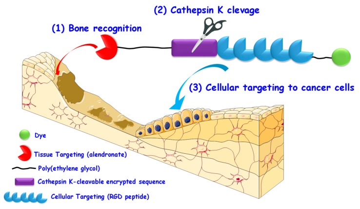
Besides targeting the particles to bone tissue, it would be desirable for the nanomedicines to be subsequently internalized only by the tumoral cells. In this sense, our group recently reported a smart approximation for the sequential targeting of bone tumors or bone metastases that could be easily implemented into any nanomedicine (Figure 4) [136].
Figure 4.
Encrypted approach for the sequential targeting of bone cancer tissue and cancer cells. (1) The presence of a bone targeting agent (alendronate) would help accumulate the nanomedicines in the bone tumor tissue; (2) Once there, the overexpressed cathepsin K would cleave a specific peptidic sequence, (3) exposing the RGD (arginine-glycine-aspartic) motif, which is able to promote the selective uptake of nanomedicine by sarcoma tumoral cells.
As observed in Figure 4, the system is composed of two targeting agents and employs PEG chains to mimic a nanocarrier. The first one is the bisphosphonate alendronate, which can bind bone tissue. Then, there is a peptidic fragment containing a cathepsin K-cleavable sequence followed by the RGD motif, which is able to promote the selective internalization in osteosarcoma cells thanks to the overexpression of αβ integrins. In this manner, the alendronate molecule would help the EPR effect to accumulate the nanomedicines in the bone tumor tissue. Once there, cathepsin K, which is overexpressed in bone tumors and bone metastases, would cleave the encrypting sequence, thereby exposing the RGD motif and triggering the preferential uptake of the nanomedicines.
As it happens with many other cancer cells, bone tumoral cells overexpress specific receptors that can be targeted using conveniently engineered MSNs. Aside from targeting MSNs to osteosarcoma [137], the RGD motif can also be employed to recognize endothelial cells, which can help MSNs target the tumor endothelium of fibrosarcoma to then eliminate the cancerous cells using multimodal therapy [138]. In this sense, folic acid can be employed to target overexpressed folate receptors in fibrosarcoma [139] and osteosarcoma cells [126]. In addition, the modification of MSNs with a glucose analog enhances their accumulation in bone tumor cells, as a consequence of their great glucose consumption due to the high metabolic demand of tumors [140]. Some surface receptors, such as the CD11c, can also be targeted using specific antibodies, which are able to trigger the selective internalization of MSNs in osteosarcoma [141].
The decoration of MSNs with proteins can also increase their cellular uptake. For instance, the lectin concanavalin A binds overexpressed sialic acid residues to promote the cellular uptake of pH-responsive MSNs in osteosarcoma cells [142]. Transferrin receptors are overexpressed in fibrosarcoma cells and, consequently, the protein transferrin can be employed to enhance the uptake of MSNs in those bone tumoral cells [124].
Besides employing active targeting moieties, MSNs can be internalized via electrostatic interactions with the negatively charged cell membrane. The positively charged surface can be shielded using PEG, which can be detached using a cleavable bond. The charge is exposed again upon application of ultrasounds, which triggers the nanoparticles uptake after the accumulation in the solid bone tumor via EPR effect [143].
3.4. Controlled Release of Therapeutics in Bone Tumors with Mesoporous Silica Nanoparticles
There are various examples of the suitability of using silica-based mesoporous nanomatrices for the delivery of antitumoral [144,145,146,147,148] or imaging agents [137,149,150] to bone cancer cells. Moreover, researchers have taken advantage of the features of the bone tumoral environment to design stimuli-responsive MSNs for the treatment of sarcomas. Among the internal stimuli, the acidic environment of the lysosomes can be employed to trigger drug release from pH-responsive polymer-coated MSNs [142] or pulsatile on-off MSNs with pore entrances that are sealed with pH-responsive nanovalves [78]. In addition, it is possible to load immunotherapy agents within the pores of pH-responsive lipid-coated MSNs for synergistic chemo-immunotherapy [151]. In addition to pH variations, the enzyme alkaline phosphatase, which is characteristic bone-related tumors, can be employed to degrade the gatekeepers of silica-based mesoporous glasses [152]. Moreover, the esterase enzymes can also be employed to cleave the nanocaps of MSNs [126].
There are some examples of the use of light to trigger drug release from MSNs in bone tumor scenarios. For instance, ultraviolet light can be employed to cleave light-responsive bonds connected to transferrin, which acts as both gatekeeper and targeting agent, triggering drug release [124]. In addition, porphyrins can be engineered as gatekeepers using a linker cleavable in the presence of singlet oxygen, which are self-produced by the porphyrin caps upon application of visible light [73].
Aside from delivering small therapeutic molecules, MSNs allow the effective delivery of proteins [153] or DNA strands [154] into bone cancer cells. There is a type of nucleic acids, small interfering RNA (siRNA), that triggers the knockdown of specific and relevant proteins, which makes them useful for the treatment of various diseases [155]. Unfortunately, siRNAs have short half-life, poor penetration through cell membranes and easily degrade upon RNase action in the organism [156]. For that reason, the use of MSNs as protective shell for these nucleic acids have been widely explored. In this sense, the polo-like kinase 1, which is an essential gene for the correct execution of cell division [157], is overexpressed in bone tumors and has been targeted with great efficacy using siRNA-loaded MSNs [158,159,160,161].
A summary of all the nanocarriers described here for bone tumors is summarized in Table 1.
Table 1.
Summary of the different silica-based nanocarriers applied for the treatment of bone tumors.
| Cell Line | Description | Reference |
|---|---|---|
| Osteosarcoma | ||
| MG-63 | MSNs loaded with ammonia borate as negative computed tomography contrast agents for the diagnosis of osteosarcoma; | [150] |
| Silica-based mesoporous glass nanospheres for the delivery of alendronate against osteosarcoma cells and osteoclasts; | [146] | |
| Silica-based mesoporous glasses with osteogenic properties for the release of alendronate against osteosarcoma cells; | [145] | |
| Eu-doped silica-based mesoporous glass nanospheres with osteogenic properties for the release of doxorubicin; | [144] | |
| Influence of the different functionalizations of MSNs on their uptake by osteosarcoma cells; | [141] | |
| KHOS | Poly-l-lysine-coated MSNs for the delivery of siRNA to knockdown polo-like kinase 1; | [159] |
| MSNs with large mesopores for the delivery of siRNA to knockdown polo-like kinase 1; | [158] | |
| Co-loading of topotecan and siRNA to knockdown polo-like kinase 1 in dendrimer-like MSNs; | [160] | |
| PEI-coated MSNs for the delivery of siRNA to knockdown polo-like kinase 1; | [161] | |
| HOS | Stimuli-responsive silica-based mesoporous glasses responsive to alkaline phosphatase overexpressed in bone tumors; | [152] |
| Dendrimer-coated MSNs for the delivery of non-viral oligonucleotides; | [154] | |
| MSNs functionalized with singlet oxygen-sensitive porphyrin caps for release of topotecan; | [73] | |
| MSNs engineered for ultrasound-induced cellular uptake through the detachment of a shielding PEG layer; | [143] | |
| Concanavalin A-targeted and pH-responsive MSNs for the delivery of doxorubicin; | [142] | |
| HTB-85 | Silica-based mesoporous glass nanospheres with osteogenic properties for the release of doxorubicin; | [147] |
| U2Os | Folic acid-targeted MSNs for enzyme-responsive release of camptothecin; | [126] |
| UMR-106 | RGD-targeted and Bi-doped MSNs for chemo-photothermal therapy and imaging; | [137] |
| Fibrosarcoma | ||
| L-929 | Ultrasound, pH and magnetically-responsive on-off gated MSNs for the delivery of doxorubicin; | [78] |
| Gd-doped MSNs for magnetic resonance imagining of fibrosarcoma; | [149] | |
| pH-responsive MSNs for the intracellular delivery of proteins; | [153] | |
| pH-responsive MSNs for combined chemo-immunotherapy; | [151] | |
| HT-1080 | Influence of MSNs size on the doxorubicin release and the uptake of the particles by fibrosarcoma cells; | [148] |
| MSNs decorated through an ultraviolet light-responsive linker with transferrin acting as gatekeeper and targeting agent; | [124] | |
| RGD-targeted MSNs for multimodal treatment of fibrosarcoma in a chicken embryo model; | [138] | |
4. Mesoporous Silica Nanoparticles for the Treatment of Bone Infection
4.1. General Concepts on Bacterial Bone Infections
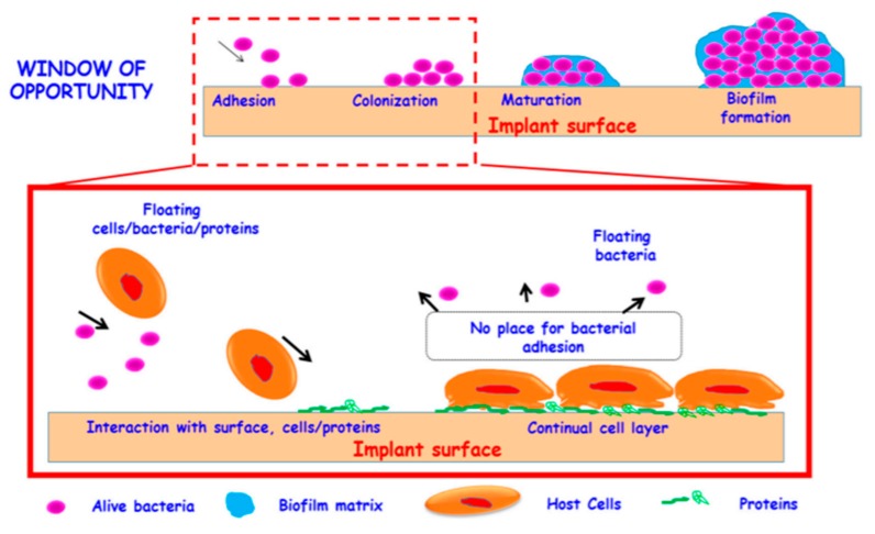
Bone infection is a major issue for health care systems and entails important socioeconomic implications [162]. The appearance of bone infections is directly related with the progressive ageing of current society and, consequently, the increased use of implantable medical devices and their potential bacterial contamination. These infections are mainly caused by Staphylococcus epidermis, Staphylococcus aureus, Escherichia coli and Pseudomonas aeruginosa [40]. Regular bacteria can be relatively easy eliminated using antibiotics. However, the inappropriate use of those antimicrobials is progressively leading to more cases of drug-resistant bacteria, which are expected to cause more than 10 million deaths by 2050 [163]. This antimicrobial resistance induces uncontrolled bacterial growth and formation of persistent biofilms. Biofilms are communities of microorganisms embedded in a self-produced polysaccharide matrix [164]. This protective matrix endows them with resistance to antibiotics and host immune systems that, otherwise, would eliminate bacteria in their planktonic state (free-floating bacteria) [165]. The biofilm—related antimicrobial resistance relies, not only on the physical hindrance of the matrix, but also on (1) the presence of bacterial and host DNA and proteins that may increase the shielding capacity of the matrix [166]; (2) the presence of bacteria with different acquired resistances and antibiotic sensitivities [167]; (3) the development of efflux pumps [168]; (4) the presence of enzymes able to degrade antimicrobials [169]; (5) the establishment of quorum sensing (bacteria-bacteria communication) [170]. The process of biofilm formation is depicted in Figure 5.
Figure 5.
Schematic representation of biofilm formation on an implant surface. The process involves four steps: (1) bacterial adhesion, (2) bacterial growth, (3) maturation and (4) biofilm formation. In addition, bacteria may leak out from the matrix and lead to bacterial dispersion. The first stages constitute a window of opportunity, in which it is still possible to prevent biofilm formation. Reproduced from [40] with permission of MDPI, 2018.
The formation of the biofilm comprises 4 steps: (1) adhesion of bacteria to the implant surface; (2) bacterial growth in multiple bacterial layers; (3) maturation; (4) final biofilm formation. In addition, bacteria detach from the biofilm to then colonize other areas and induce further infections [171]. As observed in Figure 5, during the first phases of biofilm formation, the individual microorganisms are floating on the implant, reversibly interacting with the surface. In consequence, these stages constitute a window of opportunity that clinicians should take advantage of to prevent irreversible biofilm formation and subsequent resistance [40].
4.2. Preventing Protein and Bacterial Adhesion and Biofilm Formation: Zwitterionic Mesoporous Silica Nanoparticles
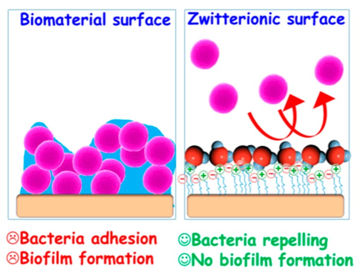
In view of the existing evidence in the previous subsection, avoiding bacterial contamination of implants constitutes a major concern. In this sense, the development of the so-called zwitterionic materials has fueled the design of antifouling nanostructured materials able to prevent protein adsorption, bacterial adhesion and biofilm formation (Figure 6).
Figure 6.
Schematic representation of bacterial colonization in standard surfaces vs. zwitterionic surfaces. Unlike in unmodified surfaces, zwitterionic materials create a hydration layer that prevents bacterial adhesion and biofilm formation. Reproduced from [40] with permission of MDPI, 2018.
Zwitterionic surfaces are characterized by an equal number of negative and positive charges, so the net charge is expected to be neutral. This neutrality leads to the formation of a hydration layer onto the surface that physically hampers adhesion and biofilm formation [172]. In fact, owing to the reduced protein adsorption, zwitterionic functionalizations have also been postulated as substitutes for PEGylation [173], which might be beneficial to overcome the growing appearance of anti-PEG antibodies [174].
The first example of mesoporous silica materials with zwitterionic behavior was reported by our group back in 2010, using SBA-15 mesoporous materials modified with randomly distributed amino and carboxylic acid short chains on the surface that resulted in significantly lower protein adhesion [175]. A similar approach using amino and phosphonate groups was recently reported, yielding MSNs with extremely low protein adsorption and excellent antibacterial properties. In addition, the nanoparticles showed great biocompatibility with preosteoblasts, assuring their biocompatibility for the treatment of bone infection [176]. Interestingly, this zwitterionic approach using two small molecules can be employed to design pH-responsive gatekeepers by taking advantage of the interaction between both short chains, which interact at physiological pH and experience repulsion forces at acid pH [177].
Aside from merging molecules with opposite charges, there are molecules that are zwitterionic in nature. In this sense, the modification of MSNs with phosphorylcholine groups yields nanoparticles showing reduced protein adsorption and able to provide sustained drug release in response to changes in pH [178]. An analogous approximation is the modification of MSNs with sulfobetaine groups to prevent protein adhesion [179]. Moreover, it is possible to polymerize this kind of zwitterionic molecules to yield polymer-coated nanoparticles with low protein binding affinity [180]. In addition, there are some amino acids that are useful for the design of this kind of surfaces. For instance, the amino acid lysine presents this behavior owing to the –NH3+/COO− pairs and has been grafted to MSNs [181] and silica-based mesoporous bioactive glasses [182], leading to reduced bacterial adhesion and biofilm formation. A similar approach consists in using the amino acid cysteine to obtain neutral surfaces, yielding MSNs with high stability in human serum [183].
4.3. Addressing Bone Infections with Mesoporous Silica Nanoparticles
Besides preventing biofilm formation, the elimination of the infection is still necessary. In this sense, it is possible to engineer multifunctional mesoporous silica nanomatrices able to prevent bacterial adhesion and biofilm formation and to release antimicrobials in a controlled manner only in infected bone tissues [184,185]. In addition, there are examples of stimuli-responsive mesoporous bioactive silica-based nanomatrices able to trigger the release only in the presence of proteolytic enzymes characteristics of infected bone tissue scenarios [152,186].
In an effort to increase the efficiency of the delivery and, consequently, a reduction of the dose, the research efforts have been headed towards the development of bacteria-targeted MSNs. In this sense, the presence of positive charges on the surface of the particles increases their affinity to the negatively charged biofilm and bacteria wall. In this manner, it is easier for the particles to diffuse into the biofilm to then interact with bacteria and exert their therapeutic effect. Examples of this approach include the use of short positively charged alkoxysilanes [187] or third-generation dendrimers, with a great number of positive charges that allow permeating the bacteria wall and inducing MSN internalization [188]. Besides using positively charged MSNs, lectins have been shown to be effective in targeting and promoting internalization of MSNs into the biofilm, as a consequence of the presence of glycan-type polysaccharides in this protective matrix. In fact, the lectin concanavalin A is able to trigger this internalization and exert antibacterial effect by itself, which is even more emphasized when loading an antibiotic in the mesopores [189].
A smart approximation to enhance the possibilities that mesoporous silica materials may offer against bone infection is the incorporation of the particles within scaffolds. In the context of bone diseases, scaffolds are materials that are intended to mimic bone tissue and contribute to its regeneration. The advantages over using bare scaffolds are increased antibiotic loading capacity or controlled drug release, among others [190]. Examples of this approximation are the incorporation of silica-based mesoporous glasses in PLGA (poly-(L-lactic-co-glycolic acid)) [191] or MSNs in porous collagen gelatin [192] for the controlled release of vancomycin against bone infection. In addition, MSNs-loaded scaffolds allow the co-delivery of therapeutic compounds. In this sense, it is possible to load cephalexin within the mesopores and vascular endothelial growth factors in the scaffold structure to achieve bacteria elimination and bone reconstruction [193].
A summary of different materials for the treatment of bone infection can be found in Table 2.
Table 2.
Summary of mesoporous silica-based materials against bone infection.
| Bacteria | Description | Reference |
|---|---|---|
| Escherichia coli | Pronase-responsive gatekeepers for levofloxacin-loaded silica-based mesoporous glasses; | [152] |
| Levofloxacin-loaded Zwitterionic MSNs with reduced protein adhesion; | [176] | |
| Lysine-coated MSNs to inhibit E. coli adhesion; | [181] | |
| Acid phosphatase-responsive gatekeepers for levofloxacin-loaded silica-based mesoporous glasses; | [186] | |
| Positively charge MSNs target the bacteria wall of E. coli; | [187] | |
| Levofloxacin-loaded MSNs coated with polycationic dendrimers destroys biofilm and internalize in bacteria; | [188] | |
| Levofloxacin-loaded MSNs decorated with concanavalin A targets and internalize the biofilm; | [189] | |
| Staphylococcus aureus | Levofloxacin-loaded Zwitterionic MSNs with reduced protein adhesion; | [176] |
| Lysine-coated zwitterionic MSNs to inhibit S. aureus adhesion and S. aureus biofilm formation; | [181] | |
| Lysine-coated zwitterionic silica-based mesoporous glasses to prevent S. aureus adhesion; | [182] | |
| Levofloxacin-loaded and positively charged MSNs targets and destroy S. aureus biofilm and bacteria; | [187] | |
| MSNs-loaded scaffolds for the co-delivery of cephalexin and vascular endothelial growth factor; | [193] | |
| Vancomycin-loaded silica-based mesoporous glasses contained in PLGA scaffolds; | [191] | |
| Vancomycin-loaded MSNs contained in collagen gelatin scaffolds; | [192] |
5. Mesoporous Silica Nanoparticles for the Treatment of Osteoporosis
5.1. General Concepts on Osteoporosis
Osteoporosis is the most frequent metabolic disease affecting bone tissue. It is characterized by reduced bone mass and microarchitectural deterioration and results in more than 9 million fractures annually worldwide (one osteoporotic fracture every 3 s) [194], with special incidence in aged women [195]. Its origin relies on the alteration of the bone remodeling process, which consists in the removal of old bone (osteoclast) to then create new one (osteoblasts). The imbalance of this process leads to reduced bone mass and, consequently, osteoporosis.
Current osteoporosis treatments, which are not fully satisfactory, are limited to anti-resorptive drugs and anabolic agents [196,197]. Anti-resorptive drugs decrease the excess of bone resorption by targeting osteoclast activity. Examples of these compounds include bisphosphonates [198], raloxifene [199] or denosumab [200]. The excess of bone resorption can be counteracted using anabolic agents, which are compounds able to stimulate bone formation. Examples of these drugs are human parathyroid hormone [201], growth factors or siRNA [202].
Unfortunately, current treatments present some drawbacks. For instance, bisphosphonates are known to induce gastric side effects or fractures after long use. Raloxifene may cause venous thromboembolism. Moreover, cases of hypocalcemia, anaphylaxis or atrial fibrillation have been associated to denosumab. In addition, anabolic agents, such as siRNA, might be easily degraded by the harsh environment present in the organism [201]. These issues might be addressed by delivering the antiosteoporotic agents specifically to the diseased bone tissues and, consequently, the use of nanoparticles seems highly appealing.
5.2. Addressing Osteoporosis with Mesoporous Silica Nanoparticles
The first example of mesoporous silica materials applied for the controlled release of anti-resorptive molecules was reported by our group back in 2006, when MCM-41 and SBA-15 materials were employed for the loading and controlled release of alendronate [203]. In this sense, the introduction of phosphorous groups in SBA-15 mesoporous silica nanomatrices enhanced the loading of alendronate and induced the formation of apatite, a component of bone, making these materials promising candidates for the treatment of osteoporosis [204]. Additional examples of anti-resorptive molecules loaded in mesoporous silica-based nanoparticles are ipriflavone [205], salmon calcitonin [206] or zolendronic acid [207], with all of them showing promising results in terms of anti-osteoclast activity and osteogenesis.
A great feature of MSNs is that they allow the loading of hydrophobic compounds, consequently enhancing their bioavailability. In this sense, they allow the incorporation within their mesopores of sparingly soluble anabolic agents able to induce bone formation. Examples are the loading of dexamethasone, which induces bone regeneration through the stimulation of bone mesenchymal stems cells [208], or estradiol, which enhances the biological functions of osteoblast and inhibits the proliferation of osteoclasts [209].
Osteostain, a C-terminal peptide from a parathyroid hormone-related protein, induces strong bone anabolism through a great stimulation of osteoblastogenesis [210]. It has been shown that osteostatin-loaded SBA-15 greatly stimulate osteoblastic growth in vitro [211]. Furthermore, these osteostatin-loaded mesoporous materials have been proved to be effective in regenerating bone defects in vivo [201,212]. In addition to osteostatin, the bone morphogenic protein-2 (BMP-2), is considered to be one of the most effective growth factors to induce osteoblast differentiation and boost bone regeneration. In this sense, MSNs are useful for the co-delivery of dexamethasone and BMP-2 to achieve great bone regeneration in vivo [213]. Moreover, the residues 73–92 of BMP-2 not only promote osteogenesis and bone regeneration but also increase the internalization of bone mesenchymal stem cells of MSNs decorated with this peptidic fragment. [214].
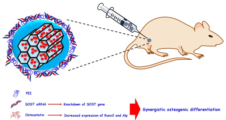
Aside from being useful for bone cancer treatment, siRNA molecules also find application in the treatment of osteoporosis. In this sense, the localized release of siRNA able to knockdown RANK from silica-based mesoporous bioactive glasses has been to shown to be highly effective in suppressing osteoclastogenesis and, consequently, osteoporosis [215]. A similar approach against osteoporosis was recently reported by our group using an in vivo model of ovariectomized mice (Figure 7).
Figure 7.
PEI-coated MSNs as anti-osteoporotic nanocarrier. Osteostatin was loaded in the mesopores and a siRNA able to knockdown the SOST gene was introduced within the polymeric mesh. The co-delivery of both therapeutic agents resulted in synergistic osteogenesis in ovariectomized mice. PEI: polyethyleneimine.
MSNs can load therapeutic compounds not only in their mesopores but also within polymeric coatings through electrostatic interaction. In this sense, Figure 5 shows MSNs carrying the anabolic agent osteostatin in the pores and a specific siRNA able to knockdown the SOST gene interacting with a PEI coating. This gene encodes the protein sclerostin, which can inhibit the Wnt/β-catenin pathway, a major signaling carrier that regulates bone development and remodeling. Based on this, the siRNA and osteostatin-loaded nanoparticles were administered to osteoporotic ovariectomized mice, showing synergistic effects on all the bone regeneration biomarkers studied [216].
There are some metal ion species known to induce osteogenesis. For instance, copper ions enhance bone density by inhibiting bone resorption, and their incorporation in mesoporous silica nanospheres has been proved to be effective in stimulating the differentiation of bone mesenchymal stem cells into the osteogenic lineage [217]. Moreover, impregnating silica-based mesoporous bioactive glasses with Ga(III) leads to the formation of apatite together with the disruption of osteoclastogenesis and early differentiation of pre-osteoblast towards osteoblastic phenotype [218]. In addition, the osteogenic ability of Zn2+ ions is enhanced when the ions are co-delivered with osteostatin from silica-based mesoporous bioactive glasses [219]. Furthermore, there are nanoparticles able to stimulate bone regeneration per se. Examples of these kind of behavior are Au nanoparticles supported on MSNs that increase the osteogenic capability of preosteoblastic cells [220] or silica-based mesoporous bioactive glasses that are capable of reducing the bone-resorbing capability of osteoclasts [221].
A summary of all the above-described materials for the treatment of osteoporosis can be found in Table 3.
Table 3.
Summary of silica-based mesoporous materials for the treatment of osteoporosis.
| Therapeutic Agent | Description | Reference |
|---|---|---|
| Anti-Resorptive Treatment | ||
| Alendronate | First example of controlled release of bisphosphonates from mesoporous silica materials (MCM-41 and SBA-15); | [203] |
| Phosphorus-containing SBA-15 mesoporous silica materials for bone regeneration and release of alendronate; | [204] | |
| Ipriflavone | Silica-based mesoporous nanospheres for the release of ipriflavone without affecting osteoblast viability; | [205] |
| Zolendronic acid | Zolendronic acid-loaded MSNs/hydroxyapatite coatings on implants with enhanced inhibition of osteoclasts activity; | [207] |
| Salmon calcitonin | MSNs for the release of salmon calcitonin with significant therapeutic effects in vivo; | [206] |
| siRNA (RANK) | Silica-based mesoporous glass nanospheres to deliver of siRNA to knockdown RANK and inhibit osteoclastogenesis; | [215] |
| Ions | Mesoporous silica-based nanospheres for the delivery of Cu ions able to inhibit osteoclastogenesis; | [217] |
| Silica-based mesoporous glasses for the release of Ga ions able to disturb osteoclastogenesis; | [218] | |
| Particle | Silica-based mesoporous glasses reduce the bone-resorbing capability of osteoclasts per se; | [221] |
| Au nanoparticles supported on MSNs increases the osteogenic capability of pre-osteoblastic cells; | [220] | |
| Anabolic Treatment | ||
| Dexamethasone | Alendronate-targeted MSNs for the delivery of dexamethasone to bone tissue; | [208] |
| Estradiol | Multilayered-coated MSNs for the delivery of estradiol from titanium substrates; | [209] |
| Osteostatin | Osteostatin-loaded SBA-15 mesoporous silica materials stimulate the growth and differentiation of osteoblasts; | [211] |
| Osteostatin-loaded SBA-15 mesoporous materials regenerate bone in a rabbit femur cavity defect; | [201] | |
| Osteostatin-loaded SBA-15 mesoporous silica materials increase the early repair response in bone after local injury; | [212] | |
| BMP-2 and dexamethasone | pH-responsive co-delivery of dexamethasone and BMP-2 protein for synergistic osteogenic effect; | [213] |
| BMP-2 derived peptide-decorated MSNs for enhanced uptake in bone mesenchymal stem cells and synergistic effect of the peptidic fragment and dexamethasone; | [214] | |
| Osteostatin and siRNA (SOST) | Enhanced osteogenic expression through MSNs co-delivering osteostatin and siRNA able to knockdown the SOST gene; | [216] |
| Zn ions and osteostatin | Co-delivery of osteogenic Zn ions and osteostatin from mesoporous silica-based glasses induces high osteogenic response; | [219] |
6. Conclusions
Bone diseases, such as bone cancer, bone infection and osteoporosis, constitute a major issue for modern societies as a consequence of their progressive ageing. Most of the current treatments present several drawbacks, leading to the deterioration of patient health and the subsequent socioeconomic impact. In this sense, the use of nanoparticles, in particular mesoporous silica-based nanoparticles, has emerged as a powerful approximation to reduce the different side effects. This type of nanoparticle presents high loading capacities, biocompatibility and can be engineered to prevent premature drug release and address the particles to the affected tissues. The different nanosystems presented here constitute reliable approximations for the treatment of bone diseases and, consequently, current research should be headed towards the effective translation of these nanomaterials into the clinic.
Acknowledgments
The authors acknowledge financial support from European Research Council through ERC-2015-AdG-694160 (VERDI) project.
Author Contributions
All authors have contributed equally. All authors have read and agreed to the published version of the manuscript.
Funding
The research was funded by the European Research Council through ERC-2015-AdG-694160 (VERDI) grant.
Conflicts of Interest
The authors declare no conflict of interest.
References
- 1.Appell D. Wired for success. Nature. 2002;419:553–555. doi: 10.1038/419553a. [DOI] [PubMed] [Google Scholar]
- 2.Serrano E., Rus G., García-Martínez J. Nanotechnology for sustainable energy. Renew. Sustain. Energy Rev. 2009;13:2373–2384. doi: 10.1016/j.rser.2009.06.003. [DOI] [Google Scholar]
- 3.Tong H., Ouyang S., Bi Y., Umezawa N., Oshikiri M., Ye J. Nano-photocatalytic Materials: Possibilities and Challenges. Adv. Mater. 2012;24:229–251. doi: 10.1002/adma.201102752. [DOI] [PubMed] [Google Scholar]
- 4.Nie S., Xing Y., Kim G.J., Simons J.W. Nanotechnology Applications in Cancer. Annu. Rev. Biomed. Eng. 2007;9:257–288. doi: 10.1146/annurev.bioeng.9.060906.152025. [DOI] [PubMed] [Google Scholar]
- 5.Wicki A., Witzigmann D., Balasubramanian V., Huwyler J. Nanomedicine in cancer therapy: Challenges, opportunities, and clinical applications. J. Control. Release. 2015;200:138–157. doi: 10.1016/j.jconrel.2014.12.030. [DOI] [PubMed] [Google Scholar]
- 6.Zhu X., Radovic-Moreno A.F., Wu J., Langer R., Shi J. Nanomedicine in the management of microbial infection—Overview and perspectives. Nano Today. 2014;9:478–498. doi: 10.1016/j.nantod.2014.06.003. [DOI] [PMC free article] [PubMed] [Google Scholar]
- 7.Doane T.L., Burda C. The unique role of nanoparticles in nanomedicine: Imaging, drug delivery and therapy. Chem. Soc. Rev. 2012;41:2885. doi: 10.1039/c2cs15260f. [DOI] [PubMed] [Google Scholar]
- 8.Bangham A.D., Standish M.M., Weissmann G. The Action of Steroids and Streptolysin S on the Permeability of Phospholipid Structures to Cations. J. Mol. Biol. 1965;13:253–259. doi: 10.1016/S0022-2836(65)80094-8. [DOI] [PubMed] [Google Scholar]
- 9.Bangham A.D., Horne R.W. Negative Staining of Phospholipids and their Structural Modification by Surface-active Agents as observed in the Electron Microscope. J. Mol. Biol. 1964;8:660–668. doi: 10.1016/S0022-2836(64)80115-7. [DOI] [PubMed] [Google Scholar]
- 10.Bozzuto G., Molinari A. Liposomes as nanomedical devices. Int. J. Nanomed. 2015;10:975–999. doi: 10.2147/IJN.S68861. [DOI] [PMC free article] [PubMed] [Google Scholar]
- 11.Kumari A., Yadav S.K., Yadav S.C. Biodegradable polymeric nanoparticles based drug delivery systems. Colloids Surfaces B Biointerfaces. 2010;75:1–18. doi: 10.1016/j.colsurfb.2009.09.001. [DOI] [PubMed] [Google Scholar]
- 12.Oerlemans C., Bult W., Bos M., Storm G., Nijsen J.F.W., Hennink W.E. Polymeric Micelles in Anticancer Therapy: Targeting, Imaging and Triggered Release. Pharm. Res. 2010;27:2569–2589. doi: 10.1007/s11095-010-0233-4. [DOI] [PMC free article] [PubMed] [Google Scholar]
- 13.Azharuddin M., Zhu G.H., Das D., Ozgur E., Uzun L., Turner A.P.F., Patra H.K. A repertoire of biomedical applications of noble metal nanoparticles. Chem. Commun. 2019;55:6964–6996. doi: 10.1039/C9CC01741K. [DOI] [PubMed] [Google Scholar]
- 14.Maiti D., Tong X., Mou X., Yang K. Carbon-Based Nanomaterials for Biomedical Applications: A Recent Study. Front. Pharmacol. 2019;9:1401. doi: 10.3389/fphar.2018.01401. [DOI] [PMC free article] [PubMed] [Google Scholar]
- 15.Manzano M., Vallet-Regí M. New developments in ordered mesoporous materials for drug delivery. J. Mater. Chem. 2010;20:5593–5604. doi: 10.1039/b922651f. [DOI] [Google Scholar]
- 16.Argyo C., Weiss V., Bra C., Bein T. Multifunctional Mesoporous Silica Nanoparticles as a Universal Platform for Drug Delivery. Chem. Mater. 2014;26:435–451. doi: 10.1021/cm402592t. [DOI] [Google Scholar]
- 17.Zhang Y., Zhi Z., Jiang T., Zhang J., Wang Z., Wang S. Spherical mesoporous silica nanoparticles for loading and release of the poorly water-soluble drug telmisartan. J. Control. Release. 2010;145:257–263. doi: 10.1016/j.jconrel.2010.04.029. [DOI] [PubMed] [Google Scholar]
- 18.Narayan R., Nayak U.Y., Raichur A.M., Garg S. Mesoporous silica nanoparticles: A comprehensive review on synthesis and recent advances. Pharmaceutics. 2018;10:118. doi: 10.3390/pharmaceutics10030118. [DOI] [PMC free article] [PubMed] [Google Scholar]
- 19.Rosenholm J.M., Mamaeva V., Sahlgren C., Lindén M. Nanoparticles in targeted cancer therapy: Mesoporous silica nanoparticles entering preclinical development stage. Nanomedicine. 2012;7:111–120. doi: 10.2217/nnm.11.166. [DOI] [PubMed] [Google Scholar]
- 20.Bukara K., Schueller L., Rosier J., Martens M.A., Daems T., Verheyden L., Eelen S., Van Speybroeck M., Libanati C., Martens J.A., et al. Ordered mesoporous silica to enhance the bioavailability of poorly water-soluble drugs: Proof of concept in man. Eur. J. Pharm. Biopharm. 2016;108:220–225. doi: 10.1016/j.ejpb.2016.08.020. [DOI] [PubMed] [Google Scholar]
- 21.Phillips E., Penate-Medina O., Zanzonico P.B., Carvajal R.D., Mohan P., Ye Y., Humm J., Gönen M., Kalaigian H., Schöder H., et al. Clinical translation of an ultrasmall inorganic optical-PET imaging nanoparticle probe. Sci. Transl. Med. 2014;6:1–10. doi: 10.1126/scitranslmed.3009524. [DOI] [PMC free article] [PubMed] [Google Scholar]
- 22.Sobczak D. Population Ageing in Europe: Facts, Implications and Policies. Publications Office of the European Union; Luxembourg: 2014. [Google Scholar]
- 23.Kresge C.T., Leonowicz M.E., Roth W.J., Vartuli J.C., Beck J.S. Ordered mesoporous molecular sieves synthesized by a liquid-crystal template mechanism. Nature. 1992;359:710–712. doi: 10.1038/359710a0. [DOI] [Google Scholar]
- 24.Yanagisawa T., Shimizu T., Kuroda K., Kato C. The preparation of alkyltrimethylammonium-kanemite complexes and their conversion to microporous materials. Bull. Chem. Soc. Jpn. 1990;63:988–992. doi: 10.1246/bcsj.63.988. [DOI] [Google Scholar]
- 25.Hoffmann F., Cornelius M., Morell J., Fröba M. Silica-based mesoporous organic-inorganic hybrid materials. Angew. Chemie Int. Ed. 2006;45:3216–3251. doi: 10.1002/anie.200503075. [DOI] [PubMed] [Google Scholar]
- 26.Vallet-Regí M., Balas F., Arcos D. Mesoporous materials for drug delivery. Angew. Chemie Int. Ed. 2007;46:7548–7558. doi: 10.1002/anie.200604488. [DOI] [PubMed] [Google Scholar]
- 27.Walcarius A., Mercier L. Mesoporous organosilica adsorbents: Nanoengineered materials for removal of organic and inorganic pollutants. J. Mater. Chem. 2010;20:4478–4511. doi: 10.1039/b924316j. [DOI] [Google Scholar]
- 28.Sangvanich T., Morry J., Fox C., Ngamcherdtrakul W., Goodyear S., Castro D., Fryxell G.E., Addleman R.S., Summers A.O., Yantasee W. Novel Oral Detoxification of Mercury, Cadmium, And Lead with Thiol-Modified Nanoporous Silica. ACS Appl. Mater. Interfaces. 2014;6:5483–5493. doi: 10.1021/am5007707. [DOI] [PMC free article] [PubMed] [Google Scholar]
- 29.Yan Z., Meng H., Shi L., Li Z., Kang P. Synthesis of mesoporous hollow carbon hemispheres as highly efficient Pd electrocatalyst support for ethanol oxidation. Electrochem. Commun. 2010;12:689–692. doi: 10.1016/j.elecom.2010.03.007. [DOI] [Google Scholar]
- 30.Serrano E., Linares N., García-Martínez J., Berenguer J.R. Sol–Gel Coordination Chemistry: Building Catalysts from the Bottom-Up. ChemCatChem. 2013;5:844–860. doi: 10.1002/cctc.201200938. [DOI] [Google Scholar]
- 31.Zhang Y., Zheng S., Zhu S., Ma J., Sun Z., Farid M. Evaluation of paraffin infiltrated in various porous silica matrices as shape-stabilized phase change materials for thermal energy storage. Energy Convers. Manag. 2018;171:361–370. doi: 10.1016/j.enconman.2018.06.002. [DOI] [Google Scholar]
- 32.Mitran R.A., Berger D., Munteanu C., Matei C. Evaluation of Different Mesoporous Silica Supports for Energy Storage in Shape-Stabilized Phase Change Materials with Dual Thermal Responses. J. Org. Chem. C. 2015;119:15177–15184. doi: 10.1021/acs.jpcc.5b02608. [DOI] [Google Scholar]
- 33.Vallet-Regí M., Rámila A., Del Real R.P., Pérez-Pariente J. A new property of MCM-41: Drug delivery system. Chem. Mater. 2001;13:308–311. doi: 10.1021/cm0011559. [DOI] [Google Scholar]
- 34.Wang Y., Zhao Q., Han N., Bai L., Li J., Liu J., Che E., Hu L., Zhang Q., Jiang T., et al. Mesoporous silica nanoparticles in drug delivery and biomedical applications. Nanomedicine Nanotechnology Biol. Med. 2015;11:313–327. doi: 10.1016/j.nano.2014.09.014. [DOI] [PubMed] [Google Scholar]
- 35.Giret S., Wong Chi Man M., Carcel C. Mesoporous-Silica-Functionalized Nanoparticles for Drug Delivery. Chem. A Eur. J. 2015;21:13850–13865. doi: 10.1002/chem.201500578. [DOI] [PubMed] [Google Scholar]
- 36.Trewyn B.G., Nieweg J.A., Zhao Y., Lin V.S. Biocompatible mesoporous silica nanoparticles with different morphologies for animal cell membrane penetration. Chem. Eng. J. 2008;137:23–29. doi: 10.1016/j.cej.2007.09.045. [DOI] [Google Scholar]
- 37.Slowing I.I., Vivero-Escoto J.L., Wu C.-W., Lin V.S. Mesoporous silica nanoparticles as controlled release drug delivery and gene transfection carriers. Adv. Drug Deliv. Rev. 2008;60:1278–1288. doi: 10.1016/j.addr.2008.03.012. [DOI] [PubMed] [Google Scholar]
- 38.Paris J.L., De la Torre P., Cabañas M.V., Manzano M., Flores A.I., Vallet-Regí M. Suicide-gene transfection of tumor-tropic placental stem cells employing ultrasound-responsive nanoparticles. Acta Biomater. 2019;83:372–378. doi: 10.1016/j.actbio.2018.11.006. [DOI] [PubMed] [Google Scholar]
- 39.Vallet-Regí M., Colilla M., Izquierdo-Barba I. Bioactive Mesoporous Silicas as Controlled Delivery Systems: Application in Bone Tissue Regeneration. J. Biomed. Nanotechnol. 2008;4:1–13. [Google Scholar]
- 40.Colilla M., Izquierdo-Barba I., Vallet-Regí M. The Role of Zwitterionic Materials in the Fight against Proteins and Bacteria. Medicines. 2018;5:125. doi: 10.3390/medicines5040125. [DOI] [PMC free article] [PubMed] [Google Scholar]
- 41.Jia Y., Zhang P., Sun Y., Kang Q., Xu J., Zhang C., Chai Y. Regeneration of large bone defects using mesoporous silica coated magnetic nanoparticles during distraction osteogenesis. Nanomed. Nanotechnol. Biol. Med. 2019;21:102040. doi: 10.1016/j.nano.2019.102040. [DOI] [PubMed] [Google Scholar]
- 42.Arcos D., Vallet-Regí M. Sol–gel silica-based biomaterials and bone tissue regeneration. Acta Biomater. 2010;6:2874–2888. doi: 10.1016/j.actbio.2010.02.012. [DOI] [PubMed] [Google Scholar]
- 43.Stöber W., Fink A., Bohn E. Controlled Growth of Monodisperse Silica Spheres in the Micron Size Range. J. Colloid Interface Sci. 1968;26:62–69. doi: 10.1016/0021-9797(68)90272-5. [DOI] [Google Scholar]
- 44.Wu S.H., Lin H.P. Synthesis of mesoporous silica nanoparticles. Chem. Soc. Rev. 2013;42:3862–3875. doi: 10.1039/c3cs35405a. [DOI] [PubMed] [Google Scholar]
- 45.Möller K., Bein T. Talented Mesoporous Silica Nanoparticles. Chem. Mater. 2017;29:371–388. doi: 10.1021/acs.chemmater.6b03629. [DOI] [Google Scholar]
- 46.Kecht J., Bein T. Oxidative removal of template molecules and organic functionalities in mesoporous silica nanoparticles by H2O2 treatment. Microporous Mesoporous Mater. 2008;116:123–130. doi: 10.1016/j.micromeso.2008.03.027. [DOI] [Google Scholar]
- 47.Wang G., Otuonye A.N., Blair E.A., Denton K., Tao Z., Asefa T. Functionalized mesoporous materials for adsorption and release of different drug molecules: A comparative study. J. Solid State Chem. 2009;182:1649–1660. doi: 10.1016/j.jssc.2009.03.034. [DOI] [Google Scholar]
- 48.Nieto A., Balas F., Manzano M., Vallet-Regí M. Functionalization degree of SBA-15 as key factor to modulate sodium alendronate dosage. Microporous Mesoporous Mater. 2008;116:4–13. doi: 10.1016/j.micromeso.2008.03.025. [DOI] [Google Scholar]
- 49.Vallet-Regí M., Manzano M., González-Calbet J.M., Okunishi E. Evidence of drug confinement into silica mesoporous matrices by STEM spherical aberration corrected microscopy. Chem. Commun. 2010;46:2956–2958. doi: 10.1039/c000806k. [DOI] [PubMed] [Google Scholar]
- 50.Moreira A.F., Dias D.R., Correia I.J. Stimuli-responsive mesoporous silica nanoparticles for cancer therapy: A review. Microporous Mesoporous Mater. 2016;236:141–157. doi: 10.1016/j.micromeso.2016.08.038. [DOI] [Google Scholar]
- 51.Gisbert-Garzarán M., Manzano M., Vallet-Regí M. pH-Responsive Mesoporous Silica and Carbon Nanoparticles for Drug Delivery. Bioengineering. 2017;4:3. doi: 10.3390/bioengineering4010003. [DOI] [PMC free article] [PubMed] [Google Scholar]
- 52.Colilla M., González B., Vallet-Regí M. Mesoporous silica nanoparticles for the design of smart delivery nanodevices. Biomater. Sci. 2013;1:114–134. doi: 10.1039/C2BM00085G. [DOI] [PubMed] [Google Scholar]
- 53.Kato Y., Ozawa S., Miyamoto C., Maehata Y., Suzuki A., Maeda T., Baba Y. Acidic extracellular microenvironment and cancer. Cancer Cell Int. 2013;13:89. doi: 10.1186/1475-2867-13-89. [DOI] [PMC free article] [PubMed] [Google Scholar]
- 54.Hu D., Li H., Wang B., Ye Z., Lei W., Jia F., Jin Q., Ren K.-F., Ji J. Surface-Adaptive Gold Nanoparticles with Effective Adherence and Enhanced Photothermal Ablation of Methicillin-Resistant Staphylococcus aureus Biofilm. ACS Nano. 2017;11:9330–9339. doi: 10.1021/acsnano.7b04731. [DOI] [PubMed] [Google Scholar]
- 55.Gisbert-Garzarán M., Lozano D., Vallet-Regí M., Manzano M. Self-Immolative Polymers as novel pH-responsive gate keepers for drug delivery. RSC Adv. 2017;7:132–136. doi: 10.1039/C6RA26771H. [DOI] [Google Scholar]
- 56.Juárez L.A., Añón E., Giménez C., Sancenón F., Martínez-Máñez R., Costero A.M., Gaviña P., Parra M., Bernardos A. Self-Immolative Linkers as Caps for the Design of Gated Silica Mesoporous Supports. Chem. A Eur. J. 2016;22:14126–14130. doi: 10.1002/chem.201602126. [DOI] [PubMed] [Google Scholar]
- 57.Muhammad F., Guo M., Qi W., Sun F., Wang A., Guo Y., Zhu G. pH-Triggered Controlled Drug Release from Mesoporous Silica Nanoparticles via Intracellular Dissolution of ZnO Nanolids. J. Am. Chem. Soc. 2011;133:8778–8781. doi: 10.1021/ja200328s. [DOI] [PubMed] [Google Scholar]
- 58.Chen X., Liu Y., Lin A., Huang N., Long L., Gang Y., Liu J. Folic acid-modified mesoporous silica nanoparticles with pH-responsiveness loaded with Amp for an enhanced effect against anti-drug-resistant bacteria by overcoming efflux pump systems. Biomater. Sci. 2018;6:1923–1935. doi: 10.1039/C8BM00262B. [DOI] [PubMed] [Google Scholar]
- 59.Costa A.G., Cusano N.E., Silva B.C., Cremers S., Bilezikian J.P. Cathepsin K: Its skeletal actions and role as a therapeutic target in osteoporosis. Nat. Rev. Rheumatol. 2011;7:447–456. doi: 10.1038/nrrheum.2011.77. [DOI] [PubMed] [Google Scholar]
- 60.Husmann K., Muff R., Bolander M.E., Sarkar G., Born W., Fuchs B. Cathepsins and osteosarcoma: Expression analysis identifies cathepsin K as an indicator of metastasis. Mol. Carcinog. 2008;47:66–73. doi: 10.1002/mc.20362. [DOI] [PubMed] [Google Scholar]
- 61.Gondi C.S., Rao J.S. Cathepsin B as a cancer target. Expert Opin. Ther. Targets. 2013;17:281–291. doi: 10.1517/14728222.2013.740461. [DOI] [PMC free article] [PubMed] [Google Scholar]
- 62.Liu Y., Ding X., Li J., Luo Z., Hu Y., Liu J., Dai L., Zhou J., Hou C., Cai K. Enzyme responsive drug delivery system based on mesoporous silica nanoparticles for tumor therapy in vivo. Nanotechnology. 2015;26:145102–145116. doi: 10.1088/0957-4484/26/14/145102. [DOI] [PubMed] [Google Scholar]
- 63.Cheng Y.J., Luo G.F., Zhu J.Y., Xu X.D., Zeng X., Cheng D.B., Li Y.M., Wu Y., Zhang X.Z., Zhuo R.X., et al. Enzyme-induced and tumor-targeted drug delivery system based on multifunctional mesoporous silica nanoparticles. ACS Appl. Mater. Interfaces. 2015;7:9078–9087. doi: 10.1021/acsami.5b00752. [DOI] [PubMed] [Google Scholar]
- 64.Zhao Q., Liu J., Zhu W., Sun C., Di D., Zhang Y., Wang P., Wang Z., Wang S. Dual-stimuli responsive hyaluronic acid-conjugated mesoporous silica for targeted delivery to CD44-overexpressing cancer cells. Acta Biomater. 2015;23:147–156. doi: 10.1016/j.actbio.2015.05.010. [DOI] [PubMed] [Google Scholar]
- 65.Traverso N., Ricciarelli R., Nitti M., Marengo B., Furfaro A.L., Pronzato M.A., Marinari U.M., Domenicotti C. Role of Glutathione in Cancer Progression and Chemoresistance. Oxid. Med. Cell. Longev. 2013:972913. doi: 10.1155/2013/972913. [DOI] [PMC free article] [PubMed] [Google Scholar]
- 66.Guo X., Cheng Y., Zhao X., Luo Y., Chen J., Yuan W.E. Advances in redox-responsive drug delivery systems of tumor microenvironment. J. Nanobiotechnol. 2018;16:74–83. doi: 10.1186/s12951-018-0398-2. [DOI] [PMC free article] [PubMed] [Google Scholar]
- 67.Li Z.-Y., Hu J.-J., Xu Q., Chen S., Jia H.-Z., Sun Y.-X., Zhuo R.-X., Zhang X.-Z. A redox-responsive drug delivery system based on RGD containing peptide-capped mesoporous silica nanoparticles. J. Mater. Chem. B. 2015;3:39–44. doi: 10.1039/C4TB01533A. [DOI] [PubMed] [Google Scholar]
- 68.Zhang B., Luo Z., Liu J., Ding X., Li J., Cai K. Cytochrome c end-capped mesoporous silica nanoparticles as redox-responsive drug delivery vehicles for liver tumor-targeted triplex therapy in vitro and in vivo. J. Control. Release. 2014;192:192–201. doi: 10.1016/j.jconrel.2014.06.037. [DOI] [PubMed] [Google Scholar]
- 69.Chen X., Sun H., Hu J., Han X., Liu H., Hu Y. Transferrin gated mesoporous silica nanoparticles for redox-responsive and targeted drug delivery. Colloids Surfaces B Biointerfaces. 2017;152:77–84. doi: 10.1016/j.colsurfb.2017.01.010. [DOI] [PubMed] [Google Scholar]
- 70.Guisasola E., Baeza A., Talelli M., Arcos D., Moros M., De La Fuente J.M., Vallet-Regí M. Magnetic-Responsive Release Controlled by Hot Spot Effect. Langmuir. 2015;31:12777–12782. doi: 10.1021/acs.langmuir.5b03470. [DOI] [PubMed] [Google Scholar]
- 71.Guisasola E., Asín L., Beola L., De La Fuente J.M., Baeza A., Vallet-Regí M. Beyond Traditional Hyperthermia: In Vivo Cancer Treatment with Magnetic-Responsive Mesoporous Silica Nanocarriers. ACS Appl. Mater. Interfaces. 2018;10:12518–12525. doi: 10.1021/acsami.8b02398. [DOI] [PubMed] [Google Scholar]
- 72.Xing R., Lin H., Jiang P., Qu F. Biofunctional mesoporous silica nanoparticles for magnetically oriented target and pH-responsive controlled release of ibuprofen. Colloids Surfaces A Physicochem. Eng. Asp. 2012;403:7–14. doi: 10.1016/j.colsurfa.2012.03.017. [DOI] [Google Scholar]
- 73.Martínez-Carmona M., Lozano D., Baeza A., Colilla M., Vallet-Regí M. A novel visible light responsive nanosystem for cancer treatment. Nanoscale. 2017;9:15967–15973. doi: 10.1039/C7NR05050J. [DOI] [PubMed] [Google Scholar]
- 74.Villaverde G., Gómez-Graña S., Guisasola E., García I., Hanske C., Liz-Marzán L.M., Baeza A., Vallet-Regí M. Targeted Chemo-Photothermal Therapy: A Nanomedicine Approximation to Selective Melanoma Treatment. Part. Part. Syst. Charact. 2018;35:1800148–1800158. doi: 10.1002/ppsc.201800148. [DOI] [Google Scholar]
- 75.Wang D., Wu S. Red-Light-Responsive Supramolecular Valves for Photocontrolled Drug Release from Mesoporous Nanoparticles. Langmuir. 2016;32:632–636. doi: 10.1021/acs.langmuir.5b04399. [DOI] [PubMed] [Google Scholar]
- 76.Paris J.L., Cabanas M.V., Manzano M., Vallet-Regí M. Polymer-Grafted Mesoporous Silica Nanoparticles as Ultrasound-Responsive Drug Carriers. ACS Nano. 2015;9:11023–11033. doi: 10.1021/acsnano.5b04378. [DOI] [PubMed] [Google Scholar]
- 77.Paris J.L., Villaverde G., Cabañas M.V., Manzano M., Vallet-Regí M. From proof-of-concept material to PEGylated and modularly targeted ultrasound-responsive mesoporous silica nanoparticles. J. Mater. Chem. B. 2018;6:2785–2794. doi: 10.1039/C8TB00444G. [DOI] [PubMed] [Google Scholar]
- 78.Lee S.F., Zhu X.M., Wang Y.X.J., Xuan S.H., You Q., Chan W.H., Wong C.H., Wang F., Yu J.C., Cheng C.H.K., et al. Ultrasound, pH and magnetically responsive crown-ether-coated core/shell nanoparticles as drug encapsulation and release systems. ACS Appl. Mater. Interfaces. 2013;5:1566–1574. doi: 10.1021/am4004705. [DOI] [PubMed] [Google Scholar]
- 79.Croissant J.G., Fatieiev Y., Almalik A., Khashab N.M. Mesoporous Silica and Organosilica Nanoparticles: Physical Chemistry, Biosafety, Delivery Strategies, and Biomedical Applications. Adv. Healthc. Mater. 2018;7:1–75. doi: 10.1002/adhm.201700831. [DOI] [PubMed] [Google Scholar]
- 80.Jokerst J.V., Lobovkina T., Zare R.N., Gambhir S.S. Nanoparticle PEGylation for imaging and therapy. Nanomedicine. 2011;6:715–728. doi: 10.2217/nnm.11.19. [DOI] [PMC free article] [PubMed] [Google Scholar]
- 81.Amoozgar Z., Yeo Y. Recent advances in stealth coating of nanoparticle drug delivery systems. Wiley Interdiscip. Rev. Nanomed. Nanobiotechnol. 2012;4:219–233. doi: 10.1002/wnan.1157. [DOI] [PMC free article] [PubMed] [Google Scholar]
- 82.Clemments A.M., Muniesa C., Landry C.C., Botella P. Effect of surface properties in protein corona development on mesoporous silica nanoparticles. RSC Adv. 2014;4:29134–29138. doi: 10.1039/C4RA03277B. [DOI] [Google Scholar]
- 83.Kramer L., Winter G., Baur B., Kuntz A.J., Kull T., Solbach C., Beer A.J., Lindén M. Quantitative and correlative biodistribution analysis of 89Zr-labeled mesoporous silica nanoparticles intravenously injected into tumor-bearing mice. Nanoscale. 2017;9:9743–9753. doi: 10.1039/C7NR02050C. [DOI] [PubMed] [Google Scholar]
- 84.He Q., Zhang Z., Gao F., Li Y., Shi J. In vivo Biodistribution and Urinary Excretion of Mesoporous Silica Nanoparticles: Effects of Particle Size and PEGylation. Small. 2011;7:271–280. doi: 10.1002/smll.201001459. [DOI] [PubMed] [Google Scholar]
- 85.Manzano M., Vallet-Regí M. Mesoporous silica nanoparticles for drug delivery. Adv. Funct. Mater. 2019:1902634. doi: 10.1002/adfm.201902634. [DOI] [Google Scholar]
- 86.Gustafson H.H., Holt-Casper D., Grainger D.W., Ghandehari H. Nanoparticle Uptake: The Phagocyte Problem. Nano Today. 2015;10:487–510. doi: 10.1016/j.nantod.2015.06.006. [DOI] [PMC free article] [PubMed] [Google Scholar]
- 87.Arvizo R.R., Miranda O.R., Moyano D.F., Walden C.A., Giri K., Robertson J.D., Rotello V.M., Reid J.M., Mukherjee P. Modulating Pharmacokinetics, Tumor Uptake and Biodistribution by Engineered Nanoparticles. PLoS ONE. 2011;6:24374. doi: 10.1371/journal.pone.0024374. [DOI] [PMC free article] [PubMed] [Google Scholar]
- 88.Paris J.L., Colilla M., Izquierdo-barba I., Manzano M., Vallet-Regí M. Tuning mesoporous silica dissolution in physiological environments: A review. J. Mater. Sci. 2017;52:8761–8771. doi: 10.1007/s10853-017-0787-1. [DOI] [Google Scholar]
- 89.Siegel R.L., Miller K.D., Jemal A. Cancer statistics, 2019. CA Cancer J. Clin. 2019;69:7–34. doi: 10.3322/caac.21551. [DOI] [PubMed] [Google Scholar]
- 90.Lewis V.O. What’s new in musculoskeletal oncology. J. Bone Jt. Surg. Ser. A. 2009;91:1546–1556. doi: 10.2106/JBJS.I.00375. [DOI] [PubMed] [Google Scholar]
- 91.Cortini M., Baldini N., Avnet S. New advances in the study of bone tumors: A lesson from the 3D environment. Front. Physiol. 2019;10:814. doi: 10.3389/fphys.2019.00814. [DOI] [PMC free article] [PubMed] [Google Scholar]
- 92.Fornetti J., Welm A.L., Stewart S.A. Understanding the Bone in Cancer Metastasis. J. Bone Miner. Res. 2018;33:2099–2113. doi: 10.1002/jbmr.3618. [DOI] [PubMed] [Google Scholar]
- 93.Weilbaecher K.N., Guise T.A., Mccauley L.K. Cancer to bone: A fatal attraction. Nat. Publ. Gr. 2011;11:411–425. doi: 10.1038/nrc3055. [DOI] [PMC free article] [PubMed] [Google Scholar]
- 94.Liu Y., Cao X. Organotropic metastasis: Role of tumor exosomes. Cell Res. 2016;26:149–150. doi: 10.1038/cr.2015.153. [DOI] [PMC free article] [PubMed] [Google Scholar]
- 95.Hoshino A., Costa-Silva B., Shen T.L., Rodrigues G., Hashimoto A., Tesic Mark M., Molina H., Kohsaka S., Di Giannatale A., Ceder S., et al. Tumour exosome integrins determine organotropic metastasis. Nature. 2015;527:329–335. doi: 10.1038/nature15756. [DOI] [PMC free article] [PubMed] [Google Scholar]
- 96.Coleman R.E. Metastatic bone disease: Clinical features, pathophysiology and treatment strategies. Cancer Treat. Rev. 2001;27:165–176. doi: 10.1053/ctrv.2000.0210. [DOI] [PubMed] [Google Scholar]
- 97.Mundy G.R. Metastasis to bone: Causes, consequences and therapeutic opportunities. Nat. Rev. Cancer. 2002;2:584–593. doi: 10.1038/nrc867. [DOI] [PubMed] [Google Scholar]
- 98.Oppenheimer S.B. Cellular basis of cancer metastasis: A review of fundamentals and new advances. Acta Histochem. 2006;108:327–334. doi: 10.1016/j.acthis.2006.03.008. [DOI] [PubMed] [Google Scholar]
- 99.Aguirre-Ghiso J.A. Models, mechanisms and clinical evidence for cancer dormancy. Nat. Rev. Cancer. 2007;7:834–846. doi: 10.1038/nrc2256. [DOI] [PMC free article] [PubMed] [Google Scholar]
- 100.Jahanban-Esfahlan R., Seidi K., Manjili M.H., Jahanban-Esfahlan A., Javaheri T., Zare P. Tumor Cell Dormancy: Threat or Opportunity in the Fight against Cancer. Cancers. 2019;11:1207. doi: 10.3390/cancers11081207. [DOI] [PMC free article] [PubMed] [Google Scholar]
- 101.Recasens A., Munoz L. Targeting Cancer Cell Dormancy. Trends Pharmacol. Sci. 2019;40:128–141. doi: 10.1016/j.tips.2018.12.004. [DOI] [PubMed] [Google Scholar]
- 102.Wyld L., Audisio R.A., Poston G.J. The evolution of cancer surgery and future perspectives. Nat. Rev. Clin. Oncol. 2015;12:115–124. doi: 10.1038/nrclinonc.2014.191. [DOI] [PubMed] [Google Scholar]
- 103.Baskar R., Lee K.A., Yeo R., Yeoh K.-W. Cancer and Radiation Therapy: Current Advances and Future Directions. Int. J. Med. Sci. 2012;9:193–199. doi: 10.7150/ijms.3635. [DOI] [PMC free article] [PubMed] [Google Scholar]
- 104.Chabner B.A., Roberts T.G. Chemotherapy and the war on cancer. Nat. Rev. Cancer. 2005;5:65–72. doi: 10.1038/nrc1529. [DOI] [PubMed] [Google Scholar]
- 105.Cho K., Wang X., Nie S., Chen Z., Shin D.M. Therapeutic nanoparticles for drug delivery in cancer. Clin. Cancer Res. 2008;14:1310–1316. doi: 10.1158/1078-0432.CCR-07-1441. [DOI] [PubMed] [Google Scholar]
- 106.Baeza A., Manzano M., Colilla M., Vallet-Regí M. Recent advances in mesoporous silica nanoparticles for antitumor therapy: Our contribution. Biomater. Sci. 2016;4:803–813. doi: 10.1039/C6BM00039H. [DOI] [PubMed] [Google Scholar]
- 107.Mura S., Nicolas J., Couvreur P. Stimuli-responsive nanocarriers for drug delivery. Nat. Mater. 2013;12:991–1003. doi: 10.1038/nmat3776. [DOI] [PubMed] [Google Scholar]
- 108.Grodzinski P., Kircher M., Goldberg M., Gabizon A. Integrating Nanotechnology into Cancer Care. ACS Nano. 2019;13:7370–7376. doi: 10.1021/acsnano.9b04266. [DOI] [PubMed] [Google Scholar]
- 109.Matsumura Y., Maeda H. A New Concept for Macromolecular Therapeutics in Cancer Chemotherapy: Mechanism of Tumoritropic Accumulation of Proteins and the Antitumor Agent Smancs. Cancer Res. 1986;46:6387–6392. [PubMed] [Google Scholar]
- 110.Martínez-Carmona M., Colilla M., Vallet-Regí M. Smart Mesoporous Nanomaterials for Antitumor Therapy. Nanomaterials. 2015;5:1906–1937. doi: 10.3390/nano5041906. [DOI] [PMC free article] [PubMed] [Google Scholar]
- 111.Fang J., Nakamura H., Maeda H. The EPR effect: Unique features of tumor blood vessels for drug delivery, factors involved, and limitations and augmentation of the effect. Adv. Drug Deliv. Rev. 2011;63:136–151. doi: 10.1016/j.addr.2010.04.009. [DOI] [PubMed] [Google Scholar]
- 112.Maeda H., Nakamura H., Fang J. The EPR effect for macromolecular drug delivery to solid tumors: Improvement of tumor uptake, lowering of systemic toxicity, and distinct tumor imaging in vivo. Adv. Drug Deliv. Rev. 2013;65:71–79. doi: 10.1016/j.addr.2012.10.002. [DOI] [PubMed] [Google Scholar]
- 113.Natfji A.A., Ravishankar D., Osborn H.M.I., Greco F. Parameters affecting the Enhanced Permeability and Retention Effect: The need for patient selection. J. Pharm. Sci. 2017;106:3179–3187. doi: 10.1016/j.xphs.2017.06.019. [DOI] [PubMed] [Google Scholar]
- 114.Liu X., Jiang J., Ji Y., Lu J., Chan R., Meng H. Targeted drug delivery using iRGD peptide for solid cancer treatment. Mol. Syst. Des. Eng. 2017;2:370–379. doi: 10.1039/C7ME00050B. [DOI] [PMC free article] [PubMed] [Google Scholar]
- 115.Ruoslahti E. Tumor penetrating peptides for improved drug delivery. Adv. Drug Deliv. Rev. 2017;110–111:3–12. doi: 10.1016/j.addr.2016.03.008. [DOI] [PMC free article] [PubMed] [Google Scholar]
- 116.Das S., Raj R. Prospects of Bacteriotherapy with Nanotechnology in Nanoparticledrug Conjugation Approach for Cancer Therapy. Curr. Med. Chem. 2016;23:1477–1494. doi: 10.2174/0929867323666160406120923. [DOI] [PubMed] [Google Scholar]
- 117.Suh S.B., Jo A., Traore M.A., Zhan Y., Coutermarsh-Ott S.L., Ringel-Scaia V.M., Allen I.C., Davis R.M., Behkam B. Nanoscale Bacteria-Enabled Autonomous Drug Delivery System (NanoBEADS) Enhances Intratumoral Transport of Nanomedicine. Adv. Sci. 2019;6:1801309. doi: 10.1002/advs.201801309. [DOI] [PMC free article] [PubMed] [Google Scholar]
- 118.Paris J.L., de la Torre P., Cabañas M.V., Manzano M., Grau M., Flores A.I., Vallet-Regí M. Vectorization of ultrasound-responsive nanoparticles in placental mesenchymal stem cells for cancer therapy. Nanoscale. 2017;9:5528–5537. doi: 10.1039/C7NR01070B. [DOI] [PubMed] [Google Scholar]
- 119.Paris J.L., de la Torre P., Manzano M., Cabañas M.V., Flores A.I., Vallet-Regí M. Decidua-derived mesenchymal stem cells as carriers of mesoporous silica nanoparticles. In vitro and in vivo evaluation on mammary tumors. Acta Biomater. 2016;33:275–282. doi: 10.1016/j.actbio.2016.01.017. [DOI] [PubMed] [Google Scholar]
- 120.Wang X., Gao J., Ouyang X., Wang J., Sun X., Lv Y. Mesenchymal stem cells loaded with paclitaxel– poly(Lactic-co-glycolic acid) nanoparticles for glioma-targeting therapy. Int. J. Nanomed. 2018;13:5231–5248. doi: 10.2147/IJN.S167142. [DOI] [PMC free article] [PubMed] [Google Scholar]
- 121.Layek B., Sadhukha T., Panyam J., Prabha S. Nano-Engineered Mesenchymal Stem Cells Increase Therapeutic Efficacy of Anticancer Drug Through True Active Tumor Targeting. Mol. Cancer Ther. 2018;17:1196–1206. doi: 10.1158/1535-7163.MCT-17-0682. [DOI] [PMC free article] [PubMed] [Google Scholar]
- 122.Lurje G., Lenz H.-J. EGFR Signaling and Drug Discovery. Oncology. 2009;77:400–410. doi: 10.1159/000279388. [DOI] [PubMed] [Google Scholar]
- 123.Villegas M.R., Baeza A., Noureddine A., Durfee P.N., Butler K.S., Agola J.O., Brinker C.J., Vallet-Regí M. Multifunctional Protocells for Enhanced Penetration in 3D Extracellular Tumoral Matrices. Chem. Mater. 2018;30:112–120. doi: 10.1021/acs.chemmater.7b03128. [DOI] [Google Scholar]
- 124.Martínez-Carmona M., Baeza A., Rodríguez-Milla M.A., García-Castro J., Vallet-Regí M. Mesoporous silica nanoparticles grafted with a light-responsive protein shell for highly cytotoxic antitumoral therapy. J. Mater. Chem. B. 2015;3:5746–5752. doi: 10.1039/C5TB00304K. [DOI] [PubMed] [Google Scholar]
- 125.Liong M., Zink J.I., Lu J., Tamanoi F., Kovochich M., Xia T., Nel A.E., Ruehm S.G. Multifunctional inorganic nanoparticles for imaging, targeting, and drug delivery. ACS Nano. 2008;2:889–896. doi: 10.1021/nn800072t. [DOI] [PMC free article] [PubMed] [Google Scholar]
- 126.Porta F., Lamers G.E.M., Morrhayim J., Chatzopoulou A., Schaaf M., den Dulk H., Backendorf C., Zink J.I., Kros A. Folic Acid-Modified Mesoporous Silica Nanoparticles for Cellular and Nuclear Targeted Drug Delivery. Adv. Healthc. Mater. 2013;2:281–286. doi: 10.1002/adhm.201200176. [DOI] [PubMed] [Google Scholar]
- 127.Lv G., Qiu L., Liu G., Wang W., Li K., Zhao X., Lin J. pH sensitive chitosan-mesoporous silica nanoparticles for targeted delivery of a ruthenium complex with enhanced anticancer effects. Dalt. Trans. 2016;45:18147–18155. doi: 10.1039/C6DT03783F. [DOI] [PubMed] [Google Scholar]
- 128.Ren W.X., Han J., Uhm S., Jang Y.J., Kang C., Kim J.H., Kim J.S. Recent development of biotin conjugation in biological imaging, sensing, and target delivery. Chem. Commun. 2015;51:10403–10418. doi: 10.1039/C5CC03075G. [DOI] [PubMed] [Google Scholar]
- 129.Russell-Jones G., Mctavish K., Mcewan J., Rice J., Nowotnik D. Vitamin-mediated targeting as a potential mechanism to increase drug uptake by tumours. J. Inorg. Biochem. 2004;98:1625–1633. doi: 10.1016/j.jinorgbio.2004.07.009. [DOI] [PubMed] [Google Scholar]
- 130.Wu X., Han Z., Schur R.M., Lu Z. Targeted Mesoporous Silica Nanoparticles Delivering Arsenic Trioxide with Environment Sensitive Drug Release for E ffective Treatment of Triple Negative Breast Cancer. ACS Biomater. Sci. 2016;2:501–507. doi: 10.1021/acsbiomaterials.5b00398. [DOI] [PubMed] [Google Scholar]
- 131.Xu H., Wang Z., Li Y., Guo Y., Zhou H., Li Y., Wu F., Zhang L., Yang X., Lu B., et al. Preparation and characterization of a dual-receptor mesoporous silica nanoparticle–hyaluronic acid–RGD peptide targeting drug delivery system. RSC Adv. 2016;6:40427–40435. doi: 10.1039/C6RA03113G. [DOI] [Google Scholar]
- 132.Schroeder A., Heller D.A., Winslow M.M., Dahlman J.E., Pratt G.W., Langer R., Jacks T., Anderson D.G. Treating metastatic cancer with nanotechnology. Nat. Rev. Cancer. 2012;12:39–50. doi: 10.1038/nrc3180. [DOI] [PubMed] [Google Scholar]
- 133.Henneman Z.J., Nancollas G.H., Ebetino F.H., Russell R.G.G., Phipps R.J. Bisphosphonate binding affinity as assessed by inhibition of carbonated apatite dissolution in vitro. J. Biomed. Mater. Res. A. 2008;85:993–1000. doi: 10.1002/jbm.a.31599. [DOI] [PMC free article] [PubMed] [Google Scholar]
- 134.Sun W., Han Y., Li Z., Ge K., Zhang J. Bone-Targeted Mesoporous Silica Nanocarrier Anchored by Zoledronate for Cancer Bone Metastasis. Langmuir. 2016;32:9237–9244. doi: 10.1021/acs.langmuir.6b02228. [DOI] [PubMed] [Google Scholar]
- 135.Sun W., Ge K., Jin Y., Han Y., Zhang H., Zhou G., Yang X., Liu D., Liu H., Liang X.J., et al. Bone-Targeted Nanoplatform Combining Zoledronate and Photothermal Therapy to Treat Breast Cancer Bone Metastasis. ACS Nano. 2019;13:7556–7567. doi: 10.1021/acsnano.9b00097. [DOI] [PubMed] [Google Scholar]
- 136.Villaverde G., Nairi V., Baeza A., Vallet-Regí M. Double Sequential Encrypted Targeting Sequence: A New Concept for Bone Cancer Treatment. Chem. A Eur. J. 2017;23:7174–7179. doi: 10.1002/chem.201605947. [DOI] [PubMed] [Google Scholar]
- 137.Lu Y., Li L., Lin Z., Li M., Hu X., Zhang Y., Peng M. Enhancing Osteosarcoma Killing and CT Imaging Using Ultrahigh Drug Loading and NIR-Responsive Bismuth Sulfide@Mesoporous Silica Nanoparticles. Adv. Healthc. Mater. 2018;7:1800602. doi: 10.1002/adhm.201800602. [DOI] [PMC free article] [PubMed] [Google Scholar]
- 138.Paris J.L., Villaverde G., Gómez-Graña S., Vallet-Regí M. Nanoparticles for Multimodal Antivascular Therapeutics: Dual Drug Release, Photothermal and Photodynamic Therapy. Acta Biomater. 2020;101:459–468. doi: 10.1016/j.actbio.2019.11.004. [DOI] [PMC free article] [PubMed] [Google Scholar]
- 139.Lin H.M., Lin H.Y., Chan M.H. Preparation, characterization, and in vitro evaluation of folate-modified mesoporous bioactive glass for targeted anticancer drug carriers. J. Mater. Chem. B. 2013;1:6147–6156. doi: 10.1039/c3tb20867b. [DOI] [PubMed] [Google Scholar]
- 140.Mehravi B., Ahmadi M., Amanlou M., Mostaar A., Ardestani M.S., Ghalandarlaki N. Conjugation of glucosamine with Gd 3+ -based nanoporous silica using a heterobifunctional ANB-NOS crosslinker for imaging of cancer cells. Int. J. Nanomed. 2013;8:3383–3394. doi: 10.2147/IJN.S44829. [DOI] [PMC free article] [PubMed] [Google Scholar]
- 141.Shahabi S., Döscher S., Bollhorst T., Treccani L., Maas M., Dringen R., Rezwan K. Enhancing Cellular Uptake and Doxorubicin Delivery of Mesoporous Silica Nanoparticles via Surface Functionalization: Effects of Serum. ACS Appl. Mater. Interfaces. 2015;7:26880–26891. doi: 10.1021/acsami.5b09483. [DOI] [PubMed] [Google Scholar]
- 142.Martínez-Carmona M., Lozano D., Colilla M., Vallet-Regí M. Lectin-Conjugated pH-Responsive Mesoporous Silica Nanoparticles for Targeted Bone Cancer Treatment. Acta Biomater. 2018;65:393–404. doi: 10.1016/j.actbio.2017.11.007. [DOI] [PubMed] [Google Scholar]
- 143.Paris J.L., Manzano M., Cabañas M.V., Vallet-Regí M. Mesoporous silica nanoparticles engineered for ultrasound-induced uptake by cancer cells. Nanoscale. 2018;10:6402–6408. doi: 10.1039/C8NR00693H. [DOI] [PubMed] [Google Scholar]
- 144.Zhang Y., Hu M., Wang X., Zhou Z., Liu Y. Design and evaluation of europium containing mesoporous bioactive glass nanospheres: Doxorubicin release kinetics and inhibitory effect on osteosarcoma MG 63 cells. Nanomaterials. 2018;8:961. doi: 10.3390/nano8110961. [DOI] [PMC free article] [PubMed] [Google Scholar]
- 145.Ravanbakhsh M., Labbaf S., Karimzadeh F., Pinna A., Houreh A.B., Nasr-Esfahani M.H. Mesoporous bioactive glasses for the combined application of osteosarcoma treatment and bone regeneration. Mater. Sci. Eng. C. 2019;104:109994. doi: 10.1016/j.msec.2019.109994. [DOI] [PubMed] [Google Scholar]
- 146.Boanini E., Panseri S., Arroyo F., Montesi M., Rubini K., Tampieri A., Covarrubias C., Bigi A. Alendronate functionalized mesoporous bioactive glass nanospheres. Materials. 2016;9:135. doi: 10.3390/ma9030135. [DOI] [PMC free article] [PubMed] [Google Scholar]
- 147.Wu C., Fan W., Chang J. Functional mesoporous bioactive glass nanospheres: Synthesis, high loading efficiency, controllable delivery of doxorubicin and inhibitory effect on bone cancer cells. J. Mater. Chem. B. 2013;1:2710–2718. doi: 10.1039/c3tb20275e. [DOI] [PubMed] [Google Scholar]
- 148.Bouchoucha M., Côté M.F., C.-Gaudreault R., Fortin M.A., Kleitz F. Size-Controlled Functionalized Mesoporous Silica Nanoparticles for Tunable Drug Release and Enhanced Anti-Tumoral Activity. Chem. Mater. 2016;28:4243–4258. doi: 10.1021/acs.chemmater.6b00877. [DOI] [Google Scholar]
- 149.Shao Y., Tian X., Hu W., Zhang Y., Liu H., He H. Biomaterials The properties of Gd2O3-assembled silica nanocomposite targeted nanoprobes and their application in MRI. Biomaterials. 2012;33:6438–6446. doi: 10.1016/j.biomaterials.2012.05.065. [DOI] [PubMed] [Google Scholar]
- 150.Meng X., Zhang H., Zhang M., Wang B., Liu Y., Wang Y. Negative CT Contrast Agents for the Diagnosis of Malignant Osteosarcoma. Adv. Sci. 2019;6:1901214. doi: 10.1002/advs.201901214. [DOI] [PMC free article] [PubMed] [Google Scholar]
- 151.Kong M., Tang J., Qiao Q., Wu T., Qi Y., Tan S., Gao X., Zhang Z. Biodegradable hollow mesoporous silica nanoparticles for regulating tumor microenvironment and enhancing antitumor efficiency. Theranostics. 2017;7:3276–3292. doi: 10.7150/thno.19987. [DOI] [PMC free article] [PubMed] [Google Scholar]
- 152.Polo L., Gómez-Cerezo N., Aznar E., Vivancos J.L., Sancenón F., Arcos D., Vallet-Regí M., Martínez-Máñez R. Molecular gates in mesoporous bioactive glasses for the treatment of bone tumors and infection. Acta Biomater. 2017;50:114–126. doi: 10.1016/j.actbio.2016.12.025. [DOI] [PubMed] [Google Scholar]
- 153.Wu X., Wu S., Yang L., Han J., Han S. Cytosolic delivery of proteins mediated by aldehyde-displaying silica nanoparticles with pH-responsive characteristics. J. Mater. Chem. 2012;22:17121–17127. doi: 10.1039/c2jm33434h. [DOI] [Google Scholar]
- 154.Martínez Á., Fuentes-Paniagua E., Baeza A., Sánchez-Nieves J., Cicuéndez M., Gõmez R., De La Mata F.J., González B., Vallet-Regí M. Mesoporous Silica Nanoparticles Decorated with Carbosilane Dendrons as New Non-viral Oligonucleotide Delivery Carriers. Chem. A Eur. J. 2015;21:15651–15666. doi: 10.1002/chem.201501966. [DOI] [PubMed] [Google Scholar]
- 155.Vaishnaw A.K., Gollob J., Gamba-Vitalo C., Hutabarat R., Sah D., Meyers R., de Fougerolles T., Maraganore J. A status report on RNAi therapeutics. Silence. 2010;1:14. doi: 10.1186/1758-907X-1-14. [DOI] [PMC free article] [PubMed] [Google Scholar]
- 156.Lee S.H., Kang Y.Y., Jang H.-E., Mok H. Current preclinical small interfering RNA (siRNA)-based conjugate systems for RNA therapeutics. Adv. Drug Deliv. Rev. 2016;104:78–92. doi: 10.1016/j.addr.2015.10.009. [DOI] [PubMed] [Google Scholar]
- 157.De Cárcer G. The Mitotic Cancer Target Polo-Like Kinase 1: Oncogene or Tumor Suppressor? Genes. 2019;10:208. doi: 10.3390/genes10030208. [DOI] [PMC free article] [PubMed] [Google Scholar]
- 158.Xiong L., Bi J., Tang Y., Qiao S.Z. Magnetic Core-Shell Silica Nanoparticles with Large Radial Mesopores for siRNA Delivery. Small. 2016;12:4735–4742. doi: 10.1002/smll.201600531. [DOI] [PubMed] [Google Scholar]
- 159.Hartono S.B., Gu W., Kleitz F., Liu J., He L., Middelberg A.P.J., Yu C., Lu G.Q., Qiao S.Z. Poly-L-lysine functionalized large pore cubic mesostructured silica nanoparticles as biocompatible carriers for gene delivery. ACS Nano. 2012;6:2104–2117. doi: 10.1021/nn2039643. [DOI] [PubMed] [Google Scholar]
- 160.Du X., Shi B., Liang J., Bi J., Dai S., Qiao S.Z. Developing functionalized dendrimer-like silica nanoparticles with hierarchical pores as advanced delivery nanocarriers. Adv. Mater. 2013;25:5981–5985. doi: 10.1002/adma.201302189. [DOI] [PubMed] [Google Scholar]
- 161.Hartono S.B., Yu M., Gu W., Yang J., Strounina E., Wang X., Qiao S., Yu C. Synthesis of multi-functional large pore mesoporous silica nanoparticles as gene carriers. Nanotechnology. 2014;25 doi: 10.1088/0957-4484/25/5/055701. [DOI] [PubMed] [Google Scholar]
- 162.Taubes G. The Bacteria Fight Back. Science. 2008;321:356–361. doi: 10.1126/science.321.5887.356. [DOI] [PubMed] [Google Scholar]
- 163.De Kraker M.E.A., Stewardson A.J., Harbarth S. Will 10 Million People Die a Year due to Antimicrobial Resistance by 2050? PLOS Med. 2016;13:e1002184. doi: 10.1371/journal.pmed.1002184. [DOI] [PMC free article] [PubMed] [Google Scholar]
- 164.Davies D. Understanding biofilm resistance to antibacterial agents. Nat. Rev. Drug Discov. 2003;2:114–122. doi: 10.1038/nrd1008. [DOI] [PubMed] [Google Scholar]
- 165.Vallet-Regí M., González B., Izquierdo-Barba I. Nanomaterials as promising alternative in the infection treatment. Int. J. Mol. Sci. 2019;20:3806. doi: 10.3390/ijms20153806. [DOI] [PMC free article] [PubMed] [Google Scholar]
- 166.Spear M. The Biofilm Challenge: Breaking Down the Walls. Plast. Surg. Nurs. 2011;31:117–120. doi: 10.1097/PSN.0b013e31822d1110. [DOI] [PubMed] [Google Scholar]
- 167.Webb R., Cutting K. Biofilm the challenge. J. Wound Care. 2014;23:519. doi: 10.12968/jowc.2014.23.11.519. [DOI] [PubMed] [Google Scholar]
- 168.Alibert S., N’gompaza Diarra J., Hernandez J., Stutzmann A., Fouad M., Boyer G., Pagès J.-M. Multidrug efflux pumps and their role in antibiotic and antiseptic resistance: A pharmacodynamic perspective. Expert Opin. Drug Metab. Toxicol. 2017;13:301–309. doi: 10.1080/17425255.2017.1251581. [DOI] [PubMed] [Google Scholar]
- 169.Nicoloff H., Andersson D.I. Indirect resistance to several classes of antibiotics in cocultures with resistant bacteria expressing antibiotic-modifying or -degrading enzymes. J. Antimicrob. Chemother. 2015;71:100–110. doi: 10.1093/jac/dkv312. [DOI] [PubMed] [Google Scholar]
- 170.Mooney J.A., Pridgen E.M., Manasherob R., Suh G., Blackwell H.E., Barron A.E., Bollyky P.L., Goodman S.B., Amanatullah D.F. Periprosthetic bacterial biofilm and quorum sensing. J. Orthop. Res. 2018;36:2331–2339. doi: 10.1002/jor.24019. [DOI] [PubMed] [Google Scholar]
- 171.Stewart P.S., William Costerton J. Antibiotic resistance of bacteria in biofilms. Lancet. 2001;358:135–138. doi: 10.1016/S0140-6736(01)05321-1. [DOI] [PubMed] [Google Scholar]
- 172.Chen S., Li L., Zhao C., Zheng J. Surface hydration: Principles and applications toward low-fouling/nonfouling biomaterials. Polymer. 2010;51:5283–5293. doi: 10.1016/j.polymer.2010.08.022. [DOI] [Google Scholar]
- 173.García K.P., Zarschler K., Barbaro L., Barreto J.A., O’Malley W., Spiccia L., Stephan H., Graham B. Zwitterionic-coated “stealth” nanoparticles for biomedical applications: Recent advances in countering biomolecular corona formation and uptake by the mononuclear phagocyte system. Small. 2014;10:2516–2529. doi: 10.1002/smll.201303540. [DOI] [PubMed] [Google Scholar]
- 174.Zhang P., Sun F., Liu S., Jiang S. Anti-PEG antibodies in the clinic: Current issues and beyond PEGylation. J. Control. Release. 2016;244:184–193. doi: 10.1016/j.jconrel.2016.06.040. [DOI] [PMC free article] [PubMed] [Google Scholar]
- 175.Colilla M., Izquierdo-Barba I., Sánchez-Salcedo S., Fierro J.L.G., Hueso J.L., Vallet-Regí M. Synthesis and characterization of zwitterionic SBA-15 nanostructured materials. Chem. Mater. 2010;22:6459–6466. doi: 10.1021/cm102827y. [DOI] [Google Scholar]
- 176.Encinas N., Angulo M., Astorga C., Colilla M., Izquierdo-Barba I., Vallet-Regí M. Mixed-charge pseudo-zwitterionic mesoporous silica nanoparticles with low-fouling and reduced cell uptake properties. Acta Biomater. 2019;84:317–327. doi: 10.1016/j.actbio.2018.12.012. [DOI] [PMC free article] [PubMed] [Google Scholar]
- 177.Khatoon S., Han H.S., Lee M., Lee H., Jung D.W., Thambi T., Ikram M., Kang Y.M., Yi G.R., Park J.H. Zwitterionic mesoporous nanoparticles with a bioresponsive gatekeeper for cancer therapy. Acta Biomater. 2016;40:282–292. doi: 10.1016/j.actbio.2016.04.011. [DOI] [PubMed] [Google Scholar]
- 178.Nechikkattu R., Park S.S., Ha C.S. Zwitterionic functionalised mesoporous silica nanoparticles for alendronate release. Microporous Mesoporous Mater. 2019;279:117–127. doi: 10.1016/j.micromeso.2018.12.022. [DOI] [Google Scholar]
- 179.Estephan Z.G., Jaber J.A., Schlenoff J.B. Zwitterion-stabilized silica nanoparticles: Toward nonstick nano. Langmuir. 2010;26:16884–16889. doi: 10.1021/la103095d. [DOI] [PubMed] [Google Scholar]
- 180.Zhu Y., Sundaram H.S., Liu S., Zhang L., Xu X., Yu Q., Xu J., Jiang S. A robust graft-to strategy to form multifunctional and stealth zwitterionic polymer-coated mesoporous silica nanoparticles. Biomacromolecules. 2014;15:1845–1851. doi: 10.1021/bm500209a. [DOI] [PubMed] [Google Scholar]
- 181.Villegas M.F., Garcia-Uriostegui L., Rodríguez O., Izquierdo-Barba I., Salinas A.J., Toriz G., Vallet-Regí M., Delgado E. Lysine-grafted MCM-41 silica as an antibacterial biomaterial. Bioengineering. 2017;4:80. doi: 10.3390/bioengineering4040080. [DOI] [PMC free article] [PubMed] [Google Scholar]
- 182.Sánchez-Salcedo S., García A., Vallet-Regí M. Prevention of bacterial adhesion to zwitterionic biocompatible mesoporous glasses. Acta Biomater. 2017;57:472–486. doi: 10.1016/j.actbio.2017.05.005. [DOI] [PubMed] [Google Scholar]
- 183.Rosen J.E., Gu F.X. Surface functionalization of silica nanoparticles with cysteine: A low-fouling zwitterionic surface. Langmuir. 2011;27:10507–10513. doi: 10.1021/la201940r. [DOI] [PubMed] [Google Scholar]
- 184.Colilla M., Martínez-Carmona M., Sánchez-Salcedo S., Ruiz-González M.L., González-Calbet J.M., Vallet-Regí M. A novel zwitterionic bioceramic with dual antibacterial capability. J. Mater. Chem. B. 2014;2:5639–5651. doi: 10.1039/C4TB00690A. [DOI] [PubMed] [Google Scholar]
- 185.Izquierdo-Barba I., Sánchez-Salcedo S., Colilla M., Feito M.J., Ramírez-Santillán C., Portolés M.T., Vallet-Regí M. Inhibition of bacterial adhesion on biocompatible zwitterionic SBA-15 mesoporous materials. Acta Biomater. 2011;7:2977–2985. doi: 10.1016/j.actbio.2011.03.005. [DOI] [PubMed] [Google Scholar]
- 186.Polo L., Gómez-Cerezo N., García-Fernández A., Aznar E., Vivancos J.L., Arcos D., Vallet-Regí M., Martínez-Máñez R. Mesoporous Bioactive Glasses Equipped with Stimuli-Responsive Molecular Gates for Controlled Delivery of Levofloxacin against Bacteria. Chem. A Eur. J. 2018;24:18944–18951. doi: 10.1002/chem.201803301. [DOI] [PubMed] [Google Scholar]
- 187.Pedraza D., Díez J., Barba I.I., Colilla M., Vallet-Regí M. Amine-functionalized mesoporous silica nanoparticles: A new nanoantibiotic for bone infection treatment. Biomed. Glas. 2018;4 doi: 10.1515/bglass-2018-0001. [DOI] [Google Scholar]
- 188.González B., Díez J., Pedraza D., Guembe M., Izquierdo-Barba I., Vallet-Regí M. Mesoporous silica nanoparticles decorated with polycationic dendrimers for infection treatment. Acta Biomater. 2018;68:261–271. doi: 10.1016/j.actbio.2017.12.041. [DOI] [PubMed] [Google Scholar]
- 189.Martínez-Carmona M., Izquierdo-Barba I., Colilla M., Vallet-Regí M. Concanavalin A-targeted mesoporous silica nanoparticles for infection treatment. Acta Biomater. 2019;96:547–556. doi: 10.1016/j.actbio.2019.07.001. [DOI] [PMC free article] [PubMed] [Google Scholar]
- 190.Vallet-Regí M., Ruiz-Hernández E. Bioceramics: From bone regeneration to cancer nanomedicine. Adv. Mater. 2011;23:5177–5218. doi: 10.1002/adma.201101586. [DOI] [PubMed] [Google Scholar]
- 191.Cheng T., Qu H., Zhang G., Zhang X. Osteogenic and antibacterial properties of vancomycin-laden mesoporous bioglass/PLGA composite scaffolds for bone regeneration in infected bone defects. Artif. Cells Nanomed. Biotechnol. 2018;46:1935–1947. doi: 10.1080/21691401.2017.1396997. [DOI] [PubMed] [Google Scholar]
- 192.Zhou X., Weng W., Chen B., Feng W., Wang W., Nie W., Chen L., Mo X., Su J., He C. Mesoporous silica nanoparticles/gelatin porous composite scaffolds with localized and sustained release of vancomycin for treatment of infected bone defects. J. Mater. Chem. B. 2018;6:740–752. doi: 10.1039/C7TB01246B. [DOI] [PubMed] [Google Scholar]
- 193.Paris J.L., Lafuente-Gómez N., Cabañas M.V., Román J., Peña J., Vallet-Regí M. Fabrication of a nanoparticle-containing 3D porous bone scaffold with proangiogenic and antibacterial properties. Acta Biomater. 2019;86:441–449. doi: 10.1016/j.actbio.2019.01.013. [DOI] [PMC free article] [PubMed] [Google Scholar]
- 194.Johnell O., Kanis J.A. An estimate of the worldwide prevalence and disability associated with osteoporotic fractures. Osteoporos. Int. 2006;17:1726–1733. doi: 10.1007/s00198-006-0172-4. [DOI] [PubMed] [Google Scholar]
- 195.Vondracek S.F., Linnebur S.A. Diagnosis and management of osteoporosis in the older senior. Clin. Interv. Aging. 2009;4:121–136. doi: 10.2147/CIA.S4965. [DOI] [PMC free article] [PubMed] [Google Scholar]
- 196.Mora-Raimundo P., Manzano M., Vallet-Regí M. Nanoparticles for the treatment of osteoporosis. AIMS Bioeng. 2017;4:259–274. [Google Scholar]
- 197.Arcos D., Boccaccini A.R., Bohner M., Díez-Pérez A., Epple M., Gómez-Barrena E., Herrera A., Planell J.A., Rodríguez-Mañas L., Vallet-Regí M. The relevance of biomaterials to the prevention and treatment of osteoporosis. Acta Biomater. 2014;10:1793–1805. doi: 10.1016/j.actbio.2014.01.004. [DOI] [PubMed] [Google Scholar]
- 198.Brown J.P., Morin S., Leslie W., Papaioannou A., Cheung A.M., Davison K.S., Goltzman D., Hanley D.A., Hodsman A., Josse R., et al. Bisphosphonates for treatment of osteoporosis: Expected benefits, potential harms, and drug holidays. Can. Fam. Physician. 2014;60:324–333. [PMC free article] [PubMed] [Google Scholar]
- 199.D’Amelio P., Isaia G.C. The use of raloxifene in osteoporosis treatment. Expert Opin. Pharmacother. 2013;14:949–956. doi: 10.1517/14656566.2013.782002. [DOI] [PubMed] [Google Scholar]
- 200.Zaheer S., LeBoff M., Lewiecki E.M. Denosumab for the treatment of osteoporosis. Expert Opin. Drug Metab. Toxicol. 2015;11:461–470. doi: 10.1517/17425255.2015.1000860. [DOI] [PMC free article] [PubMed] [Google Scholar]
- 201.Trejo C.G., Lozano D., Manzano M., Doadrio J.C., Salinas A.J., Dapía S., Gómez-Barrena E., Vallet-Regí M., García-Honduvilla N., Buján J., et al. The osteoinductive properties of mesoporous silicate coated with osteostatin in a rabbit femur cavity defect model. Biomaterials. 2010;31:8564–8573. doi: 10.1016/j.biomaterials.2010.07.103. [DOI] [PubMed] [Google Scholar]
- 202.Tokatlian T., Segura T. siRNA applications in nanomedicine. WIREs Nanomed. Nanobiotechnol. 2010;2:305–315. doi: 10.1002/wnan.81. [DOI] [PMC free article] [PubMed] [Google Scholar]
- 203.Balas F., Manzano M., Horcajada P., Vallet-Regí M. Confinement and controlled release of bisphosphonates on ordered mesoporous silica-based materials. J. Am. Chem. Soc. 2006;128:8116–8117. doi: 10.1021/ja062286z. [DOI] [PubMed] [Google Scholar]
- 204.Colilla M., Izquierdo-Barba I., Vallet-Regí M. Phosphorus-containing SBA-15 materials as bisphosphonate carriers for osteoporosis treatment. Microporous Mesoporous Mater. 2010;135:51–59. doi: 10.1016/j.micromeso.2010.06.010. [DOI] [Google Scholar]
- 205.Casarrubios L., Gómez-Cerezo N., Feito M.J., Vallet-Regí M., Arcos D., Portolés M.T. Incorporation and effects of mesoporous SiO2-CaO nanospheres loaded with ipriflavone on osteoblast/osteoclast cocultures. Eur. J. Pharm. Biopharm. 2018;133:258–268. doi: 10.1016/j.ejpb.2018.10.019. [DOI] [PubMed] [Google Scholar]
- 206.Yu P., Chen Y., Wang Y., Liu Y., Zhang P., Guo Q., Li S., Xiao H., Xie J., Tan H., et al. Pentapeptide-decorated silica nanoparticles loading salmon calcitonin for in vivo osteoporosis treatment with sustained hypocalcemic effect. Mater. Today Chem. 2019;14:100189. doi: 10.1016/j.mtchem.2019.08.008. [DOI] [Google Scholar]
- 207.Zhu M., Zhu Y., Ni B., Xie N., Lu X., Shi J., Zeng Y., Guo X. Mesoporous silica nanoparticles/hydroxyapatite composite coated implants to locally inhibit osteoclastic activity. ACS Appl. Mater. Interfaces. 2014;6:5456–5466. doi: 10.1021/am405013t. [DOI] [PubMed] [Google Scholar]
- 208.Ren H., Chen S., Jin Y., Zhang C., Yang X., Ge K., Liang X.J., Li Z., Zhang J. A traceable and bone-targeted nanoassembly based on defect-related luminescent mesoporous silica for enhanced osteogenic differentiation. J. Mater. Chem. B. 2017;5:1585–1593. doi: 10.1039/C6TB02552H. [DOI] [PubMed] [Google Scholar]
- 209.Hu Y., Cai K., Luo Z., Jandt K.D. Layer-by-layer assembly of β-estradiol loaded mesoporous silica nanoparticles on titanium substrates and its implication for bone homeostasis. Adv. Mater. 2010;22:4146–4150. doi: 10.1002/adma.201000854. [DOI] [PubMed] [Google Scholar]
- 210.Esbrit P., Alcaraz M.J. Current perspectives on parathyroid hormone (PTH) and PTH-related protein (PTHrP) as bone anabolic therapies. Biochem. Pharmacol. 2013;85:1417–1423. doi: 10.1016/j.bcp.2013.03.002. [DOI] [PubMed] [Google Scholar]
- 211.Lozano D., Manzano M., Carlos J., Salinas A.J., Vallet-Regí M., Gómez-barrena E., Esbrit P. Osteostatin-loaded bioceramics stimulate osteoblastic growth and differentiation. Acta Biomater. 2010;6:797–803. doi: 10.1016/j.actbio.2009.08.033. [DOI] [PubMed] [Google Scholar]
- 212.Lozano D., Trejo C.G., Gómez-Barrena E., Manzano M., Doadrio J.C., Salinas A.J., Vallet-Regí M., García-Honduvilla N., Esbrit P., Buján J. Osteostatin-loaded onto mesoporous ceramics improves the early phase of bone regeneration in a rabbit osteopenia model. Acta Biomater. 2012;8:2317–2323. doi: 10.1016/j.actbio.2012.03.014. [DOI] [PubMed] [Google Scholar]
- 213.Gan Q., Zhu J., Yuan Y., Liu H., Qian J., Li Y., Liu C. A dual-delivery system of pH-responsive chitosan-functionalized mesoporous silica nanoparticles bearing BMP-2 and dexamethasone for enhanced bone regeneration. J. Mater. Chem. B. 2015;3:2056–2066. doi: 10.1039/C4TB01897D. [DOI] [PubMed] [Google Scholar]
- 214.Zhou X., Feng W., Qiu K., Chen L., Wang W., Nie W., Mo X., He C. BMP-2 Derived Peptide and Dexamethasone Incorporated Mesoporous Silica Nanoparticles for Enhanced Osteogenic Differentiation of Bone Mesenchymal Stem Cells. ACS Appl. Mater. Interfaces. 2015;7:15777–15789. doi: 10.1021/acsami.5b02636. [DOI] [PubMed] [Google Scholar]
- 215.Kim T., Singh R.K., Kang M.S., Kim J., Kim H.-W. Inhibition of osteoclastogenesis through siRNA delivery with tunable mesoporous bioactive nanocarriers. Acta Biomater. 2016;29:352–364. doi: 10.1016/j.actbio.2015.09.035. [DOI] [PubMed] [Google Scholar]
- 216.Mora-Raimundo P., Lozano D., Manzano M., Vallet-Regí M. Nanoparticles to Knockdown Osteoporosis-Related Gene and Promote Osteogenic Marker Expression for Osteoporosis Treatment. ACS Nano. 2019;13:5451–5464. doi: 10.1021/acsnano.9b00241. [DOI] [PMC free article] [PubMed] [Google Scholar]
- 217.Shi M., Chen Z., Farnaghi S., Friis T., Mao X., Xiao Y., Wu C. Copper-doped mesoporous silica nanospheres, a promising immunomodulatory agent for inducing osteogenesis. Acta Biomater. 2016;30:334–344. doi: 10.1016/j.actbio.2015.11.033. [DOI] [PubMed] [Google Scholar]
- 218.Gómez-cerezo N., Verron E., Montouillout V., Fayon F., Lagadec P., Bouler J.M., Bujoli B. The response of pre-osteoblasts and osteoclasts to gallium containing mesoporous bioactive glasses. Acta Biomater. 2018;76:333–343. doi: 10.1016/j.actbio.2018.06.036. [DOI] [PubMed] [Google Scholar]
- 219.Rebeca P., Sanchez-salcedo S., Lozano D., Heras C., Esbrit P., Vallet-Regí M., Salinas A.J. Osteogenic Effect of ZnO-Mesoporous Glasses Loaded with Osteostatin. Nanomaterials. 2018;8 doi: 10.3390/nano8080592. [DOI] [PMC free article] [PubMed] [Google Scholar]
- 220.Liang H., Jin C., Ma L., Feng X., Deng X., Wu S., Liu X., Yang C. Accelerated Bone Regeneration by Gold-Nanoparticle-Loaded Mesoporous Silica through Stimulating Immunomodulation. ACS Appl. Mater. Interfaces. 2019;11:41758–41769. doi: 10.1021/acsami.9b16848. [DOI] [PubMed] [Google Scholar]
- 221.Gómez-Cerezo N., Casarrubios L., Morales I., Feito M.J., Vallet-Regí M., Arcos D., Portolés M.T. Effects of a mesoporous bioactive glass on osteoblasts, osteoclasts and macrophages. J. Colloid Interface Sci. 2018;528:309–320. doi: 10.1016/j.jcis.2018.05.099. [DOI] [PubMed] [Google Scholar]


