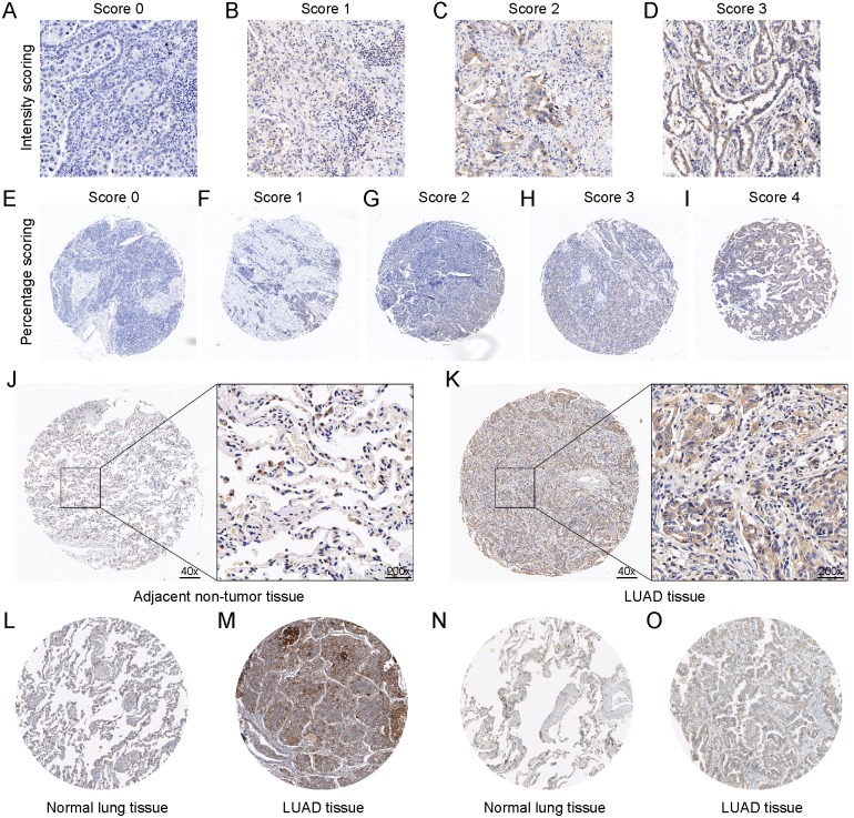
Figure 2. The protein expression of DSG2 in LUAD tissues and non-tumor tissues.
The scoring system of the tissue microarray was displayed. A final histological overall score was calculated by the multiplication of the intensity score (A–D) and percentage score (E–I). Representative images of IHC staining of DSG2 in 60 adjacent non-tumor tissues (J) and 62 LUAD tissues (K). DSG2 protein expression was searched from the Human Protein Atlas database. CAB025122 antibody was used in normal lung tissue (L) and LUAD tissue (M). HPA004896 antibody was used in normal lung tissue (N) and LUAD tissue (O).

