A 58 year-old man presented with 6 months of fatigue, weight loss, and progressive weakness. On examination he had intrinsic hand and right lower extremity weakness, which became widespread after 6 months, resulting in grade 3–4 strength in most limb muscles. There was no history of trauma, statin use, heavy exertion, or myalgias. Sensation and reflexes were normal. Bulbar abnormalities were absent. Nerve conduction studies are shown in table 1. Needle electromyography (EMG) showed fibrillation potentials in proximal and distal muscles of the right upper and lower extremities, and cervical, thoracic, and lumbar paraspinal muscles. There were fasciculation potentials in the soleus, biceps femoris, deltoid and lumbar paraspinal muscles. All muscles had high amplitude, long duration, polyphasic motor unit potentials with decreased recruitment. Initial creatine kinase (CK) was 353 units/liter (Ref 0–200 U/L), rising to 1,905 U/L at six months. Weakness progressed over the following year, causing repeated falls and inability to perform activities of daily living.
Table 1:
Nerve conduction studies at 6 months
| Sensory | |||||
| Nerve/site | Peak Latency (ms) | Amplitude (uV) | Ref Amplitude (uV) | Onset velocity (m/s) | Ref Velocity (m/s) |
| Right median- digit II | |||||
| Mid palm | 2.9 | 43.7 | > 45 | 41.5 | > 45 |
| Wrist | 4.5 | 14.3 | > 13 | 45.3 | > 45 |
| Right ulnar-digit V | |||||
| Wrist | 3.7 | 17.9 | > 13 | 49.7 | > 45 |
| Right sural-ankle | |||||
| Calf | 5.1 | 8 | > 5 | 36.9 | > 40 |
| Motor studies | |||||
| Nerve/site | Latency (ms) | Ref Latency (ms) | Amplitude (mV) | Ref Amplitude (mV) | Velocity (Ref Velocity m/s) |
| Right median-APB | |||||
| Wrist | 6.1 | < 4.2 | 3.7 | > 4.5 | |
| Elbow | 12.1 | 3.2 | > 4.5 | 39.2 (> 43) | |
| Right ulnar-ADM | |||||
| Wrist | 3.2 | < 3.9 | 6.7 | > 4.5 | |
| Below elbow | 7.2 | 6.6 | > 4.5 | 58.1 (> 50) | |
| Above elbow | 10.3 | 6.5 | > 4.5 | 56.9 (> 50) | |
| Right peroneal-EDB | |||||
| Ankle | 4.5 | < 5 | 1.8 | > 2.5 | |
| Fib head | 14.8 | 1.7 | > 2.5 | 38 (> 40) | |
| Pop fossa | 17.3 | 1.6 | 38 (> 39) | ||
| Right tibial-AH | |||||
| Ankle | 4.5 | < 6 | 2.2 | > 2.5 | |
Ms: millisecond
uV: microvolt
m/s: meters/second
The patient underwent muscle biopsy of the left vastus lateralis that showed marked variation in fiber size and shape with clusters of small angulated fibers on hematoxylin and eosin stain. ATPase staining revealed fiber type grouping and esterase stain showed dark cytoplasmic staining of the small angulated fibers, indicating denervation. There was no fiber necrosis, inflammation, or ragged red fibers. By 7 months from initial presentation, the patient demonstrated pathologically brisk reflexes meeting El Escorial criteria for probable amyotrophic lateral sclerosis (ALS)1 and a CK level of 1,699 U/L.
We aimed to determine the distribution of CK levels in ALS patients using the publicly available Pooled Resource Open-Access ALS Clinical Trials (PRO-ACT) database which contains over 8 million data points for 8,600 ALS patients previously enrolled in clinical trials.2 SPSS v25 (IBM, Armonk, NY) was used for all analyses.
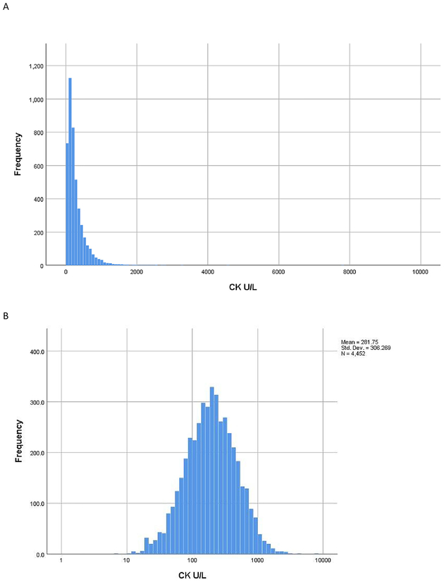
4,452 unique subjects with ALS had 39,249 CK tests [initial visit overall mean (SD) = 282 (306) U/L, median (IQ range) = 195 (106–353) U/L; all CKs from all visits mean (SD) = 280 (282) U/L, median (IQ range) = 195 (106–354) U/L]. Values were logarithmically distributed (Figure 1). 51.4% of patients had normal CK, and 97% had CK less than 1,000 U/L. 0.3% had CK over 2,000 U/L. CK was not correlated with age (Spearman’s rho, n=4,276, r=0.02, p=0.20), weight (n=2,404, r=0.02, p=0.34), riluzole use (mean rank 2,065 for 2,847 patients on riluzole; 2,104 for 1,307 patients not on riluzole; p=0.34), or sex (mean rank 2,244 for 1,709 women; 2,743 for 2,743 men; p=0.47).
Figure 1:
A. Arithmetic distribution of creatine kinase in 4,452 ALS patients. B. Logarithmic distribution of creatine kinase in 4,452 ALS patients
While most patients with ALS have normal or mildly elevated CK, some individuals have levels over 2,000 U/L - values not found on a literature search.3,4,5,6 ALS has been mistaken for polymyositis due to high CK, resulting in unnecessary steroid use.6 Possibly, unpublished cases exist. Elevated aspartate aminotransferase (AST) is seen with muscle breakdown, correlating with CK levels, and can lead to extensive studies including liver biopsy in patients with muscle disease. In the ALS population, elevated AST may be inappropriately attributed to riluzole toxicity.7, 8
Evidence is conflicting whether CK is predictive of disease progression in ALS.3,4,5,9 CK correlates with muscle loss and cramping, which is hypothesized to lead to muscle breakdown.4, 10 CK also corresponds to the degree of abnormal spontaneous activity on EMG in ALS, suggesting that high CK results from neurogenic denervation of muscles and altered membrane permeability.4 Using this PRO-ACT database, studies found that baseline CK does not predict prognosis2, but the rate of change does, such that CK declines more rapidly in subjects with faster clinical progression.9
In summary, we highlight the wide distribution of CK in ALS. Elevated CK alone should not prompt muscle biopsy unless clinical data raises questions regarding an isolated motor neuron disorder. Future studies are needed to better elucidate the clinical and pathologic significance of CK in ALS.
Abbreviations
- CK
Creatine kinase
- U/L
Units/Liter
- ALS
amyotrophic lateral sclerosis
- PRO-ACT
Pooled Resource Open-Access ALS Clinical Trials
- SPSS
Statistical Package for the Social Sciences
Footnotes
We confirm that we have read the Journal’s position on issues involved in ethical publication and affirm that this report is consistent with those guidelines.
We did not perform any research on human or animal subjects as a part of this article. All information regarding our patient has been de-identified.
None of the authors has any conflicts of interest to disclose.
References
- 1.Brooks BR. El escorial World Federation of Neurology criteria for the diagnosis of amyotrophic lateral sclerosis. Journal of the Neurological Sciences 1994; 124: 96–107. [DOI] [PubMed] [Google Scholar]
- 2.Atassi N, et al. The PRO-ACT database. Neurology 2014; 83: 1719–1725 [DOI] [PMC free article] [PubMed] [Google Scholar]
- 3.Rafiq MK, Lee E, Bradburn M, McDermott CJ, Shaw PJ. Creatine kinase enzyme level correlates positively with serum creatinine and lean body mass, and is a prognostic factor for survival in amyotrophic lateral sclerosis. Eur J Neurol. 2016; 23: 1071–1078. [DOI] [PubMed] [Google Scholar]
- 4.Tai H et al. Creatine kinase level and its relationship with quantitative electromyographic characteristics in amyotrophic lateral sclerosis. Clinical Neurophysiology. 2018; 129: 926–930. [DOI] [PubMed] [Google Scholar]
- 5.Chahin N and Sorenson EJ Serum creatine kinase levels in spinobulbar muscular atrophy and amyotrophic lateral sclerosis. Muscle Nerve. 2009; 40: 126–129 [DOI] [PubMed] [Google Scholar]
- 6.Harrington TM, Cohen MD, Bartleson JD and Ginsburg WW Elevation of creatine kinase in amyotrophic lateral sclerosis. Arthritis & Rheumatism. 1983; 26: 201–205. [DOI] [PubMed] [Google Scholar]
- 7.Weibrecht K, Dayno M, Darling C, Bird SB. Liver aminotransferases are elevated with rhabdomyolysis in the absence of significant liver injury. J Med Toxicol. 2010;6(3):294–300. doi: 10.1007/s13181-010-0075-9 [DOI] [PMC free article] [PubMed] [Google Scholar]
- 8.Mathur T 1, Manadan AM, Thiagarajan S, Hota B, Block JA. Serum transaminases are frequently elevated at time of diagnosis of idiopathic inflammatory myopathy and normalize with creatine kinase. J Clin Rheumatol. 2014. April;20(3):130–2. doi: 10.1097/RHU.0000000000000038. [DOI] [PubMed] [Google Scholar]
- 9.Ong ML, Tan PF, Holbrook JD. Predicting functional decline and survival in amyotrophic lateral sclerosis. PLoS ONe. 2017; 13: 1–16. 7 [DOI] [PMC free article] [PubMed] [Google Scholar]
- 10.Gibson SB, Kasarskis EJ, Hu N, et al. Relationship of creatine kinase to body composition, disease state, and longevity in ALS. Amyotroph Lateral Scler Frontotemporal Degener. 2015;16(7–8):473–477. doi: 10.3109/21678421.2015.1062516 8 [DOI] [PMC free article] [PubMed] [Google Scholar]


